. 2024 Aug 31;15(1):7574.
doi: 10.1038/s41467-024-52045-7.

Structural basis for CCR6 modulation by allosteric antagonists

Affiliations

Structural basis for CCR6 modulation by allosteric antagonists

David Jonathan Wasilko et al. Nat Commun. .

Abstract

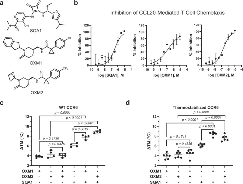
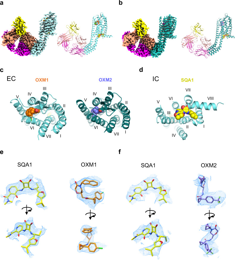
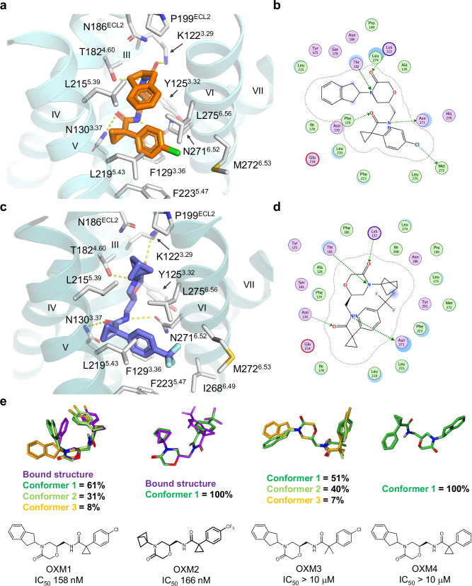
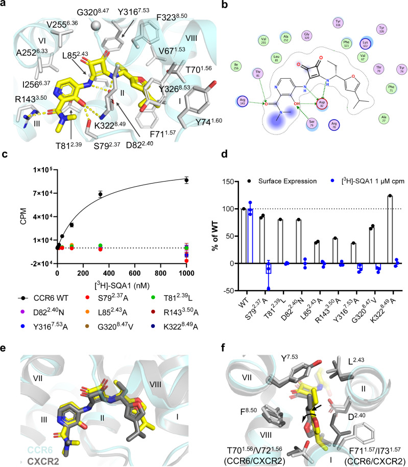
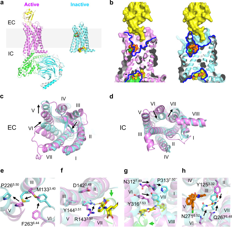
The CC chemokine receptor 6 (CCR6) is a potential target for chronic inflammatory diseases. Previously, we reported an active CCR6 structure in complex with its cognate chemokine CCL20, revealing the molecular basis of CCR6 activation. Here, we present two inactive CCR6 structures in ternary complexes with different allosteric antagonists, CCR6/SQA1/OXM1 and CCR6/SQA1/OXM2. The oxomorpholine analogues, OXM1 and OXM2 are highly selective CCR6 antagonists which bind to an extracellular pocket and disrupt the receptor activation network. An energetically favoured U-shaped conformation in solution that resembles the bound form is observed for the active analogues. SQA1 is a squaramide derivative with close-in analogues reported as antagonists of chemokine receptors including CCR6. SQA1 binds to an intracellular pocket which overlaps with the G protein site, stabilizing a closed pocket that is a hallmark of inactive GPCRs. Minimal communication between the two allosteric pockets is observed, in contrast to the prevalent allosteric cooperativity model of GPCRs. This work highlights the versatility of GPCR antagonism by small molecules, complementing previous knowledge of CCR6 activation, and sheds light on drug discovery targeting CCR6.


Conflict of interest statement

All authors were either employees of Pfizer Inc. or were employees of Sosei Heptares at the time the work was performed.

Figures

Fig. 1
Fig. 1. SQA and OXM analogues inhibit CCL20-mediated activation of CCR6 and display additive binding stabilization to the receptor.
a Chemical structures of SQA1, OXM1 and OXM2. b Inhibition of CCL20-mediated CCR6+ human T-cell chemotaxis demonstrated by SQA1 (pIC50 = 6.7 ± 0.8), OXM1 (pIC50 = 6.8 ± 0.2), and OXM2 (pIC50 = 6.78 ± 0.11). Data are presented as mean ± s.d. from n = 3 independent experiments. c,d Thermal shifts of c, WT and d, thermostabilized CCR6 Nα7.1 from Apo to liganded conditions as indicated. Data are presented as mean ± s.d. from n = 4 (WT) and n = 6 (Nα7.1) independent experiments. p value presented in c and d is two-sided from unpaired t test, p < 0.05 was considered significant. Source data are provided as a Source Data file.
Fig. 2
Fig. 2. Overall structures of CCR6 in complex with OXM and SQA analogues.
a,b Cryo-EM map (left) and model (right) of CCR6 complex with anti-BRIL Fab and anti-Fab Nb bound to a OXM1 (orange carbon spheres) and SQA1 (yellow carbon spheres), or b OXM2 (slate carbon spheres) and SQA1 (yellow carbon spheres). Protein is coloured by subunit as follows: Fab heavy chain, magenta; Fab light chain, salmon; Nb, yellow; CCR6, aquamarine in a and deep teal in b. c Extracellular (EC) view of CCR6 bound to OXM analogues, OXM1 (left) and OXM2 (right). d Intracellular (IC) view of CCR6 bound to the SQA analogue, SQA1. Both c and d use the same colour code as a and b. e SQA1, OXM1, and f SQA1, OXM2 fits into the cryo-EM density maps of the two structures from two viewing angles.
Fig. 3
Fig. 3. Structures of OXM analogues bound to CCR6 and comparison to solution conformations.
a Binding pocket of OXM1 (orange carbon sticks). b Protein-ligand interaction diagram for OXM1 prepared with Molecular Operating Environment (MOE). c Binding pocket of OXM2 (slate carbon sticks). d Protein-ligand interaction diagram for OXM2 prepared with MOE. In a and c, CCR6 protein is shown in aquamarine and deep teal ribbons, respectively. Side chains interacting with the small molecules are shown as white carbon sticks. Hydrogen bonds are highlighted by yellow dashed lines. In b and d, OXM pocket residues are presented as follows: polar residues in pink, hydrophobic residues in green, acidic residues with a red contour ring, basic residues with a blue contour ring. Green dotted arrows indicate hydrogen and halogen bonds mediated by side chains that contribute to ligand binding. Ligand atoms exposed to environment are shaded in blue according to degrees of exposure, scaled by size. Light-blue halos around residues indicate the degree of interaction with ligand, scaled by size. The dotted contour around the ligand reflects steric room for methyl substitution. e Chemical structures of different OXM analogues and their solution conformations were determined by NMR using residual dipolar couplings (RDC). Three most populated conformations are illustrated by dark green, light green, and orange carbon sticks, respectively. The bound conformations of OXM1 and OXM2 are shown as purple carbon sticks in the overlays for comparison. The IC50 value of each OXM analogue determined in the CCL20-mediated human CCR6+ T-cell chemotaxis assay is shown under the corresponding chemical structure. Solution conformer library files of the OXM analogues are provided as Source Data files.
Fig. 4
Fig. 4. SQA1 binds to an intracellular pocket.
a Structure of SQA1 (yellow carbon sticks) bound to CCR6 (aquamarine ribbon) from the higher-resolution CCR6/SQA1/OXM1 cryo-EM structure. Side chains interacting with SQA1 are shown by white carbon sticks. Hydrogen bonds are highlighted by yellow dashed lines. b Protein-ligand interaction diagram for SQA1 prepared with MOE. Definitions of the schematic representation are same as that in Fig. 3b, d. c Saturation binding curves of [3H]-SQA1 on CCR6 WT and mutants as indicated. Binding of [3H]-SQA1 on WT provided a KD of 250 ± 22 nM. Data shown as mean ± s.d. are from n = 3 independent experiments. d Expression are confirmed for all mutants transiently expressed in HEK293 cells. Normalized surface expression of mutants to WT are shown by black circles and columns. CPM (counts per minute) of [3H]-SQA1 measured at 1,000 nM ligand concentration from c normalized to WT are shown as mean ± s.d. from n = 3 independent experiments (blue circle and columns). e Overlay of CCR6 and CXCR2 (PDB ID 6LFL) inactive structures bound by SQA1. f Major differences of the binding pockets in CCR6 and CXCR2 SQA1-bound structures. Side chains of pocket residues are shown in sticks. In e and f, CXCR2 and SQA1 from 6LFL are shown by dark grey ribbon and dark grey sticks. CCR6 structure is shown by the same cartoon representations as in a. Source data are provided as a Source Data file.
Fig. 5
Fig. 5. Different mechanisms of allosteric antagonism of CCR6 by OXM and SQA.
a Overall structures of the active CCR6 (magenta, left, PDB ID 6WWZ) in complex with its cognate chemokine ligand CCL20 (yellow) and a heterotrimeric G protein coloured by subunit (Gαo, green, Gβ, cyan, and Gγ, light grey), and the inactive CCR6 (aquamarine, right) in complex with SQA1 (yellow carbon spheres) and OXM1 (orange carbon spheres). The light grey box highlights the location of cell membranes. b Overlays of CCL20, OXM1, and SQA1 bound structures onto the active (left) and the inactive (right) CCR6 structures. Structures are presented in the same colour codes as in a. Blue lines depict pockets revealed by the structures. c–d, Structural overlays of the active (magenta) and inactive (aquamarine) CCR6 structures from c EC and d IC views. eh, Conformational changes of conserved functional motifs, e P2265.50-M1333.40-F2636.44, f D1423.49-R1433.50-Y1443.51, g N3127.49-P3137.50-x-x-Y3167.53, and h Y1253.32-Q2676.48-N2716.52 revealed by structural overlay of active (magenta) and inactive (aquamarine) CCR6. In ch significant TM movements and side chain conformational changes from active to inactive states are highlighted by black arrows.

References

    1. Hughes, C. E. & Nibbs, R. J. B. A guide to chemokines and their receptors. FEBS J.285, 2944–2971 (2018). 10.1111/febs.14466 - DOI - PMC - PubMed
    1. Bonecchi, R. & Graham, G. J. Atypical chemokine receptors and their roles in the resolution of the inflammatory response. Front Immunol.7, 224 (2016). 10.3389/fimmu.2016.00224 - DOI - PMC - PubMed
    1. Murdoch, C. & Finn, A. Chemokine receptors and their role in inflammation and infectious diseases. Blood95, 3032–3043 (2000). 10.1182/blood.V95.10.3032 - DOI - PubMed
    1. Chow, M. T. & Luster, A. D. Chemokines in cancer. Cancer Immunol. Res. 2, 1125–1131 (2014). 10.1158/2326-6066.CIR-14-0160 - DOI - PMC - PubMed
    1. Murphy, P. M. et al. International union of pharmacology. XXII. Nomenclature for chemokine receptors. Pharm. Rev.52, 145–176 (2000). - PubMed

LinkOut - more resources