. 2024 Aug 31;14(1):20278.
doi: 10.1038/s41598-024-71159-y.

Pleiotropy in FOXC1-attributable phenotypes involves altered ciliation and cilia-dependent signaling

Affiliations

Pleiotropy in FOXC1-attributable phenotypes involves altered ciliation and cilia-dependent signaling

Serhiy Havrylov et al. Sci Rep. .

Abstract

Alterations to cilia are responsible for a wide range of severe disease; however, understanding of the transcriptional control of ciliogenesis remains incomplete. In this study we investigated whether altered cilia-mediated signaling contributes to the pleiotropic phenotypes caused by the Forkhead transcription factor FOXC1. Here, we show that patients with FOXC1-attributable Axenfeld-Rieger Syndrome (ARS) have a prevalence of ciliopathy-associated phenotypes comparable to syndromic ciliopathies. We demonstrate that altering the level of Foxc1 protein, via shRNA mediated inhibition, CRISPR/Cas9 mutagenesis and overexpression, modifies cilia length in vitro. These structural changes were associated with substantially perturbed cilia-dependent signaling [Hedgehog (Hh) and PDGFRα], and altered ciliary compartmentalization of the Hh pathway transcription factor, Gli2. Consistent with these data, in primary cultures of murine embryonic meninges, cilia length was significantly reduced in heterozygous and homozygous Foxc1 mutants compared to controls. Meningeal expression of the core Hh signaling components Gli1, Gli3 and Sufu was dysregulated, with comparable dysregulation of Pdgfrα signaling evident from significantly altered Pdgfrα and phosphorylated Pdgfrα expression. On the basis of these clinical and experimental findings, we propose a model that altered cilia-mediated signaling contributes to some FOXC1-induced phenotypes.

Keywords: Axenfeld–Rieger syndrome; FOXC1; Hedgehog; PDGFRα signaling; Primary cilia.


Conflict of interest statement

The authors declare no competing interests.

Figures

Fig. 1
Fig. 1
Pleiotropic and variable phenotypes are consistent with ciliary dysfunction. (A) Skeletal phenotypes present in the cohort of patients with FOXC1 mutation or copy number variation include scoliosis and pre-axial polydactyly (arrowhead); the duplicated second phalanx of the left thumb was previously surgically removed. The mutation present in this individual (p.D117Tfs64) is predicted to result in loss of two thirds of the FOXC1 protein. (B) CNS phenotypes include cerebellar vermis hypoplasia, posterior fossa enlargement, and lateral ventricular dilation: example from an individual with a missense mutation (p.S82T). Images showing normal cerebellar and ventricular morphology, are outlined in green. (C) Ocular anomalies include irregular iris sphincter width (red arrow), posterior embryotoxon extending through 3 clock hours (green arrow), and asymmetric irides. Normal iris anatomy for comparison (green box).
Fig. 2
Fig. 2
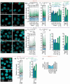
Altered levels of Foxc1 are associated with changes to cilia length in three cell types. (A) Representative images of Arl13b/γ-tubulin ciliary staining in NIH3T3 cells expressing a Foxc1-targeting shRNA, or with Foxc1 overexpression (OE) (non-targeting shRNA control: non-trg.; vector controls: pLKO.1, pLXSH). These reveal shortening of the cilium (red) with shRNA inhibition, and the converse with OE. (B) Quantification of cilia length as fold change relative to vector controls from independent experiments (shRNA: n = 6; OE: n = 7). (C) Prevalence of short (blue bars; length ≤ lower quartile of controls) and long cilia (green bars; length ≥ median of controls); see Supplemental Fig. 1 for details. (DF) In IMCD3 cells, Foxc1 knock-down and OE respectively reduce and increase cilia length; graphs depict fold change in cilia length and prevalence of short and long cilia (shRNA, OE: n = 1). (GI) In ATDC5 cells, the decreased cilia length induced by CRISPR mutagenesis of Foxc1 is attributable to an increased proportion of cells with short cilia (n = 4). (J) In NIH3T3 cells, Foxc1 protein levels (relative to WT) are reduced ~ 2 fold (40–61%) by Foxc1-targeting shRNAs (n = 1). [Statistical analyses: boxplots—Dunn’s (post hoc Kruskal–Wallis) test; barplots—nested ANOVA (C shRNA), one-way ANOVA (C Foxc1 OE, I)].
Fig. 3
Fig. 3
Altered levels of Foxc1 induce aberrant ciliary Hedgehog signaling. (AD) In NIH3T3 cells, quantitative Western Immunoblots demonstrate that shRNA inhibition of Foxc1 decreases the basal level of Gli1 expression, while Foxc1 overexpression has a converse effect. (B,C) Reduced Foxc1 expression leads to twofold decrease in Gli1 protein levels [measured in serum-starved cells stimulated with 400 nM Smoothened agonist (SAG), for 20 h; n = 1 for each of the four shRNAs]. (D) Increased Foxc1 expression results in ~ 2 fold increase in the basal level of Gli1 protein (n = 4). (E) In immortalised E16.5 chondrocytes, that express high levels of Foxc1, Foxc1 shRNA inhibition decreased basal level of Gli1 mRNA (n = 3). (F) Cells overexpressing Foxc1 exhibit increased basal levels of Gli1, Gli2 and Ptch1 mRNA (n = 3). (G) Immunoblots demonstrate that overexpressing Foxc1 induces faster accumulation of Gli1 protein in serum-starved NIH3T3 cells. Quantification reveals significantly increased Gli1 protein levels for Foxc1 OE at 3 and 6 h (n = 3). [Statistical analyses: Tukey HSD test post hoc one-way (AF) or two-way ANOVA (G)].
Fig. 4
Fig. 4
Foxc1 alters the dynamics of Hh signaling and enhances Gli2 accumulation at the ciliary tip. (A) Immunoblots demonstrate that overexpressing Foxc1 induces faster accumulation of Gli1. Quantification in serum-starved Gli2-mGFP-expressing NIH3T3 cells reveals substantially higher Gli1 protein levels with Foxc1 OE: 14-, 20- and 26-fold increases vs non-treated wild-type control at 0, 3 and 6 h (n = 1). Note the progressive accumulation of Gli1 protein in response to SAG treatment. (B) Images illustrate the increased accumulation of Gli2-mGFP at the axonemal tips of Foxc1 OE cells, including after stimulation with SAG. Orientation of primary cilia, from basal body (γ-tubulin staining) to axonemal tip (Gli2-mGFP), is depicted by the red arrow. (C) Axonemal tip Gli2-mGFP signal intensity, expressed as fold change relative to pLXSH control, is significantly increased by Foxc1 OE. The prevalence of high and low Gli2-mGFP signal intensity in 7 independent experiments is also significantly altered compared to control [high Gli2-mGFP signal intensity ≥ upper quartile, green bar; low ≤ lower quartile of control, blue]. [Statistical analysis: boxplots—Dunn’s (post hoc Kruskal–Wallis) test; barplots—Tukey HSD post hoc one-way ANOVA].
Fig. 5
Fig. 5
Altered levels of Foxc1 impact PDGFRα signaling. (AC) Foxc1 shRNA inhibition significantly reduced the level of total Pdgfrα, and auto-phosphorylated pY754-Pdgfrα in serum-starved NIH3T3 cells. This effect was observed both in the presence and absence of Pdgf-AA ligand stimulation (n = 1 for each of the four shRNAs). (DF) Foxc1 overexpression induced comparable reductions in total Pdgfrα, and pY754-Pdgfrα levels, indicative of impaired cilia-mediated Pdgf signaling (n = 3). [Statistical analyses: Tukey HSD post hoc one-way (B,C) or two-way (E,F) ANOVA. Statistical analysis in panels (B) and (C) shows data pooled for all shRNAs].
Fig. 6
Fig. 6
Reduced cilia length in meningeal cells cultured from Foxc1 mutant embryos. (A) Representative images of Arl13b/γ-tubulin staining in primary meningeal cell cultures from Foxc1ΔTg(EIIa-cre) and wild-type sibling E15.5 embryos. Note the shortening of the cilium (red), most apparent in the homozygous mutant (Foxc1ΔΔ) compared to controls. (B) Quantification of cilia length in primary cultures of meninges isolated from 4 litters comprising 6 Foxc1+/+, 16 Foxc1+/Δ and 5 Foxc1Δ/Δ embryos. (C) Mean cilia length is reduced by 8% and 10% in Foxc1+/Δ and Foxc1Δ/Δ relative to wild-type cultures. This is attributable to a decreased proportion of longer cilia, as demonstrated by the barplot on the right depicting the prevalence of short (blue bars; length ≤ lower quartile of controls) and long cilia (green bars; length ≥ median of controls). [Statistical analysis: one-way ANOVA].
Fig. 7
Fig. 7
Dysregulated Hedgehog signaling in the meninges of Foxc1 mutant embryos. (AC) Western immunoblot analysis demonstrates that meningeal expression of core Hh components is dysregulated by Foxc1 mutation. (A) Gli1 protein expression is decreased in Foxc1+/Δ heterozygotes, and almost entirely lost in Foxc1Δ/Δ meninges. (B) Foxc1 mutation is associated with increased expression of full length Gli3, and its increased processing into the C-terminally truncated repressor form (Gli3R). (C) Expression and phosphorylation of Sufu, a core Hh pathway inhibitor, is also increased with Foxc1 mutation. [Quantification above each gel: fold change in signal, normalized to actin, relative to wild-type. Samples pooled from n = 18 embryos (7 Foxc1+/+; 8 Foxc1+/Δ, 3 Foxc1Δ/Δ].
Fig. 8
Fig. 8
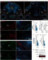
Decreased Pdgfrα expression in the meninges of Foxc1 mutant embryos. (A) At E14.5, Pdgfrα immunofluorescence is decreased in the forebrain meninges of Foxc1-null embryos. (B) This is particularly apparent in the meninges lining the anterior cranial fossa [insets from (A)]. (C,D) The mean Pdgfrα signal intensity in the forebrain meninges is decreased by 50% (P = 2.6 × 10−4). Consistently, RNA-sequencing demonstrates significantly reduced meningeal Pdgfra mRNA expression [27% mean FPKM decrease, q = 6.4 × 10−5], while in contrast Pdgfrb expression is unaltered. (E) E14.5 Foxc1/ embryos exhibited comparable reductions in forebrain meningeal pY754 Pdgfrα expression. (F) Quantification confirms the significant decrease in the mean signal intensity of the active phosphorylated form of Pdgfrα, and comparable reductions in the level of the tight junction protein Zo1, relative to wild-type embryos. (H) Western immunoblots of meningeal tissue lysates reveal similar reductions in total Pdgfrα levels to those observed by immunofluorescence (replicates as in Fig. 7). [Panels 8C, F, G: 6 Foxc1+/+, 6 Foxc1/ embryos; 8D: 5 Foxc1+/+, 4 Foxc1Δ/Δ embryos]. [Mgs meninges, LV lateral ventricle, ChP choroid plexus, FC mesenchymal condensation forming falx cerebri. Statistical analyses: one-way ANOVA; FPKM Fragments Per Kilobase of transcript, per Million mapped reads].
Fig. 9
Fig. 9
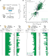
Similar transcriptional changes in the meninges of Foxc1 and Pdgf pathway mutant mice. (A) Experimental design for analysis of differentially expressed genes in the meninges of Foxc1Δ/Δ embryos (4 Foxc1Δ/Δ, 5 Foxc1+/+) and mice with severely perturbed Pdgfrα signaling (Pdgfc/; PdgfraGFP/+). (B) The majority of genes dysregulated in the Pdgf mutant are similarly up- or down-regulated in Foxc1 mutant embryos; a correlation that extends to 1193 of 1324 genes; correlation coefficient ρ = 0.77, P = 2.2 × 10−16. (C) Restricting analysis to the 50 most upregulated, and 50 most downregulated genes, makes the similar differential expression evident: more than 80% of these most dysregulated genes in Pdgfc/; PdgfraGFP/+ mice are also significantly dysregulated in the meninges of Foxc1Δ/Δ embryos.

References

    1. Katsanis, N. et al. Triallelic inheritance in Bardet–Biedl syndrome, a Mendelian recessive disorder. Science293(5538), 2256–2259 (2001). 10.1126/science.1063525 - DOI - PubMed
    1. Zaghloul, N. A. et al. Functional analyses of variants reveal a significant role for dominant negative and common alleles in oligogenic Bardet–Biedl syndrome. Proc. Natl. Acad. Sci. USA107(23), 10602–10607 (2010). 10.1073/pnas.1000219107 - DOI - PMC - PubMed
    1. Huangfu, D. et al. Hedgehog signalling in the mouse requires intraflagellar transport proteins. Nature426(6962), 83–87 (2003). 10.1038/nature02061 - DOI - PubMed
    1. Schneider, L. et al. PDGFRalphaalpha signaling is regulated through the primary cilium in fibroblasts. Curr. Biol.15(20), 1861–1866 (2005). 10.1016/j.cub.2005.09.012 - DOI - PubMed
    1. Simons, M. et al. Inversin, the gene product mutated in nephronophthisis type II, functions as a molecular switch between Wnt signaling pathways. Nat. Genet.37(5), 537–543 (2005). 10.1038/ng1552 - DOI - PMC - PubMed

MeSH terms

Substances

Supplementary concepts