An official website of the United States government
The .gov means it’s official.
Federal government websites often end in .gov or .mil. Before
sharing sensitive information, make sure you’re on a federal
government site.
The site is secure.
The https:// ensures that you are connecting to the
official website and that any information you provide is encrypted
and transmitted securely.
PARP inhibitors (PARPi) show selective efficacy in tumors with homologous recombination repair (HRR)-defects but the activation mechanism of HRR pathway in PARPi-treated cells remains enigmatic. To unveil it, we searched for the mediator bridging PARP1 to ATM pathways by screening 211 human ubiquitin-related proteins. We discovered TRIM44 as a crucial mediator that recruits the MRN complex to damaged chromatin, independent of PARP1 activity. TRIM44 binds PARP1 and regulates the ubiquitination-PARylation balance of PARP1, which facilitates timely recruitment of the MRN complex for DSB repair. Upon exposure to PARPi, TRIM44 shifts its binding from PARP1 to the MRN complex via its ZnF UBP domain. Knockdown of TRIM44 in cells significantly enhances the sensitivity to olaparib and overcomes the resistance to olaparib induced by 53BP1 deficiency. These observations emphasize the central role of TRIM44 in tethering PARP1 to the ATM-mediated repair pathway. Suppression of TRIM44 may enhance PARPi effectiveness and broaden their use even to HR-proficient tumors.
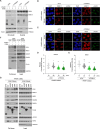
Screening of ubiquitin-related proteins (URPs) at damaged chromatin. ( A ) Schematic illustrating…
Figure 1.
Screening of ubiquitin-related proteins (URPs) at damaged chromatin. (A) Schematic illustrating the FokI-screening process for 211 GFP-tagged human URPs. (B) Co-localization of GFP-tagged URPs and mCherry-FokI nuclease at DSB sites. Representative images indicate mCherry-FokI-positive human URPs associated with DDR (left panel). URPs are categorized as either previously uncharacterized (red; 16 out of 211) or known (black; 11 out of 211) in their association with DDR (right panel). Nuclei were counterstained with DAPI. (C) Identification of DDR-related URPs possessing evolutionarily conserved motifs. (D) Classification of the 24 URPs based on regulation by either PARP1 or ATM activity. (E–G) Visualization of TRIMs' accumulation at DNA break sites post-laser microirradiation. U2OS cells, transfected with GFP-TRIMs underwent laser microirradiation. Pre-treatments included control DMSO (E), PARP1 inhibitor PJ34 at 5 μM (F), and ATM inhibitor KU55933 at 10 μM (G) for 1 h before microirradiation. Cells were fixed using 4% paraformaldehyde 10 min post-microirradiation. The co-localization frequency of GFP-TRIMs with DNA damage marker γH2AX at the laser-irradiated regions is depicted in each panel. Scale bars, 5 μm.
Figure 2.
Domain analysis of TRIM44 binding…
Figure 2.
Domain analysis of TRIM44 binding to PARP1. ( A ) U2OS 2–6–5 cells…
Figure 2.
Domain analysis of TRIM44 binding to PARP1. (A) U2OS 2–6–5 cells were transfected with GFP-TRIM44 and treated with 4-OHT (1 μM) and shield1 (1 μM) for 4 h to induce DNA damage. After induction, cells were fixed stained with TRIM44. (B) U2OS 2–6–5 cells were transfected with GFP-TRIM44, pretreated with DMSO, ATM inhibitor or PARP inhibitor for 1 h and treated with 4-OHT (1 μM) and shield1 (1 μM) for 4 h to induce DNA damage. After induction, cells were fixed. (C) 293T cells were transfected empty vector or SFB-TRIM44. Cells were treated with zeocin (100 μg/ml) for 1 h to induce DSBs. Cells were lysed, subjected to Streptavidin beads pulldown assay, and analyzed by immunoblotting with the indicated antibodies. (D) Schematic illustration of domain structure of human TRIM44. (E) 293T cells were transfected with the indicated combinations of SFB-tagged TRIM44 truncated mutants or SFB vector. Cells were treated with zeocin (300 μg/ml) for 1 h to induce DSBs. Cells were lysed, subjected to Streptavidin beads pull-down assay, and analyzed by immunoblotting with the indicated antibodies. (F) U2OS 2–6–5 cells were transfected with GFP-TRIM44 WT and GFP-TRIM44 ΔGR and treated with 4-OHT (1 μM) and shield1 (1 μM) for 4 h to induce DNA damage. After DSBs induction, cells were fixed. Fixed cells image (upper) and graph (lower). Scale bar, 10 μm. Student's t-test was used for statistical analysis.
Figure 3.
Correlation between PARP1 hyperactivation and…
Figure 3.
Correlation between PARP1 hyperactivation and its ubiquitination. ( A ) U2OS cells were…
Figure 3.
Correlation between PARP1 hyperactivation and its ubiquitination. (A) U2OS cells were transfected with the indicated siRNAs and pretreated with DMSO and PARP inhibitor (AG14361). Cells were treated with H2O2 (1 mM) for indicated time-points to induce DNA damage and whole-cell extracts were analyzed by immunoblotting with the indicated antibodies. (B) U2OS cells were transfected with indicated siRNAs and subjected to laser microirradiation. After damaged, cells were fixed and co-stained with PAR and γH2AX. Scale bar, 10 μm. Student's t-test was used for statistical analyses. (C) U2OS TRIM44 KO cells were transfected with the indicated combinations of SFB-tagged TRIM44 truncated mutants. Cells were treated with H2O2 (1 mM) for indicated time-points to induce DNA damage and whole-cell extracts were analyzed by immunoblotting with the indicated antibodies. (D) U2OS cells were transfected with the indicated siRNAs and HA-tagged ubiquitin. Cells were treated with H2O2 (1 mM) for indicated time-points to induce DNA damage and immunoprecipitated (IP) with a PARP1 antibody. Cells were analyzed by immunoblotting with the indicated antibodies. (E) U2OS cells were transfected with the indicated combinations of GFP-tagged TRIM44 truncated mutants and HA-tagged ubiquitin. Cells were treated with H2O2 (1 mM) for 5min to induce DNA damage and immunoprecipitated (IP) with a PARP1 antibody. Cells were analyzed by immunoblotting with the indicated antibodies. (F) U2OS cells were transfected with the indicated siRNAs and HA-tagged ubiquitin. Cells were treated with H2O2 (1 mM) for indicated time to induce DNA damage and immunoprecipitated (IP) with a PARP1 antibody. Cells were analyzed by immunoblotting with the indicated antibodies.
Figure 4.
Effect of TRIM44 on the…
Figure 4.
Effect of TRIM44 on the accumulation of the MRN complex to DNA lesions.…
Figure 4.
Effect of TRIM44 on the accumulation of the MRN complex to DNA lesions. (A) U2OS cells were co-transfected with indicated siRNAs and pcDNA or GFP-TRIM44 WT. Cells were treated with zeocin (300 μg/ml) and subjected to whole-cell extraction. (B) U2OS TRIM44 WT or KO cells were treated with zeocin (300 μg/ml) for 1 h to induce DSBs and subjected to chromatin fraction. (C) U2OS TRIM44 KO (KO) cells were transfected with the indicated combinations of SFB-tagged TRIM44 truncated mutants and SFB empty vector. Cells were treated with zeocin (300 μg/ml) for 1 h to induce DSBs. Cells were lysed, subjected to chromatin fraction, and analyzed by immunoblotting with the indicated antibodies. (D and E) U2OS cells were transfected with indicated siRNAs and subjected to laser microirradiation. After damaged, cells were fixed and co-stained with MRE11 and γH2AX. Scale bar, 10 μm.
Figure 5.
Effect of TRIM44 on DSB…
Figure 5.
Effect of TRIM44 on DSB repair. ( A ) U2OS cells were transfected…
Figure 5.
Effect of TRIM44 on DSB repair. (A) U2OS cells were transfected with indicated siRNAs and treated with H2O2 for 1h to induce DNA damage. After induction, cells were release with fresh medium for 0 hr, 3 hr, 6 hr and 12 hr and proceeded to comet assay (upper) and western blotting (lower). (B) Analysis of homologous recombination repair efficiency. The effect of TRIM44 depletion was assessed in DR-GFP-U2OS cells. Statistical significance was determined using one-way ANOVA, followed by Dunnett's multiple comparisons test. Data represent mean ± s.e.m. of six independent experiments. **** P < 0.0001. (C) U2OS cells were transfected with indicated siRNAs and treated with various dose of phleomycin (0, 0.5, 1 and 2 μg/ml) for clonogenic survival assay. Graph (left) and colony (right). Student's t-test was used for statistical analysis.
Figure 6.
Interaction of TRIM44 with the…
Figure 6.
Interaction of TRIM44 with the MRN complex in the presence of PARPi. ( …
Figure 6.
Interaction of TRIM44 with the MRN complex in the presence of PARPi. (A) U2OS cells were transfected with the indicated combinations of SFB-tagged TRIM44 truncated mutants and SFB empty vector (EV). Cells were pretreated with DMSO or PARP inhibitor (AG14361) for 1hr and treated with zeocin (300 μg/ml) for 1 h to induce DSBs. Cells were lysed, subjected to chromatin fraction, and analyzed by immunoblotting with the indicated antibodies. (B and C) U2OS cells were transfected with indicated siRNAs. Cells were pretreated with DMSO or PARP inhibitor (AG14361) for 1hr and subjected to laser microirradiation. After damaged, cells were fixed and co-stained with MRE11, NBS1 and γH2AX. Scale bar, 10 μm. (D) Quantification of (B). Student's t-test was used for statistical analysis. (E) Quantification of (C). Student's t-test was used for statistical analysis. (F) 293T cells were transfected with SFB empty vector or SFB-tagged TRIM44 WT and pretreated with DMSO or PARP inhibitor (AG14361) for 1 hr. Cells were treated with zeocin (300 μg/ml) for 1 h to induce DSBs. Cells were lysed, subjected to Streptavidin beads pull-down, and analyzed by immunoblotting with the indicated antibodies. (G) 293T cells were transfected with SFB empty vector or SFB-tagged TRIM44 truncated mutants and pretreated with PARP inhibitor (AG14361) for 1 h. Cells were treated with zeocin (300 μg/ml) for 1 h to induce DSBs. Cells were lysed, subjected to streptavidin beads pull-down, and analyzed by immunoblotting with the indicated antibodies.
Figure 7.
Effect of TRIM44 on the…
Figure 7.
Effect of TRIM44 on the sensitivity to PARP inhibitors. ( A and B …
Figure 7.
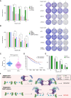
Effect of TRIM44 on the sensitivity to PARP inhibitors. (A and B) U2OS cells were transfected with indicated siRNAs and treated with various dose of olaparib (0, 0.5, 1 and 2 μM) for clonogenic survival assay. Graph (left) and colony (right). Student's t-test was used for statistical analysis. (C) Expression level of TRIM44 in kidney renal clear cell carcinoma (KIRC). (D) Kaplan-Meier plot for TRIM44 in KIRC (http://www.oncodb.org/). (E) HEK293 cells were transfected with indicated siRNAs and treated with various dose of olaparib or phleomycin (0, 0.5, 1 and 2 μM) for clonogenic survival assay. Student's t-test was used for statistical analysis. (F) Functional mechanism of TRIM44 at the DNA lesions in the presence of PARPi.
Khanna K.K., Jackson S.P.. DNA double-strand breaks: signaling, repair and the cancer connection. Nat. Genet. 2001; 27:247–254.
-
PubMed
Krenning L., van den Berg J., Medema R.H.. Life or death after a break: what determines the choice?. Mol. Cell. 2019; 76:346–358.
-
PubMed
Perina D., Mikoc A., Ahel J., Cetkovic H., Zaja R., Ahel I.. Distribution of protein poly(ADP-ribosyl)ation systems across all domains of life. DNA Repair (Amst.). 2014; 23:4–16.
-
PMC
-
PubMed
Ray Chaudhuri A., Nussenzweig A. The multifaceted roles of PARP1 in DNA repair and chromatin remodelling. Nat. Rev. Mol. Cell Biol. 2017; 18:610–621.
-
PMC
-
PubMed
Slade D., Dunstan M.S., Barkauskaite E., Weston R., Lafite P., Dixon N., Ahel M., Leys D., Ahel I.. The structure and catalytic mechanism of a poly(ADP-ribose) glycohydrolase. Nature. 2011; 477:616–620.
-
PMC
-
PubMed