. 2024 Sep 2;22(1):188.
doi: 10.1186/s12915-024-01987-x.

Identification of a novel DNA oxidative damage repair pathway, requiring the ubiquitination of the histone variant macroH2A1.1

Affiliations

Identification of a novel DNA oxidative damage repair pathway, requiring the ubiquitination of the histone variant macroH2A1.1

Khalid Ouararhni et al. BMC Biol. .

Abstract

Background: The histone variant macroH2A (mH2A), the most deviant variant, is about threefold larger than the conventional histone H2A and consists of a histone H2A-like domain fused to a large Non-Histone Region responsible for recruiting PARP-1 to chromatin. The available data suggest that the histone variant mH2A participates in the regulation of transcription, maintenance of heterochromatin, NAD+ metabolism, and double-strand DNA repair.

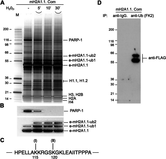
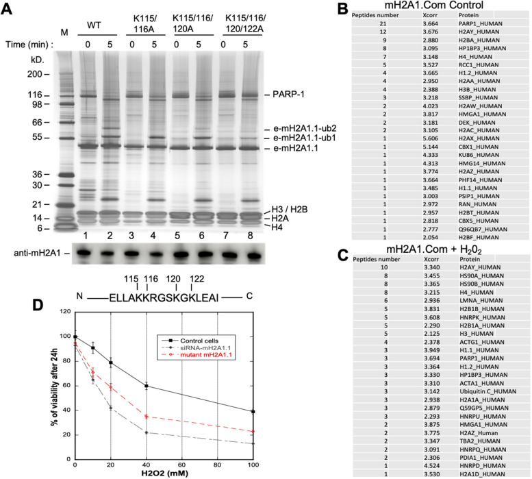
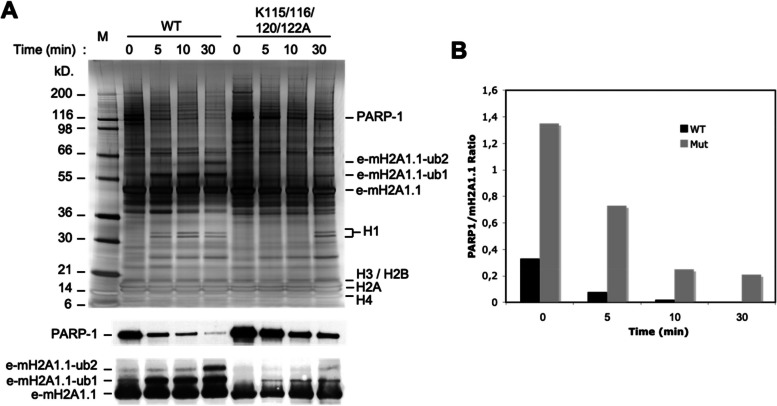
Results: Here, we describe a novel function of mH2A, namely its implication in DNA oxidative damage repair through PARP-1. The depletion of mH2A affected both repair and cell survival after the induction of oxidative lesions in DNA. PARP-1 formed a specific complex with mH2A nucleosomes in vivo. The mH2A nucleosome-associated PARP-1 is inactive. Upon oxidative damage, mH2A is ubiquitinated, PARP-1 is released from the mH2A nucleosomal complex, and is activated. The in vivo-induced ubiquitination of mH2A, in the absence of any oxidative damage, was sufficient for the release of PARP-1. However, no release of PARP-1 was observed upon treatment of the cells with either the DNA alkylating agent MMS or doxorubicin.

Conclusions: Our data identify a novel pathway for the repair of DNA oxidative lesions, requiring the ubiquitination of mH2A for the release of PARP-1 from chromatin and its activation.

Keywords: Chromatin; DNA repair; Histone variant; PARP-1; mH2A.


Conflict of interest statement

The authors declare that they have no competing interests.

Figures

Fig. 1
Fig. 1
The presence of mH2A is essential for the repair of DNA damage and cell survival upon treatment with H2O2. A Control (transfected with scrambled siRNA), siRNA resistant (SR) and siRNA mH2A1 transfected cells were treated with H2O2, allowed to recover for the indicated times, and were then subjected to alkaline Comet assay. B Western blot analysis of total cell extract prepared from control and mH2A1 siRNA treated cells. The blot was first probed with anti-mH2A1 and then with an anti-H2B antibody as a control for equal protein loading. C Quantification of the Comet assay data. Values represent means and standard deviations for ~ 300 cells from 3 independent experiments. D The removal of nuclear poly-ADP ribose foci induced after oxidative DNA damage is dependent on the presence of mH2A1. Control and mH2A1 siRNA transfected HeLa cells were treated with H2O2, allowed to recover for the indicated times and then fixed and immunostained with anti-poly(ADP-ribose) antibody. E Quantification of the data presented in (D). The means of two independent experiments are presented. For each time point no less than 100 cells were analyzed. F mH2A1 is required for cell survival after oxidative stress. Control and mH2A1 siRNA transfected HeLa cells were treated with H2O2 at the indicated concentrations. The percentage cell survival was measured 24 h after the H2O2 treatment. The means of 3 different experiments are shown
Fig. 2
Fig. 2
Upon H2O2 treatment, mH2A.1.1 is ubiquitinted and PARP-1 is both released from the e-mH2A1.1 nucleosome complex and activated. A Silver staining e-mH2A1.1 nucleosome complex (run on SDS PAGE) isolated from either control (-) or H2O2 ( +) treated HeLa cells stably expressing e-mH2A1.1. The proteins identified by mass spectrometry are indicated. Lane M, molecular mass markers. The masses of the different protein markers are indicated. The lower part of the Figure shows the Western blot of e-mH2A1.1 nucleosomal complexes isolated from control (-) and H2O2 ( +) treated e-mH2A1.1 stable HeLa cell lines. The blot was first probed with anti-PARP-1 antibody and then, to visualize e-mH2A1.1, it was probed with anti-HA antibody. B Western blot quantification of mH2A1.1 expression level in untagged and epitope tagged HeLa cells. * indicates the ubiquitinated form of mH2A1. C Kinetics of PARP-1 auto-ADP-ribosylation after oxidative DNA damage in control and mH2A1 siRNA transfected HeLa cells. Cells were treated with H2O2 and after recovery for the indicated times, the cell lysates were run on SDS PAGE, transferred and the blots were probed with either anti-poly(ADP-ribose) antibody (upper panel) or with anti-macroH2A1 (middle panel) or anti-H2A antibodies (lower panel), respectively
Fig. 3
Fig. 3
mH2A is ubiquitinated upon oxidative stress. A Stable HeLa cell lines expressing e-mH2A1.1 were treated with H2O2 and allowed recovering for the times indicated. The e-mH2A1.1 nucleosomal complex was immunopurified, run on a gel containing SDS and proteins were identified by mass spectroscopy. The positions of PARP-1, non-modified and mono(ub1)- and bi-ubiquitinated(ub2) e-mH2A1.1, histone H1 and core histones are indicated. B Western blot of the e-mH2A.1.1 complex. The blot was first revealed with anti-PARP-1 antibody and then with anti-HA antibody for visualization of mH2A1.1. C Aminoacid sequence of mH2A1.1 encompassing AA 109–132. The positions of the two identified by mass spectrometry ubiquitination sites (I) and (II), corresponding to lysine residues 115 and 120, are indicated. D Western blot analysis of the e-mH2A.1.1 complex immunoprecipitated either with anti-IgG or with anti-ubiquitin FK2, and blotted with an anti-FLAG antibody
Fig. 4
Fig. 4
The release of PARP-1 from the mH2A1 nucleosomal complex is affected by mutations of the mH2A ubiquitination sites. Stable HeLa cell lines expressing e-mH2A1.1 bearing lysine (K) to alanine (A) mutations either at positions 115 and 116, or at 115, 116 and 120 or at 115, 116, 120 and 122, were established. They were treated with H2O2 and allowed recovering for 5 min (at time point zero (0), the cells were treated with H2O2 and not allowed to recover). Then the e-mH2A1.1 mutated nucleosome complexes were isolated, run on a SDS PAGE and the proteins and e-mH2A1.1 ubiquitinated species were identified by mass spectrometry. The positions of the identified proteins were indicated at the right part of the figure. M, protein molecular mass markers. The molecular weights of the markers are indicated. In the middle of the figure is shown a Western blot against mH2A1. In the lower part of the figure is shown the aminoacid sequence of mH2A1.1 encompassing AA 112–124. The positions of the lysines, which were mutated to alanines, are indicated. (The percentage cell survival was measured 24 h after the H2O2 treatment. The means of 3 different experiments are shown. B Mass spectrometry table of the e-mH2A1.1 complex purified from untreated HeLa cells. C Mass spectrometry table of the e-mH2A1.1 complex purified from H202 treated HeLa cells. D Control HeLa cells, mH2A1 siRNA transfected HeLa cells and e-mH2A1.1 K115/116/120/122A mutant HeLa cells were treated with H2O2 at the indicated concentrations
Fig. 5
Fig. 5
Time course of PARP-1 release from mH2A chromatin, induced after treatment with H2O2. Stable cell lines expressing either wild type mH2A1.1 or four lysine to alanine mutated (K115/116/120/122A) mH2A1.1 were treated with H2O2 and allowed recovering for the times indicated. The mH2A1.1 complexes were then purified, run on SDS PAGE and the proteins were identified by mass spectrometry. The positions of the identified proteins are indicated. The lower part of the figure shows the Western blot analysis of respective e-mH2A1.1 nucleosome complexes. The blot was first probed with anti-PARP-1 antibody and then, to visualize e-mH2A1.1, it was probed with anti-HA antibody. B Quantification of the Western blot data shown in (A). The PARP-1/mH2A1.1 ratio for both WT (black) and mutated (grey) mH2A1.1 nucleosomal complexes is presented
Fig. 6
Fig. 6
Ubiquitination of mH2A is sufficient to release PARP-1 from mH2A chromatin. A Treatment with NEM of stable HeLa cell lines expressing mH2A1.1 results in ubiquitination of mH2A1.1 and release of PARP-1 from the mH2A1 nucleosomal complex. Stable HeLa cell lines expressing mH2A1.1 were treated with 5 mM NEM for 30 min and mH2A1.1 nucleosomal complex was purified and run on a SDS PAGE. The protein bands were identified by mass spectrometry. The positions of PARP-1, non-modified and mono- and bi-ubiquitinated e-mH2A1.1 as well as histone H1 and the core histones are indicated. The Western blot of the respective e-mH2A1.1 complexes is shown in the lower part of the gel. The blot was first probed with anti-PARP-1 antibody and then, to visualize e-mH2A1.1, it was probed with anti-HA antibody. B Schematic presentation of the mechanism of PARP-1 release from the mH2A chromatin upon treatment with H2O2

References

    1. van Holde K. Chromatin. Berlin: Springer-Verlag KG; 1988.
    1. Latrick CM, Marek M, Ouararhni K, Papin C, Stoll I, Ignatyeva M, Obri A, Ennifar E, Dimitrov S, Romier C, et al. Molecular basis and specificity of H2A.Z-H2B recognition and deposition by the histone chaperone YL1. Nat Struct Mol Biol. 2016;23(4):309–16. 10.1038/nsmb.3189 - DOI - PubMed
    1. Obri A, Ouararhni K, Papin C, Diebold ML, Padmanabhan K, Marek M, Stoll I, Roy L, Reilly PT, Mak TW, et al. ANP32E is a histone chaperone that removes H2A.Z from chromatin. Nature. 2014;505(7485):648–53. 10.1038/nature12922 - DOI - PubMed
    1. Drane P, Ouararhni K, Depaux A, Shuaib M, Hamiche A. The death-associated protein DAXX is a novel histone chaperone involved in the replication-independent deposition of H3.3. Genes Dev. 2010;24(12):1253–65. 10.1101/gad.566910 - DOI - PMC - PubMed
    1. Shuaib M, Ouararhni K, Dimitrov S, Hamiche A. HJURP binds CENP-A via a highly conserved N-terminal domain and mediates its deposition at centromeres. Proc Natl Acad Sci U S A. 2010;107(4):1349–54. 10.1073/pnas.0913709107 - DOI - PMC - PubMed

LinkOut - more resources