Injury to the palmar supporting structures of the fetlock alters limb stiffness and fetlock angle
- PMID: 39219092
- PMCID: PMC11982416
- DOI: 10.1111/evj.14409
Injury to the palmar supporting structures of the fetlock alters limb stiffness and fetlock angle
Abstract
Background: In vivo measurement of limb stiffness and conformation provides a non-invasive proxy assessment of superficial digital flexor tendon (SDFT) and suspensory ligament (SL) function. Here, we compared it in fore and hindlimbs and after injury.
Objectives: To compare the limb stiffness and conformation in forelimbs and hindlimbs, changes with age, and following injury to the SDFT and SL.
Study design: Retrospective cohort study.
Methods: Limb stiffness was calculated using floor scales and an electrogoniometer taped to the dorsal fetlock. The fetlock angle and weight were simultaneously recorded five times with the limb weight-bearing and when the opposite limb was picked up (increased load). Limb stiffness of both limbs was calculated from the gradient of the regression line of angle versus load. Fetlock angle when the weight was zero was extrapolated from the graph and used as a measure of conformation. Limb stiffness was measured in uninjured forelimbs (n = 42 limbs), hindlimbs (n = 19 limbs), forelimbs with SDFT injury (n = 18) and hindlimbs with SL injury (n = 5).
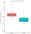
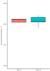
Results: Limb stiffness correlated with weight in forelimbs as shown previously (p < 0.001) but also in hindlimbs (p = 0.006). When normalised to the horse's weight (503 kg, IQR 471.5-560), forelimb stiffness was significantly higher (22.3 [±4.5] × 10-3 degree-1) than for the hindlimb (16.4 [±4.0] × 10-3 degree-1; p < 0.001). While there were no significant differences between forelimb and hindlimb conformation in unaffected or SDFT injury, both limb stiffness and conformation was significantly greater in limbs with SL injury (p = 0.009 and p = 0.002, respectively).
Main limitations: Small sample size, lack of clinical data including lameness and quantification of injuries.
Conclusions: Injury to the forelimb SDFT does not alter limb stiffness or conformation in the long-term, while hindlimb SL injury simultaneously increases limb stiffness and fetlock angle, suggesting an increase in SL length following injury.
Hintergrund: Die in vivo Messung der Steifigkeit und Konformation der Gliedmaßen bietet eine nicht‐invasive Möglichkeit zur Beurteilung der Funktion der oberflächlichen Beugesehne (SDFT) und des Fesselträgers (SL). Diese wurde bei Vorder‐ und Hintergliedmaßen sowie nach einer Verletzung verglichen.
Ziele: Der Vergleich der Steifigkeit und Konformation der Gliedmaßen bei Vorder‐ und Hintergliedmaßen, Veränderungen im Alter und nach Verletzungen der SDFT und SL.
Studiendesign: Retrospektive Kohortenstudie.
Methoden: Die Steifigkeit der Gliedmaßen wurde mithilfe von Bodenwaagen und einem an das dorsale Fesselgelenk geklebten Elektrogoniometer berechnet. Der Fesselwinkel und das Gewicht wurden gleichzeitig fünfmal erfasst, sowohl mit tragender Gliedmaße als auch bei Anheben der gegenüberliegenden Gliedmaße (erhöhte Belastung). Die Steifigkeit der Gliedmaßen beider Beine wurde aus der Steigung der Regressionslinie von Winkel zu Last berechnet. Der Fesselwinkel bei einem Gewicht von null wurde aus dem Diagramm extrapoliert und als Maß für die Konformation verwendet. Die Steifigkeit der Gliedmaßen wurde bei unverletzten Vordergliedmaßen (n = 42 Gliedmaßen), Hintergliedmaßen (n = 19 Gliedmaßen), Vordergliedmaßen mit SDFT‐Verletzung (n = 18) und Hintergliedmaßen mit SL‐Verletzung (n = 5) gemessen.
Resultate: Die Steifigkeit der Gliedmaßen korrelierte mit dem Gewicht bei den Vordergliedmaßen, wie zuvor gezeigt (p < 0,001), aber auch bei den Hintergliedmaßen (p = 0,006). Nach Normierung auf das Gewicht des Pferdes (503 kg, IQR 471,5–560) war die Steifigkeit der Vordergliedmaßen signifikant höher (22,3 [±4,5] × 10−3 Grad‐1) als die der Hintergliedmaßen (16,4 [±4,0] × 10−3 Grad‐1; p < 0,001). Während es keine signifikanten Unterschiede zwischen der Konformation von Vorder‐ und Hintergliedmaßen bei unbeeinträchtigten oder SDFT‐verletzten Tieren gab, waren sowohl die Steifigkeit als auch die Konformation bei Gliedmaßen mit SL‐Verletzung signifikant größer (p = 0,009 bzw. p = 0,002). WICHTIGSTE EINSCHRÄNKUNGEN: Kleine Stichprobengröße, Mangel an klinischen Daten, einschließlich Lahmheit und Quantifizierung der Verletzungen.
Zusammenfassung: Eine Verletzung der oberflächlichen Beugesehne (SDFT) der Vordergliedmaße verändert die Steifigkeit oder Konformation der Gliedmaße langfristig nicht, während eine Verletzung des Fesselträgers (SL) der Hintergliedmaße gleichzeitig die Steifigkeit der Gliedmaße und den Fesselwinkel erhöht, was auf eine Verlängerung des Fesselträgers nach der Verletzung hindeutet.
Keywords: fetlock conformation; horse; ligament; limb stiffness; superficial digital flexor tendon; suspensory ligament; tendon.
© 2024 The Author(s). Equine Veterinary Journal published by John Wiley & Sons Ltd on behalf of EVJ Ltd.
Conflict of interest statement
The authors declare no conflicts of interest.
Figures


Similar articles
-
The relationship between in vivo limb and in vitro tendon mechanics after injury: a potential novel clinical tool for monitoring tendon repair.Equine Vet J. 2011 Jul;43(4):418-23. doi: 10.1111/j.2042-3306.2010.00303.x. Epub 2010 Sep 29. Equine Vet J. 2011. PMID: 21496076
-
Comparison of healing in forelimb and hindlimb surgically induced core lesions of the equine superficial digital flexor tendon.Vet Comp Orthop Traumatol. 2014;27(5):358-65. doi: 10.3415/VCOT-13-11-0136. Epub 2014 Jul 31. Vet Comp Orthop Traumatol. 2014. PMID: 25078543
-
In vivo measurements of flexor tendon and suspensory ligament forces during trotting using the thoroughbred forelimb model.J Equine Sci. 2014;25(1):15-22. doi: 10.1294/jes.25.15. Epub 2014 Apr 22. J Equine Sci. 2014. PMID: 24834009 Free PMC article.
-
Prevalence of superficial digital flexor tendonitis and suspensory desmitis in Japanese Thoroughbred flat racehorses in 1999.Equine Vet J. 2004 May;36(4):346-50. doi: 10.2746/0425164044890580. Equine Vet J. 2004. PMID: 15163043
-
A review of tendon injury: why is the equine superficial digital flexor tendon most at risk?Equine Vet J. 2010 Mar;42(2):174-80. doi: 10.2746/042516409X480395. Equine Vet J. 2010. PMID: 20156256 Review.
References
-
- Kasashima Y, Takahashi T, Smith RKW, Goodship AE, Kuwano A, Ueno T, et al. Prevalence of superficial digital flexor tendonitis and suspensory desmitis in Japanese Thoroughbred flat racehorses in 1999. Equine Vet J. 2004;36:346–350. - PubMed
-
- Ely ER, Avella CS, Price JS, Smith RKW, Wood JLN, Verheyen KLP. Descriptive epidemiology of fracture, tendon and suspensory ligament injuries in National Hunt racehorses in training. Equine Vet J. 2009;41:372–378. - PubMed
-
- Williams RB, Harkins LS, Hammond CJ, Wood JLN. Racehorse injuries, clinical problems and fatalities recorded on British racecourses from flat racing and National Hunt racing during 1996, 1997 and 1998. Equine Vet J. 2001;33:478–486. - PubMed
-
- Avella CS, Ely ER, Verheyen KLP, Price JS, Wood JLN, Smith RKW. Ultrasonographic assessment of the superficial digital flexor tendons of National Hunt racehorses in training over two racing seasons. Equine Vet J. 2009;41:449–454. - PubMed
-
- Biewener AA, Roberts TJ. Muscle and tendon contributions to force, work, and elastic energy savings: a comparative perspective. Exerc Sport Sci Rev. 2000;28:99–107. - PubMed
MeSH terms
LinkOut - more resources
Full Text Sources

