

Three-Dimensional Heart Modeling of Hypertrophic Obstructive Cardiomyopathy for In Situ Patient-Specific Simulation to Optimize Septal Myectomy
- PMID: 39219341
- PMCID: PMC11613515
- DOI: 10.1177/15569845241273538
Three-Dimensional Heart Modeling of Hypertrophic Obstructive Cardiomyopathy for In Situ Patient-Specific Simulation to Optimize Septal Myectomy
Abstract
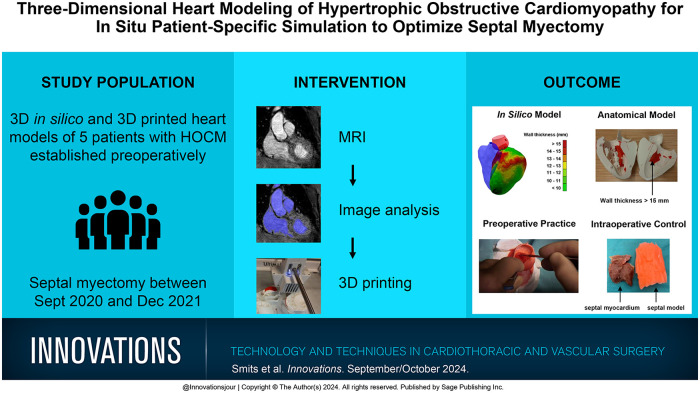
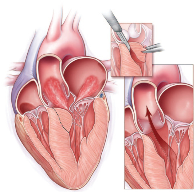
Objective: Hypertrophic obstructive cardiomyopathy (HOCM) develops in at least 1 out of 715 young adults. Patients who are refractory to medical therapy qualify for septal myectomy. Due to anatomy, serious complications such as ventricular septal defect and heart block may occur. Establishing cardiovascular magnetic resonance (CMR)-based 3-dimensional (3D) models as part of preoperative planning and training has the potential to decrease procedure-related complications and improve results.
Methods: CMR images were used to segment cardiac structures. Left ventricular wall thickness was calculated and projected on top of the in silico model. A 3D model was printed with a red layer indicating a wall thickness exceeding 15 mm and used for preoperative resection planning and patient counseling. To provide preoperative patient-specific in situ simulation, the planned resection volume was replaced with silicone in a second model. For perioperative quality control, resected silicone was compared with resected myocardial tissue. The impact of the models was evaluated descriptively through consultation of both the cardiothoracic surgeon and patients and through patient outcomes.
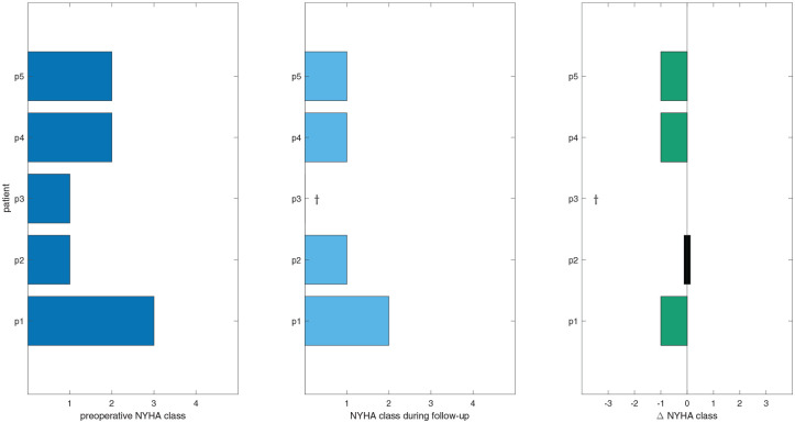
Results: Three-dimensional in silico and 3D-printed heart models of 5 patients were established preoperatively. Since the introduction of the models in October 2020, the surgeon feels better prepared, more confident, and less difficulty with making decisions. In addition, patients feel better informed preoperatively.
Conclusions: Using 3D heart models optimized preoperative planning and training, intraoperative quality control, and patient consultation. Reduction of procedure-related complications and clinical outcome should be studied in larger cohorts.
Keywords: 3D printing; Morrow procedure; in situ simulation; magnetic resonance imaging; simulation-based training; virtual surgical planning.
Conflict of interest statement
Declaration of Conflicting InterestsThe authors declared no potential conflicts of interest with respect to the research, authorship, and/or publication of this article.
Figures


Similar articles
-
Role of Preoperative Cardiovascular Magnetic Resonance in Planning Ventricular Septal Myectomy in Patients With Obstructive Hypertrophic Cardiomyopathy.Am J Cardiol. 2019 May 1;123(9):1517-1526. doi: 10.1016/j.amjcard.2019.01.041. Epub 2019 Feb 8. Am J Cardiol. 2019. PMID: 30791998
-
Virtual and real septal myectomy using 3-dimensional printed models.Interact Cardiovasc Thorac Surg. 2018 May 1;26(5):881-882. doi: 10.1093/icvts/ivx410. Interact Cardiovasc Thorac Surg. 2018. PMID: 29281016
-
Development of a 3-D printing-based cardiac surgical simulation curriculum to teach septal myectomy.J Thorac Cardiovasc Surg. 2018 Sep;156(3):1139-1148.e3. doi: 10.1016/j.jtcvs.2017.09.136. Epub 2017 Nov 6. J Thorac Cardiovasc Surg. 2018. PMID: 30029780
-
Role of cardiac magnetic resonance (CMR) in planning ventricular septal myomectomy in patients with hypertrophic obstructive cardiomyopathy (HOCM).J Card Surg. 2022 Dec;37(12):4186-4189. doi: 10.1111/jocs.17090. Epub 2022 Nov 22. J Card Surg. 2022. PMID: 36434805 Review.
-
Post-operative management of hypertrophic obstructive cardiomyopathy.Asian Cardiovasc Thorac Ann. 2022 Jan;30(1):57-63. doi: 10.1177/02184923211069189. Epub 2022 Feb 15. Asian Cardiovasc Thorac Ann. 2022. PMID: 35167344 Review.
References
-
- Ommen SR, Mital S, Burke MA, et al.. 2020 AHA/ACC guideline for the diagnosis and treatment of patients with hypertrophic cardiomyopathy: a report of the American College of Cardiology/American Heart Association Joint Committee on Clinical Practice Guidelines. Circulation 2020; 142: e558–e631. - PubMed
-
- Maron BJ, Gardin JM, Flack JM, et al.. Prevalence of hypertrophic cardiomyopathy in a general population of young adults. Echocardiographic analysis of 4111 subjects in the CARDIA Study. Coronary Artery Risk Development in (Young) Adults. Circulation 1995; 92: 785–789. - PubMed
-
- Maron MS, Olivotto I, Zenovich AG, et al.. Hypertrophic cardiomyopathy is predominantly a disease of left ventricular outflow tract obstruction. Circulation 2006; 114: 2232–2239. - PubMed
-
- Semsarian C, Ingles J, Maron MS, et al.. New perspectives on the prevalence of hypertrophic cardiomyopathy. J Am Coll Cardiol 2015; 65: 1249–1254. - PubMed
MeSH terms
LinkOut - more resources
Full Text Sources
Medical
