Review
. 2024 Sep 2;20(17):e1062-e1075.
doi: 10.4244/EIJ-D-23-00840.

Aortic regurgitation: from mechanisms to management

Affiliations
Review

Aortic regurgitation: from mechanisms to management

Andreas Baumbach et al. EuroIntervention. .

Abstract

Aortic regurgitation (AR) is a common clinical disease associated with significant morbidity and mortality. Investigations based largely on non-invasive imaging are pivotal in discerning the severity of disease and its impact on the heart. Advances in technology have contributed to improved risk stratification and to our understanding of the pathophysiology of AR. Surgical aortic valve replacement is the predominant treatment. However, its use is limited to patients with an acceptable surgical risk profile. Transcatheter aortic valve implantation is an alternative treatment. However, this therapy remains in its infancy, and further data and experience are required. This review article on AR describes its prevalence, mechanisms, diagnosis and treatment.


Conflict of interest statement

A. Baumbach has received speaker fees from Abbott, Medtronic, and MicroPort; and has been a proctor for JenaValve. K.P. Patel has received an unrestricted research grant from Edwards Lifesciences. V. Delgado has received speaker fees from Edwards Lifesciences, GE HealthCare, Novartis, and Philips; and has received consulting fees from Edwards Lifesciences, Novo Nordisk, and Merck & Co. T.K. Rudolph has been a proctor and medical advisor for JenaValve; she has received speaker fees from Edwards Lifesciences, Boston Scientific, Medtronic, and JenaValve. H. Treede has been a proctor and advisor for JenaValve. A.R. Tamm has been a proctor for the Trilogy system.

Figures

Figure 1
Figure 1. Mechanisms of aortic regurgitation.
Adapted with permission from Boodhwani et al. AR: aortic regurgitation; SCA: subcommissural annuloplasty; STJ: sinotubular junction
Figure 2
Figure 2. Anatomy of the aortic valve.
Images (A), (B) and (C) show the transthoracic and transoesophageal echocardiograms and computed tomography of a patient with a bicuspid aortic valve with 2 commissures and 2 cusps. Images (D), (E) and (F) show the transthoracic and transoesophageal echocardiograms and computed tomography of a patient with a bicuspid aortic valve with 2 commissures and 3 cusps, 2 of which are fused by a fusion raphe (arrow).
Figure 3
Figure 3. Assessment of aortic root and ascending aorta dimensions.
Measurement of the diameter of the aortic root at the level of the sinus of Valsalva (a), sinotubular junction (b) and the ascending aorta (c) on a transthoracic echocardiogram (A) and a transoesophageal echocardiogram (B). On computed tomography angiography (C), from the 3-volume acquisition, the multiplanar reformation planes can be aligned to obtain the cross-sectional area of the ascending aorta (black double arrowhead in D). E) An example of a cine cardiac magnetic resonance acquisition and measurement (white double arrowhead) of the ascending aorta in a patient with severe aortic regurgitation.
Figure 4
Figure 4. Measurement of the aortic annulus.
Aortic annulus measurement with 3-dimensional echocardiography (A) and computed tomography (B). The multiplanar reformation planes are aligned across the aortic annulus bisecting the nadir points of the aortic cusps. The aortic annulus area is then planimetered (dotted line).
Figure 5
Figure 5. Assessment of aortic regurgitation with echocardiography.
The following parameters should be taken into consideration to assess the severity of aortic regurgitation: the ratio between the width of the regurgitant jet (double white arrowhead) and the diameter of the left ventricular outflow tract on colour M-mode of the parasternal long-axis view (A), the vena contracta as measured on the colour Doppler image acquired from the apical 5-chamber view (double black arrowhead; B), and the dense signal of the regurgitant jet on a continuous wave Doppler image where the pressure half-time can be measured (C, dotted line). D) Diastolic flow reversal obtained with pulsed wave Doppler from the suprasternal view (white arrow). Three-dimensional colour Doppler transoesophageal echocardiography permits the measurement of the anatomical regurgitant orifice area (E, dotted encircling line) by aligning the multiplanar reformation planes across the vena contracta.
Figure 6
Figure 6. Phenotypes of bicuspid aortic valves and related repair techniques.
BAV: bicuspid aortic valve
Figure 7
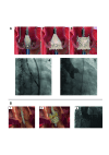
Figure 7. Transfemoral transcatheter heart valves.
A) J-Valve prosthesis (JC Medical). B) Trilogy prosthesis (JenaValve).
Figure 8
Figure 8. Deployment of transcatheter heart valves.
A) Deployment of the Trilogy THV: 1. Locators align the THV with the native cusps; 2. Inflow is deployed, with limited protrusion; 3. Outflow is deployed: locators “clip” onto native leaflets, forming a seal and stable securement; 4. THV with locators spread during implantation, seating the locators in the sinuses; 5. Valve after implantation with secure anchoring and no paravalvular regurgitation. B) Deployment of the J-Valve THV: 1. Anchor rings grasp the native leaflets in correct anatomical alignment; 2. The THV is deployed, anchoring in the annulus; 3. The THV after implantation with full expansion and no aortic regurgitation. THV: transcatheter heart valve
Central illustration
Central illustration. Aortic regurgitation – its evaluation, risk stratification and treatment.
A) The evaluation and risk stratification of AR, with the key strengths of each imaging modality in red. B) The main treatment options. AR: aortic regurgitation; CMR: cardiac magnetic resonance imaging; CT: computed tomography; Echo: echocardiography; LV: left ventricle; LVESD: left ventricular end-systolic diameter; TAVI: transcatheter aortic valve implantation

References

    1. Singh JP, Evans JC, Levy D, Larson MG, Freed LA, Fuller DL, Lehman B, Benjamin EJ. Prevalence and clinical determinants of mitral, tricuspid, and aortic regurgitation (the Framingham Heart Study). Am J Cardiol. 1999;83:897–902. - PubMed
    1. d’Arcy JL, Coffey S, Loudon MA, Kennedy A, Pearson-Stuttard J, Birks J, Frangou E, Farmer AJ, Mant D, Wilson J, Myerson SG, Prendergast BD. Large-scale community echocardiographic screening reveals a major burden of undiagnosed valvular heart disease in older people: the OxVALVE Population Cohort Study. Eur Heart J. 2016;37:3515–22. - PMC - PubMed
    1. Boodhwani M, de Kerchove, Glineur D, Poncelet A, Rubay J, Astarci P, Verhelst R, Noirhomme P, El Khoury. Repair-oriented classification of aortic insufficiency: impact on surgical techniques and clinical outcomes. J Thorac Cardiovasc Surg. 2009;137:286–94. - PubMed
    1. Carpentier A. Cardiac valve surgery--the “French correction”. J Thorac Cardiovasc Surg. 1983;86:323–37. - PubMed
    1. Evangelista A, Sitges M, Jondeau G, Nijveldt R, Pepi M, Cuellar H, Pontone G, Bossone E, Groenink M, Dweck MR, Roos-Hesselink JW, Mazzolai L, van Kimmenade, Aboyans V, Rodríguez-Palomares J. Multimodality imaging in thoracic aortic diseases: a clinical consensus statement from the European Association of Cardiovascular Imaging and the European Society of Cardiology working group on aorta and peripheral vascular diseases. Eur Heart J Cardiovasc Imaging. 2023;24:e65–85. - PubMed

MeSH terms

LinkOut - more resources