

Unusual Evidence of Fat Embolism in the Subclavian Vein Detected by High-Intensity Transient Signals
- PMID: 39220350
- PMCID: PMC11363887
- DOI: 10.14797/mdcvj.1395
Unusual Evidence of Fat Embolism in the Subclavian Vein Detected by High-Intensity Transient Signals
Abstract
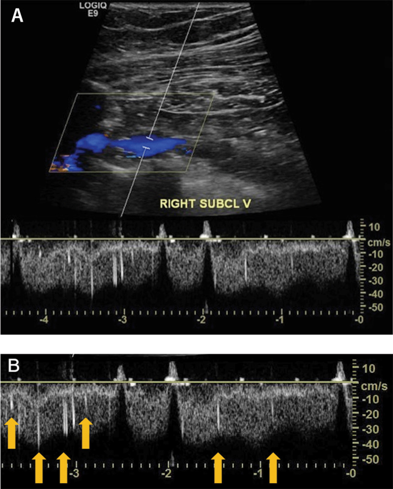
Fat emboli may occur in patients after traumatic fractures or orthopedic procedures; however, their clinical detection is a very rare finding. Here, we describe a 77-year-old female who was admitted to the emergency department with a fracture of the right humerus. We diagnosed fat embolism after an ultrasound of the right subclavian vein. The embolism was detected by high-intensity transient signals present on the spectral Doppler. While these signals are well known for microembolization in transcranial Doppler, to our knowledge this is the first case report in the medical literature to observe and describe high-intensity transient signals seen in the upper extremities by spectral Doppler. Although it is unusual to detect a fat embolism in transit, we believe clinicians should be aware of this finding, particularly when evaluating high-risk patients.
Keywords: fat embolism; fat embolism syndrome; high-intensity transient signals; spectral Doppler; subclavian vein; venous duplex ultrasound.
Copyright: © 2024 The Author(s).
Conflict of interest statement
The authors have no competing interests to declare.
Figures

Similar articles
-
Fat embolism syndrome from an isolated humerus fracture.J Orthop Trauma. 1997 Feb-Mar;11(2):141-4. doi: 10.1097/00005131-199702000-00016. J Orthop Trauma. 1997. PMID: 9057154 No abstract available.
-
Transcranial Doppler detection of cerebral fat emboli and relation to paradoxical embolism: a pilot study.Circulation. 2011 May 10;123(18):1947-52. doi: 10.1161/CIRCULATIONAHA.110.950634. Epub 2011 Apr 25. Circulation. 2011. PMID: 21518982 Clinical Trial.
-
Perioperative Detection of Cerebral Fat Emboli From Bone Using High-Frequency Doppler Ultrasound.Ultrasound Med Biol. 2025 Jan;51(1):138-148. doi: 10.1016/j.ultrasmedbio.2024.09.017. Epub 2024 Oct 21. Ultrasound Med Biol. 2025. PMID: 39438224
-
Fat embolism syndrome: a complication of orthopaedic trauma.Orthop Nurs. 1998 Mar-Apr;17(2):41-3, 46, 58. Orthop Nurs. 1998. PMID: 9601398 Review.
-
Fat embolism syndrome following percutaneous vertebroplasty: a case report.Spine J. 2014 Apr;14(4):e1-5. doi: 10.1016/j.spinee.2013.09.021. Epub 2013 Oct 12. Spine J. 2014. PMID: 24314905 Review.
References
-
- Von Bergmann E. Ein fall todlicher fettenbolic. Berl Klin Wochenscher 1873;10:385.
-
- Herndon JH, Bechtol CO, Crickenberger DP. Fat embolism during total hip replacement. A prospective study. J Bone Joint Surg Am. 1974. Oct;56(7):1350–62. PMID: 4447670 - PubMed
-
- Husebye EE, Lyberg T, Røise O. Bone marrow fat in the circulation: clinical entities and pathophysiological mechanisms. Injury. 2006. Oct;37 Suppl 4:S8–18. doi: - PubMed
Publication types
MeSH terms
LinkOut - more resources
Full Text Sources
