. 2024 Jul 25;32(3):101305.
doi: 10.1016/j.omtm.2024.101305. eCollection 2024 Sep 12.

Characterization of residual microRNAs in AAV vector batches produced in HEK293 mammalian cells and Sf9 insect cells

Affiliations

Characterization of residual microRNAs in AAV vector batches produced in HEK293 mammalian cells and Sf9 insect cells

Magalie Penaud-Budloo et al. Mol Ther Methods Clin Dev. .

Abstract

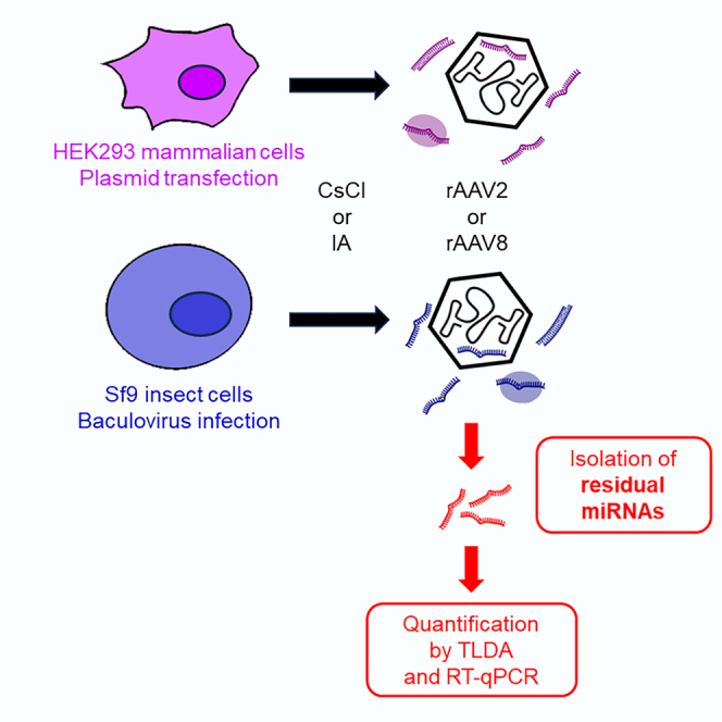
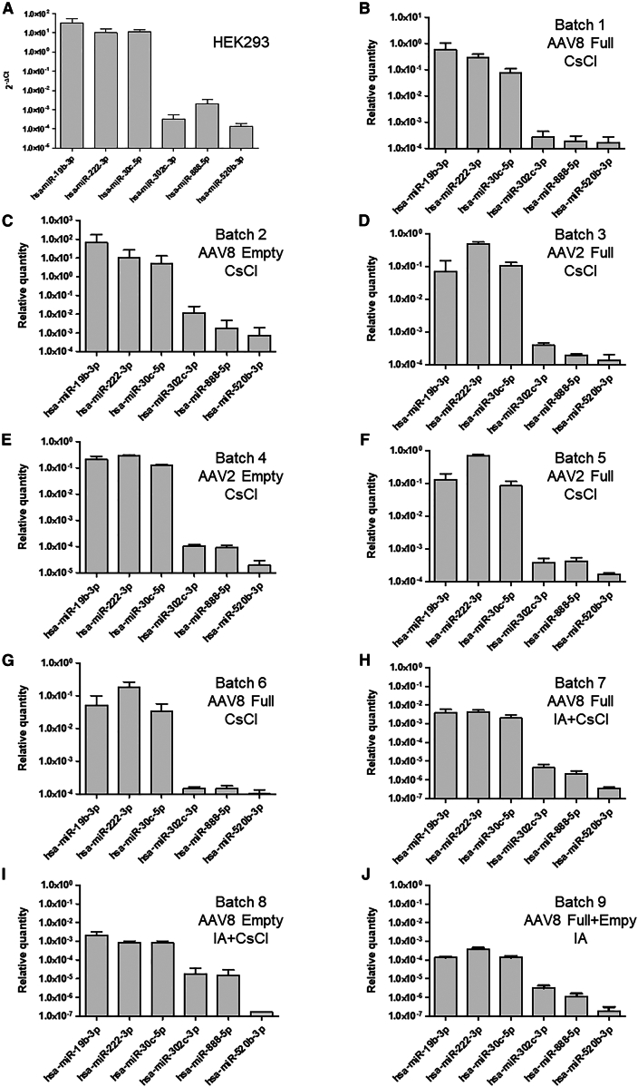
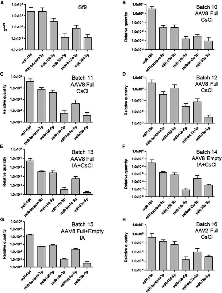
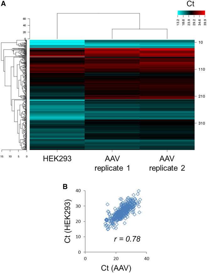
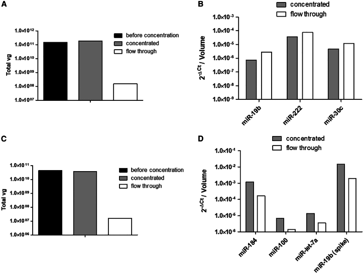
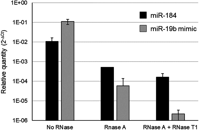
With more than 130 clinical trials and 8 approved gene therapy products, adeno-associated virus (AAV) stands as one of the most popular vehicles to deliver therapeutic DNA in vivo. One critical quality attribute analyzed in AAV batches is the presence of residual DNA, as it could pose genotoxic risks or induce immune responses. Surprisingly, the presence of small cell-derived RNAs, such as microRNAs (miRNAs), has not been investigated previously. In this study, we examined the presence of miRNAs in purified AAV batches produced in mammalian or in insect cells. Our findings revealed that miRNAs were present in all batches, regardless of the production cell line or capsid serotype (2 and 8). Quantitative assays indicated that miRNAs were co-purified with the recombinant AAV particles in a proportion correlated with their abundance in the production cells. The level of residual miRNAs was reduced via an immunoaffinity chromatography purification process including a tangential flow filtration step or by RNase treatment, suggesting that most miRNA contaminants are likely non-encapsidated. In summary, we demonstrate, for the first time, that miRNAs are co-purified with AAV particles. Further investigations are required to determine whether these miRNAs could interfere with the safety or efficacy of AAV-mediated gene therapy.

Keywords: AAV vectors; HEK293 mammalian cells; Sf9 insect cells; bioproduction; gene therapy; microRNA; quality control; residual nucleic acids.


Conflict of interest statement

M.P.-B. and E.A. are inventors of patents related to AAV gene therapy licensed to biopharma companies.

Figures

None
Graphical abstract
Figure 1
Figure 1
Small RNA content of AAV vector batches Total RNA was extracted from HEK293 mammalian cells (A), HEK293-derived rAAV (batch 17) (B), Sf9 insect cells (C), and Sf9-derived rAAV (batch 21) (D). AAV8-GFP vectors (shown in B and D) were purified by CsCl density gradient ultracentrifugation. Electropherograms were obtained after capillary electrophoresis on an Agilent Small RNA chip (6–150 nucleotides) with the miRNA region set to the range 15–35 nucleotides (area between the two green lines). miRNA, microRNA; tRNA, transfer RNA; rRNA, ribosomal RNA. The scale of the x axis is displayed in nucleotides [nt], and the y axis represents fluorescence units [FU].
Figure 2
Figure 2
Quantification of miRNAs in HEK293-derived AAV vector batches MiRNAs were quantified by RT-qPCR in HEK293 cell extract (A) and in AAV vector batches produced in HEK293 mammalian cells (B–J). AAV8 full (B) and empty (C) particles were purified by CsCl, as well as AAV2 full (D) and empty (E) particles. Quantification was performed from duplicate lots of full AAV2 (F) and full AAV8 (G) purified by CsCl. AAV8 full (H) and empty (I) particles were purified from the same bulk by IA followed by CsCl gradient ultracentrifugation. Batch 9 corresponds to an AAV8 purified by IA (J). The same cassette (cytomegalovirus [CMV] promoter and enhanced green fluorescent protein [eGFP] transgene) was used for all AAV vectors. MicroRNAs were extracted in triplicate and quantified in duplicate by SYBR Green-based RT-qPCR. The relative quantity of each miRNA was determined using the ΔCt method. For AAV, data were normalized to account for culture and batch volumes.
Figure 3
Figure 3
Quantification of miRNAs in Sf9-derived AAV vector batches MiRNAs were quantified by RT-qPCR from Sf9 cell extract (A) and in rAAV produced in Sf9 insect cells (B–H). Three batches of AAV8-GFP (B–D) and one batch of AAV2-GFP (H) purified by CsCl were tested. AAV8 full (E) and empty (F) particles were purified from the same bulk by IA followed by CsCl gradient ultracentrifugation. Batch 15 corresponds to an AAV8 purified by IA (G). The same cassette (CMV-eGFP) was used for all AAV vectors. MicroRNAs were extracted in triplicate and quantified in duplicate by SYBR Green-based RT-qPCR. The relative quantity of each miRNA was calculated using the ΔCt method. For AAV, data were normalized to account for culture and batch volumes.
Figure 4
Figure 4
Comparison of miRNA content in HEK293-producing cells and AAV vector by TLDA Human miRNAs were quantified in HEK293 cells and an AAV8 vector batch (n = 2). AAV particles were purified by CsCl gradient ultracentrifugation. Results are represented as a heatmap (A) and a Ct correlation graph (B).
Figure 5
Figure 5
Filtration assay on Amicon centrifugal filters AAV8-GFP were produced in HEK293 (A and B) or in Sf9 cells (C and D) and purified by the CsCl gradient method. AAV vectors were loaded on a centrifugal filter of 100 kDa MWCO that does not allow the AAV particles to pass through. A synthetic miR-19 was spiked before filtration in the Sf9-derived AAV sample, as control of free miRNA. Vector genomes (A and C) were quantified by qPCR before concentration, and in the concentrated (C) and flowthrough (FT) fractions. The quantity of residual miRNAs (B and D) was quantified by RT-qPCR in the C and FT fractions after centrifugation.
Figure 6
Figure 6
Removal of miRNA by RNase pretreatment of rAAV batches RNase digestion was realized in duplicate on a CsCl-purified AAV8-GFP batch produced in Sf9 cells. RT-qPCR was performed targeting miR-184 (black) and miR-19b mimic (gray) that was spiked before RNase treatment. The miRNA relative quantity was calculated using the SIC by the ΔCt method for 200 μL of AAV.

References

    1. Muhuri M., Maeda Y., Ma H., Ram S., Fitzgerald K.A., Tai P.W., Gao G. Overcoming innate immune barriers that impede AAV gene therapy vectors. J. Clin. Invest. 2021;131 - PMC - PubMed
    1. Costa-Verdera H., Unzu C., Valeri E., Adriouch S., González Aseguinolaza G., Mingozzi F., Kajaste-Rudnitski A. Understanding and Tackling Immune Responses to Adeno-Associated Viral Vectors. Hum. Gene Ther. 2023;34:836–852. - PubMed
    1. Duan D. Lethal immunotoxicity in high-dose systemic AAV therapy. Mol. Ther. 2023;31:3123–3126. - PMC - PubMed
    1. Ghauri M.S., Ou L. AAV Engineering for Improving Tropism to the Central Nervous System. Biology. 2023;12:186. - PMC - PubMed
    1. Penaud-Budloo M., François A., Clément N., Ayuso E. Pharmacology of Recombinant Adeno-associated Virus Production. Mol. Ther. Methods Clin. Dev. 2018;8:166–180. - PMC - PubMed