. 2024 Jul 25;10(8):1594-1608.
doi: 10.1021/acscentsci.4c00506. eCollection 2024 Aug 28.

Epi-Cyclophellitol Cyclosulfate, a Mechanism-Based Endoplasmic Reticulum α-Glucosidase II Inhibitor, Blocks Replication of SARS-CoV-2 and Other Coronaviruses

Affiliations

Epi-Cyclophellitol Cyclosulfate, a Mechanism-Based Endoplasmic Reticulum α-Glucosidase II Inhibitor, Blocks Replication of SARS-CoV-2 and Other Coronaviruses

Melissa Thaler et al. ACS Cent Sci. .

Abstract

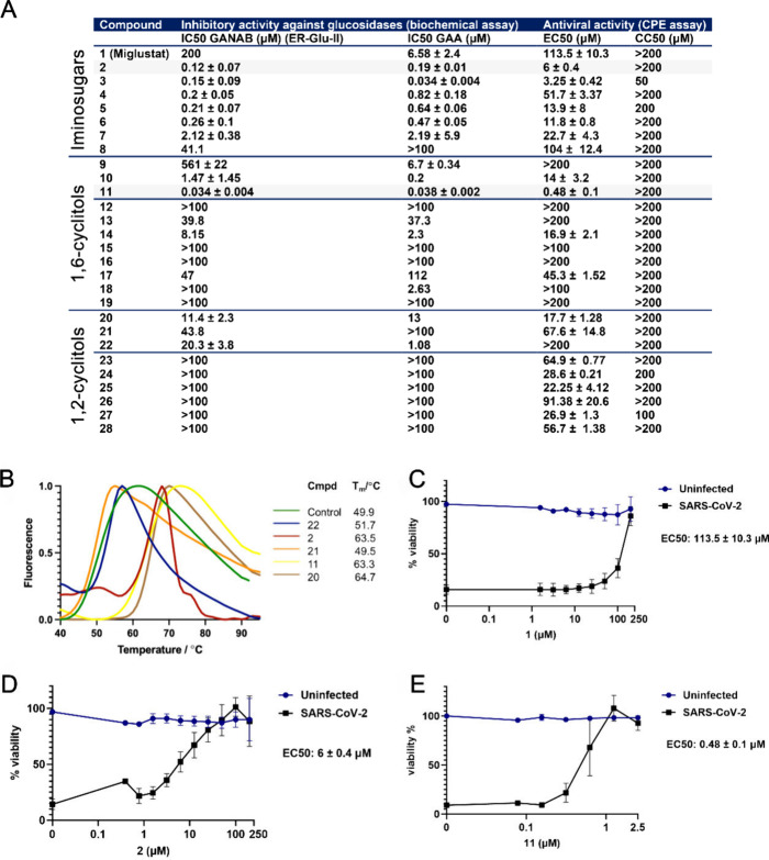
The combined inhibition of endoplasmic reticulum (ER) α-glucosidases I and II has been shown to inhibit replication of a broad range of viruses that rely on ER protein quality control. We found, by screening a panel of deoxynojirimycin and cyclitol glycomimetics, that the mechanism-based ER α-glucosidase II inhibitor, 1,6-epi-cyclophellitol cyclosulfate, potently blocks SARS-CoV-2 replication in lung epithelial cells, halting intracellular generation of mature spike protein, reducing production of infectious progeny, and leading to reduced syncytium formation. Through activity-based protein profiling, we confirmed ER α-glucosidase II inhibition in primary airway epithelial cells, grown at the air-liquid interface. 1,6-epi-Cyclophellitol cyclosulfate inhibits early pandemic and more recent SARS-CoV-2 variants, as well as SARS-CoV and MERS-CoV. The reported antiviral activity is comparable to the best-in-class described glucosidase inhibitors, all competitive inhibitors also targeting ER α-glucosidase I and other glycoprocessing enzymes not involved in ER protein quality control. We propose selective blocking ER-resident α-glucosidase II in a covalent and irreversible manner as a new strategy in the search for effective antiviral agents targeting SARS-CoV-2 and other viruses that rely on ER protein quality control.


Conflict of interest statement

The authors declare no competing financial interest.

Figures

Figure 1
Figure 1
(A) Schematic of N-glycan processing of newly synthesized proteins in the ER lumen. Folding of nascent proteins in the ER is promoted by the calnexin–calreticulin cycle (CNX–CRT cycle), which relies on glycan trimming by ER α-Glu II (ER-II). (B) Focused library of 28 iminosugars and cyclitol subjects of the here-presented studies.
Figure 2
Figure 2
ER α-Glu II inhibitory potency correlates with reduction of SARS-CoV-2 mediated cytopathic effect in cell culture. (A) IC50 values of compounds in in vitro enzyme activity assays with ER α-Glu II and GAA, and EC50 and CC50 values of compounds determined by CPE reduction assays with SARS-CoV-2. (B) Thermal shift profile of preincubated ER α-Glu II with inhibitors. (C–E) SARS-CoV-2 CPE reduction assay dose–response curves of (C) Miglustat 1, (D) naphthyl-deoxynojirimycin 2, and (E) cyclosulfate 11. n = 3 independent experiments. The viability of uninfected compound-treated cells was established by MTS assay in parallel. Mean ± SEM values are shown. The 50% inhibitory concentration (EC50) values were determined by nonlinear regression with GraphPad Prism 6.
Figure 3
Figure 3
Spectrum of activity of 1,6-epi-cyclophellitol cyclosulfate 11 and iminosugars 1 and 2 against various coronaviruses. (A, B) Viral load reduction assay on H1299/ACE2 cells with SARS-CoV-2 (MOI 1) in the presence of compounds 1 or 11. (A) Infectious virus titer and (B) extracellular viral RNA copy numbers were quantified by plaque assay and RT-qPCR, respectively. Uninfected compound-treated cells were assessed by MTS assay in parallel to measure cytotoxicity of the compounds. n = 3 independent experiments. Mean ± SEM values are shown. (C) The specific infectivity of treated (using 1.5 μM of compound 11) and untreated samples was calculated by dividing the infectious virus titer (PFU/mL) by the viral RNA copy number (copies/mL). Viral load reduction assays with (D) SARS-CoV-2 variants in H1299/ACE2 cells, (E) SARS-CoV in Vero E6 cells, (F) MERS-CoV in HuH-7 cells, and (G) HCoV-229E in H1299/ACE2 cells (all with MOI 1), and treatment with 1, 2, or 11. Supernatant was harvested at 16 hpi to quantify infectious progeny by plaque assay. n = 3 independent experiments. Uninfected compound-treated cells were measured by MTS assay in parallel to assess the cytotoxicity of the compounds. Mean ± SEM values are shown. Statistical analysis was conducted using one-way ANOVA, and significant differences are indicated by ∗, p < 0.05.
Figure 4
Figure 4
Reduction of SARS-CoV-2 infection in primary bronchial epithelial cells is consistent with inhibition of active ER α-glucosidase II. (A) Viral load reduction assay in ALI-PBEC. Supernatant was harvested at 48 hpi to quantify infectious progeny by plaque assay. n = 3 independent experiments. Mean ± SEM values are shown. Statistical analysis was conducted using one-way ANOVA, and significant differences are indicated by ∗, p < 0.05. (B) The viability of uninfected compound-treated cells was measured by LDH release assay in parallel, to assess the cytotoxicity of the compounds. Mean ± SEM values are shown. (C) Following compound treatment, cells were lysed and the lysate at pH 7.0 was treated with activity-based probe (ABP) 29 to assess cellular retaining α-glucosidase activities in a competitive activity-based protein profiling experiment. A representative gel of three independent experiments (with two biological replicates/ALI-PBEC inserts each) is shown. (D) Schematic representation of ABP labeling. Part of the figure in (D) was adapted from ref (52), and part was generated using Servier Medical Art, provided by Servier, licensed under a Creative Commons Attribution 3.0 unported license. Figure S4 shows the Gelcode Blue stained gel of (C), which demonstrated that equal amounts of protein were loaded.
Figure 5
Figure 5
1,6-epi-Cyclophellitol cyclosulfate 11 inhibits SARS-CoV-2 replication and syncytium formation by reducing intracellular spike protein levels and processing. (A) Virucidal activity assay in which SARS-CoV-2 was incubated with compound 11 or 70% ethanol (as control) for 1 h at RT, and (remaining) infectious progeny was quantified by plaque assay. n = 2 independent experiments. Mean ± SEM values are shown. Statistical analysis was conducted using one-way ANOVA, and significant differences are indicated by ∗, p < 0.05. (B) H1299/ACE2 cells were infected with SARS-CoV-2 (MOI 3) and treated with 11 from 1 hpi until harvesting at the indicated time points. Intracellular viral RNA copies were quantified by RT-qPCR. n = 3 independent experiments. (C, D) Plaque reduction assay was performed with 1 h infection and incubation for 3 days until cells were fixed and stained with crystal violet. Cells were treated with 5 μM compound 11, either before infection (pretreatment), during infection, or after infection (postinfection) in the overlay. Treatment with RDV in the overlay was used as a control. n = 2 independent experiments. Mean ± SEM values are shown. (E) Western blot analysis of viral S protein in the medium and cell lysates of untreated (UNT) or compound 11 treated (2 μM) H1299/ACE2 cells that were infected with SARS-CoV-2 (MOI 2) and analyzed at 10 hpi using an S2-specific antibody. The medium was spiked with ovalbumin (Ova) as a recovery control and was concentrated before a sample corresponding to ∼250 μL of the original medium volume was analyzed. α-Tubulin was used as a loading control for cell lysates. (F) H1299/ACE2 cells were infected with SARS-CoV-2 (MOI 0.1), fixed at 10 hpi, and the viral S protein and ER marker PDI were visualized by immunofluorescence microscopy. Cells were stained with human anti-SARS-CoV-2 S protein antibody (green), mouse anti-PDI antibody for ER staining (red), and Hoechst for visualizing nuclei (blue). White arrows indicate colocalization of S with PDI. Images are representative of n = 2 independent experiments.

Similar articles

Cited by

References

    1. Feng T.; Zhang J.; Chen Z.; Pan W.; Chen Z.; Yan Y.; Dai J. Glycosylation of viral proteins: Implication in virus-host interaction and virulence. Virulence 2022, 13, 670–683. 10.1080/21505594.2022.2060464. - DOI - PMC - PubMed
    1. O’Keefe S.; Roebuck Q. P.; Nakagome I.; Hirono S.; Kato A.; Nash R.; High S. Characterizing the selectivity of ER α-glucosidase inhibitors. Glycobiology 2019, 29, 530–542. 10.1093/glycob/cwz029. - DOI - PMC - PubMed
    1. Hebert D. N.; Foellmer B.; Helenius A. Glucose trimming and reglucosylation determine glycoprotein association with calnexin in the endoplasmic reticulum. Cell 1995, 81, 425–33. 10.1016/0092-8674(95)90395-X. - DOI - PubMed
    1. Kostova Z.; Wolf D. H. For whom the bell tolls: protein quality control of the endoplasmic reticulum and the ubiquitin-proteasome connection. EMBO J. 2003, 22, 2309–2317. 10.1093/emboj/cdg227. - DOI - PMC - PubMed
    1. Gruters R. A.; Neefjes J. J.; Tersmette M.; de Goede R. E.; Tulp A.; Huisman H. G.; Miedema F.; Ploegh H. L. Interference with HIV-induced syncytium formation and viral infectivity by inhibitors of trimming glucosidase. Nature 1987, 330, 74–7. 10.1038/330074a0. - DOI - PubMed

LinkOut - more resources