

Evaluation of primary teeth root canal orifices with naked eye and using magnifying loupes - An in vivo study
- PMID: 39220757
- PMCID: PMC11362793
- DOI: 10.1016/j.jobcr.2024.08.001
Evaluation of primary teeth root canal orifices with naked eye and using magnifying loupes - An in vivo study
Abstract
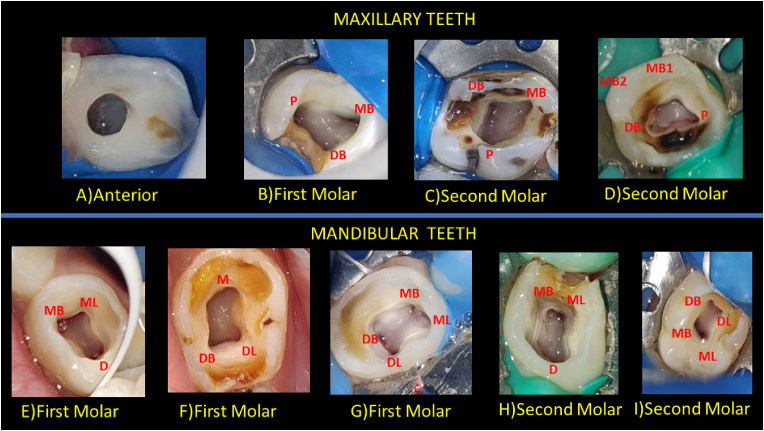
Background: Knowledge of the anatomy and morphology of root canal orifices and variations are vital elements affecting treatment outcomes.
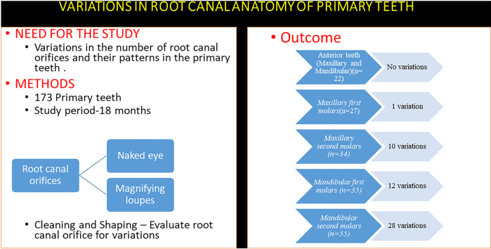
Aim: The objective of this study was to evaluate variations in the number of root canal orifices and their patterns in primary teeth, as identified by both the naked eye and under magnifying loupes.
Materials and methods: Total of 173 primary teeth was scheduled for pulpectomy over a period of 18 months. Two examiners assessed the number and pattern of the root canal orifices. After access cavity preparation, the operator recorded the number of root canal orifices with naked eye, and examiner recorded the same using magnifying loupes (3.5×). After cleaning and shaping, the same protocol was used. Collected data were statistically analyzed using SPSS version 23.0 and compared using a paired t-test.
Results: The overall variation in the in the identification of root canal orifices between the naked eye and magnifying loupes (3.005 ± 0.971) was statistically significant after access cavity preparation (P ≤ 0.05).
Conclusion: Magnifying loupes significantly enhances the determination of the number and pattern of root canal orifices in primary teeth. Therefore, the application of magnifying loupes is essential for accurately assessing variations in root canal orifices in primary dentition.
Keywords: Magnification; Magnifying loupes; Primary teeth; Root canal anatomy; Variation.
© 2024 The Authors.
Conflict of interest statement
The research is original, not under publication consideration elsewhere, and free of conflict of interest.
Figures
References
-
- Grindefjord M., Dahllöf G., Modéer T. Caries development in children from 2.5 to 3.5 years of age: a longitudinal study. Caries Res. 1995;29(6):449–454. - PubMed
-
- Dugas N.N., Lawrence H.P., Teplitsky P., Friedman S. Quality of life and satisfaction outcomes of endodontic treatment. J Endod. 2002;28(12):819–827. - PubMed
-
- Ahmed H.M. Anatomical challenges, electronic working length determination and current developments in root canal preparation of primary molar teeth. Int Endod J. 2013;46(11):1011–1022. - PubMed
-
- Salama F.S., Anderson R.W., McKnight-Hanes C.M., Barenie J.T., Myers D.R. Anatomy of primary incisor and molar root canals. Pediatr Dent. 1992;14(2):117–118. - PubMed
LinkOut - more resources
Full Text Sources
