Case Reports
. 2024 Aug 6;19(10):4583-4598.
doi: 10.1016/j.radcr.2024.07.005. eCollection 2024 Oct.

Strategic multidisciplinary management of pelvic carcinosarcoma: Emphasizing advanced diagnostic imaging and staged surgical interventions

Affiliations
Case Reports

Strategic multidisciplinary management of pelvic carcinosarcoma: Emphasizing advanced diagnostic imaging and staged surgical interventions

Mena Louis et al. Radiol Case Rep. .

Abstract

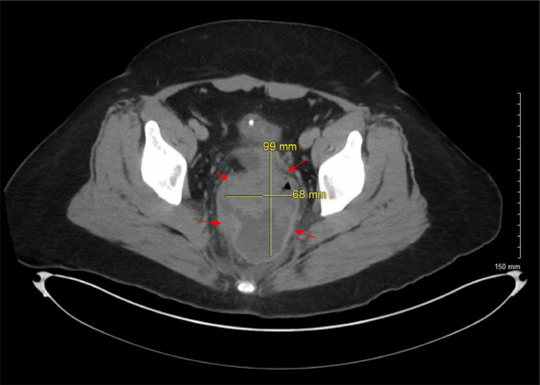
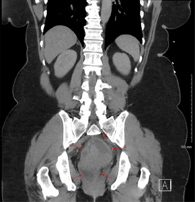
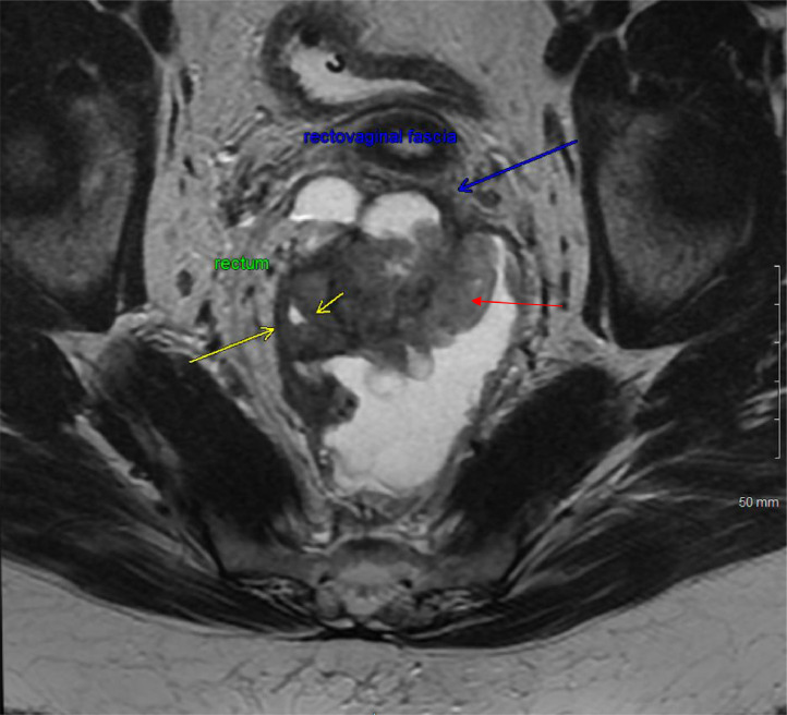
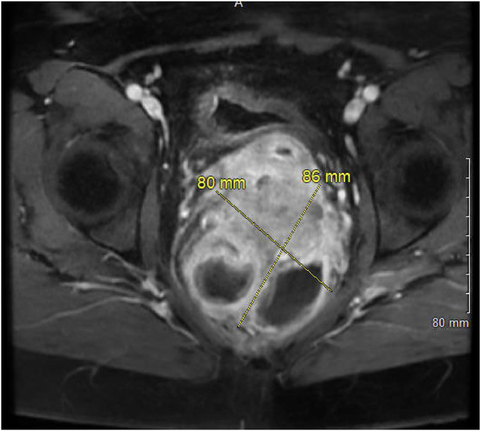
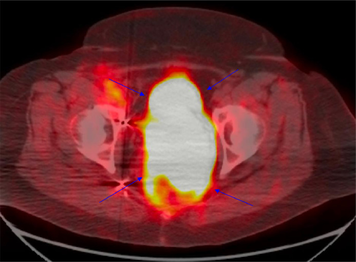
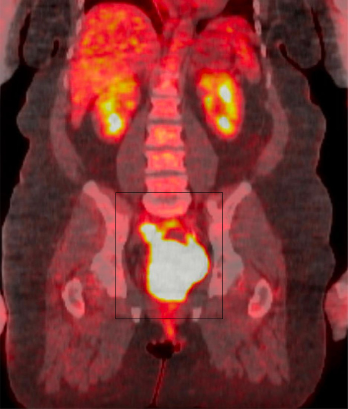
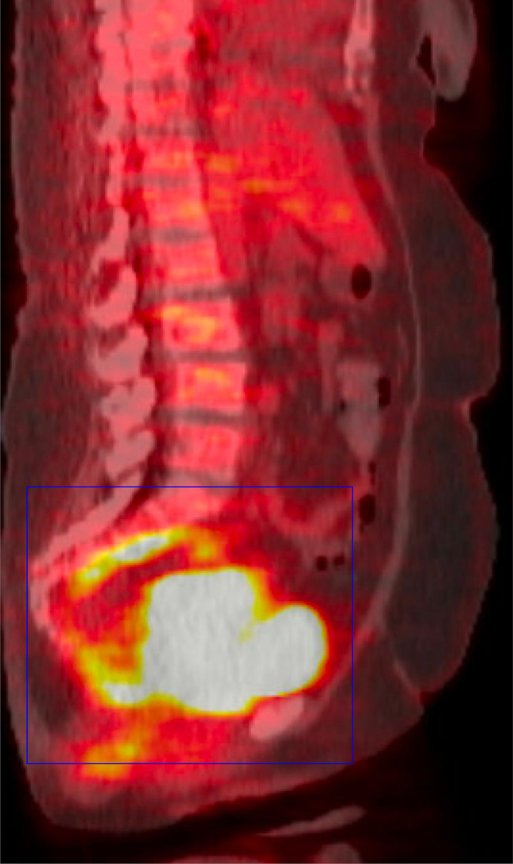
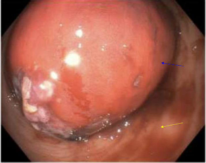
Pelvic carcinosarcoma is an aggressive malignancy with significant diagnostic and management hurdles due to its complex vascularity and potential for extensive local invasion. A 59-year-old female presented with severe abdominal pain and significant weight loss, leading to the discovery of a large, complex pelvic mass through CT scans, MRI, and PET CT, suggesting aggressive malignancy. Initial management included a robotic laparoscopic proximal sigmoid loop colostomy to alleviate obstruction. Significant vascularity led to consultations with Vascular Surgery and subsequent preoperative embolization. Definitive surgery involved a supralevator posterior exenteration for en bloc resection of the vagina, mass, and sigmoid colon, combined with a low anterior resection and an omental J flap in anticipation of potential postoperative radiation therapy. This case underscores the importance of integrated imaging and staged surgical interventions in managing pelvic carcinosarcoma, emphasizing a multidisciplinary approach to optimize outcomes and minimize complications.

Keywords: Advanced imaging; Biopsy; J flap; MRI; Multimodal imaging; Oncological diagnosis; PET CT; Pelvic carcinosarcoma; Posthysterectomy.


Figures

Fig 1
Fig. 1
Axial view CT of the abdomen and pelvis with IV contrast reveals a complex, centrally located mass in the pelvis that displays both cystic and solid characteristics. Marked by red arrows, the mass's ambiguous nature and central pelvic position raise concerns about a possible neoplastic process.
Fig 2
Fig. 2
Coronal view CT of the abdomen and PELVIS with and without IV contrast showcases a complex mass centrally located in the pelvis, exhibiting both cystic and solid components. Highlighted by red arrows, this mass presents an ambiguous origin but prompts concerns for a neoplastic process.
Fig 3
Fig. 3
MRI of the rectum with and without contrast in a coronal view displays a heterogeneously enhancing mixed solid and cystic lesion in the extraperitoneal pelvic region, marked by red arrow. This lesion, predominantly centered in the retro-rectal space, exerts pressure on the rectum, pushing it anterolaterally as indicated by yellow arrows, causing mass effect and probable luminal narrowing of the mid rectum. Additionally, as shown by blue arrow, the lesion is pushing the rectovaginal fascia anteriorly with possible extension into the lower posterior wall of the vagina.
Fig 4
Fig. 4
Axial LAVA Flex MRI reveals a heterogeneously enhancing lesion in the extraperitoneal pelvic region, centered predominantly in the retro-rectal space. This mixed solid and cystic lesion is captured in detail, illustrating its complex structure and positioning relative to critical pelvic anatomy.
Fig 5
Fig. 5
FDG PET CT Whole Body, axial view, shows a posterior pelvic mass exhibiting marked FDG uptake, indicated by blue arrows. This significant metabolic activity suggests high cellular activity, often associated with aggressive neoplastic processes.
Fig 6
Fig. 6
FDG PET CT Whole Body, coronal view, displays a posterior pelvic mass highlighted by a black box, showing marked FDG uptake. This significant uptake indicates intense metabolic activity within the mass, which is characteristic of aggressive neoplastic processes.
Fig 7
Fig. 7
FDG PET CT Whole Body in sagittal view showcases a posterior pelvic mass, highlighted by a blue box, which exhibits marked FDG uptake. This significant uptake suggests active metabolic processes typically associated with malignant tissue, providing essential information on the mass's behavior and aggressiveness.
Fig 8
Fig. 8
Colonoscopy image showing an obstructing rectal mass, indicated by a blue arrow, surrounded by normal mucosa, highlighted with a yellow arrow. This visualization contrasts the abnormal mass with the surrounding healthy tissue, providing a clear view of the extent and nature of the obstruction within the rectal lumen.
Unlabelled image
Fig. A
This image highlights tumor cells positively stained brown for cytokeratin 7 (CK7), indicated by a black arrow. The presence of CK7 is critical for distinguishing between gynecologic and nongynecologic tumors, as it is typically expressed in tissues like the breast, endometrium, lung, and ovary but absent in lower gastrointestinal cancers. This image, taken at 20x magnification, highlights the diagnostic utility of CK7 in identifying the tissue origin of tumors.
Unlabelled image
Fig. B
Displayed at 20x magnification, the tumor cells show positive staining for paired box gene 8 (PAX8), marked by a black arrow, suggesting a Mullerian origin. Pax 8 is expressed in gynecologic malignancies, thyroid cancer, and renal cell carcinoma.
Unlabelled image
Fig. C
This H&E-stained section at 20x magnification reveals the tumor's dual nature, comprising both carcinomatous (black star) and sarcomatous (blue circle) components. The carcinomatous areas depict poorly differentiated malignant epithelial cells, while the sarcomatous regions contain mesenchymal elements, illustrating the complex histopathological landscape of carcinosarcomas.
Unlabelled image
Fig. D
Highlighting sarcomatous features (blue circle) at 20x magnification, this H&E-stained image provides a close-up view of the tumor's mesenchymal elements, contributing to understanding its aggressive behavior.
Unlabelled image
Fig. E
At 10x magnification, this image captures carcinoma metastasis into a lymph node. It contrasts carcinomatous components (black star) with normal lymph node tissue (red triangle), emphasizing the tumor's invasive potential.
Unlabelled image
Fig. F
Displayed at 20x magnification, this H&E stained section shows the interplay between carcinomatous (black star) and sarcomatous (blue circle) features, providing a detailed view of the tumor's complex cellular composition.
Fig 9
Fig. 9
The pelvic angiogram postembolization depicts successful targeting of the right uterine artery (red arrow), right vaginal artery (black arrow), and left uterine artery (blue arrow). This strategic intervention is crucial for reducing the blood supply to the pelvic mass, potentially minimizing bleeding during subsequent surgical procedures, and reducing tumor viability.

Similar articles

References

    1. Kitazono I, Akahane T, Kobayashi Y, Yanazume S, Tabata K, Tasaki T, et al. Pelvic carcinosarcoma showing a diverse histology and hierarchical gene mutation with a common pole mutation to endometrial endometroid carcinoma: a case report. Int J Surg Pathol. 2022;30(8):891–899. doi: 10.1177/10668969221088880. - DOI - PubMed
    1. Elshaikh MA, Modh A, Jhingran A, Biagioli MC, Coleman RL, Gaffney DK, et al. Executive summary of the American Radium Society® appropriate use criteria for management of uterine carcinosarcoma. Gynecol Oncol. 2020;158(2):460–466. doi: 10.1016/j.ygyno.2020.04.683. - DOI - PubMed
    1. Singh R. Review literature on uterine carcinosarcoma. J Cancer Res Ther. 2014;10(3):461–468. doi: 10.4103/0973-1482.138197. - DOI - PubMed
    1. Arend R, Doneza JA, Wright JD. Uterine carcinosarcoma. Curr Opin Oncol. 2011;23(5):531–536. doi: 10.1097/CCO.0b013e328349a45b. - DOI - PubMed
    1. Cowdell I, Smyth SL, Eltawab S, Soleymani Majd H. Radical abdomino-pelvic surgery in the management of uterine carcinosarcoma with concomitant para-aortic lymphadenopathy metastasising from anal carcinoma. BMJ Case Rep. 2022;15(11):e252233. doi: 10.1136/bcr-2022-252233. - DOI - PMC - PubMed

Publication types

LinkOut - more resources