Case Reports
. 2024 Aug 7;19(10):4604-4609.
doi: 10.1016/j.radcr.2024.07.077. eCollection 2024 Oct.

External auditory canal involvement by nasopharyngeal carcinoma via eustachian tube spread: A case report

Affiliations
Case Reports

External auditory canal involvement by nasopharyngeal carcinoma via eustachian tube spread: A case report

Won Young Yoon et al. Radiol Case Rep. .

Abstract

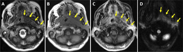
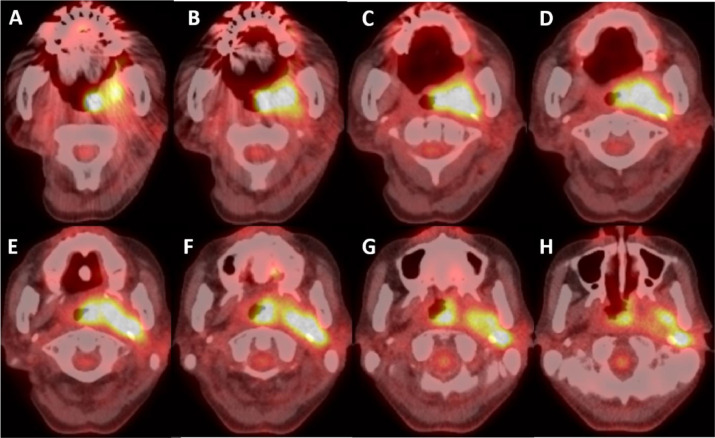
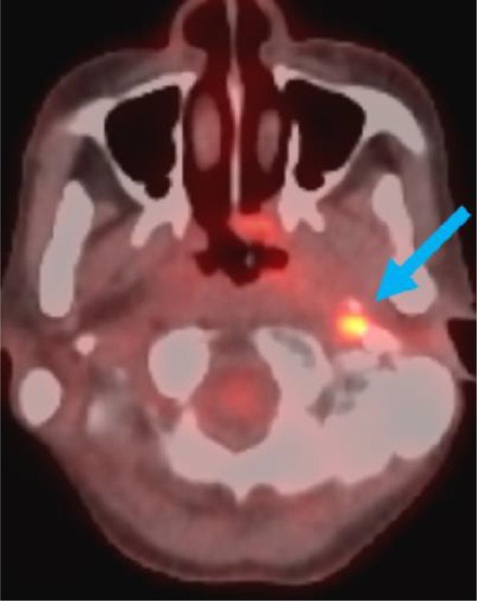
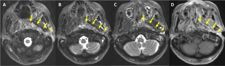
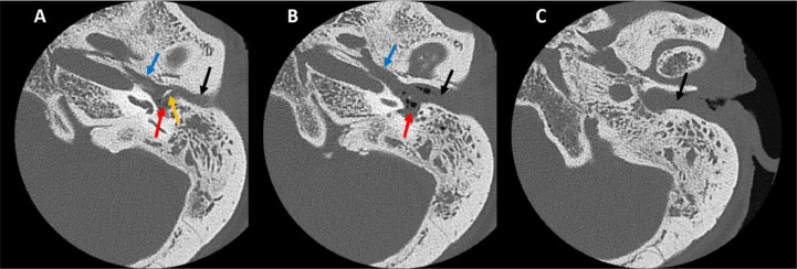
We present the imaging findings of a 44-year-old female patient who was diagnosed with nasopharyngeal carcinoma (NPC) extending from the nasopharynx to the external auditory canal (EAC) through the Eustachian tube (ET). The patient presented with a left neck submandibular lump on initial presentation that showed NPC upon fine needle aspiration, leading to chemoradiotherapy. Despite treatment, the patient experienced multiple relapses and later presented with aural symptoms, including left ear pain, foul-smelling drainage, and trismus on recurrence, and was subsequently diagnosed through biopsy. CT, MRI, and PET-CT scans revealed an extensive infiltrative nasopharyngeal mass extending into the left ET, involving the EAC. This rare case highlights the importance of considering the extension of NPC into the EAC as a potential etiology in patients who present with aural symptoms.

Keywords: Biopsy; Diagnosis; Discharge; Eustachian tube; External auditory canal; Magnetic resonance imaging; Management; Nasopharyngeal carcinoma; Otalgia; Positron emission tomography–computed tomography.


Figures

Fig 1
Fig. 1
Representative axial T2-weighted (A), T1-weighted (B), postcontrast T1-weighted (C), and diffusion weighted (D) MR images show extensive infiltrative left nasopharyngeal mass (arrows) that slightly cross the midline and on multiple images is seen to involve the left oropharynx, and left parapharyngeal, parotid, and masticator spaces. There is also involvement of left V3 nerve and the skull base, including the clivus, left greater wing of the sphenoid, and left pterygoid plate (not shown here).
Fig 2
Fig. 2
Axial PET-CT (sequential inferior to superior slices: A to H) of the head and neck shows intense FDG uptake of the left nasopharyngeal mass in a similar distribution to MRI findings in Fig. 1.
Fig 3
Fig. 3
Axial PET-CT shows hypermetabolic activity within the left posterior nasopharynx and Eustachian tube (arrow).
Fig 4
Fig. 4
Axial T2-weighted (sequential inferior to superior slices: A-C) and postcontrast T1-weighted (D) MR images show a recurrent mass in the left nasopharynx extends along the left Eustachian tube to the left middle ear and external auditory canal (arrows).
Fig 5
Fig. 5
Thin-slice axial CT images of the left temporal bone using bone algorithm and viewed in bone windows (sequential superior to inferior slices: A-C). There is a widened bony segment of the Eustachian tube (blue arrow), near-complete opacification of the left middle ear cavity (red arrow), likely disruption of middle ear ossicles (orange arrow), and external auditory canal opacification (black arrow).

Similar articles

References

    1. Yu MC, Yuan JM. Epidemiology of nasopharyngeal carcinoma. Semin Cancer Biol. 2002;12(6):421–429. - PubMed
    1. Zhang LF, Li YH, Xie SH, et al. Incidence trend of nasopharyngeal carcinoma from 1987 to 2011 in Sihui County, Guangdong Province, South China: an age-period-cohort analysis. Chin J Cancer. 2015;34(8):350–357. - PMC - PubMed
    1. Wei KR, Zheng RS, Zhang SW, et al. Nasopharyngeal carcinoma incidence and mortality in China, 2013. Chin J Cancer. 2017;36(1):90. - PMC - PubMed
    1. Lee AW, Foo W, Law SC, et al. Nasopharyngeal carcinoma: presenting symptoms and duration before diagnosis. Hong Kong Med J. 1997;3(4):355–361. - PubMed
    1. Lekskul A, Thanomteeranant S, Tangtammaruk P, et al. Isolated sixth nerve palsy as a first presentation of nasopharyngeal carcinoma: a case series. Int Med Case Rep J. 2021;14:801–808. - PMC - PubMed

Publication types

LinkOut - more resources