

Case Reports
doi: 10.1016/j.radcr.2024.07.060.
eCollection 2024 Oct.
Radiology of malignant degeneration of pilonidal sinus: Report of a case and review of the literature
Affiliations
- PMID: 39220790
- PMCID: PMC11363716
- DOI: 10.1016/j.radcr.2024.07.060
Item in Clipboard
Case Reports
Radiology of malignant degeneration of pilonidal sinus: Report of a case and review of the literature
Radiol Case Rep.
.
Abstract
Pilonidal sinus disease is a frequent and recurrent pathology in young adults, with a male predominance, while malignant transformation of the pilonidal sinus is a rare complication, it occurs in 0.1% of patients, with a poor prognosis. Early surgical removal of the primary lesion remains the best treatment. We report a case of malignant transformation of pilonidal disease into squamous cell carcinoma.
Keywords: Malignant transformation; Pilonidal sinus; Squamous cell carcinoma.
© 2024 The Authors. Published by Elsevier Inc. on behalf of University of Washington.
Figures

Axial and coronal sections of a pelvic MRI (A, B) show intergluteal mass with irregular and budding contours, intensely and heterogeneously enhanced after contrast, extending towards the sacrum without rectal extension (arrow). Axial section of a pelvic MRI (A, C) showing left inguinal adenopathy, heterogeneously enhanced after contrast, delineating central necrotic areas (Asterix).
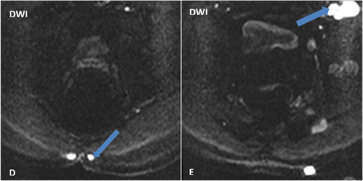
Axial section of MRI pelvic (D, E) showing diffusion hypersignal of the intergluteal mass and inguinal adenopathies (arrow), diffusion restriction suggests the tumoral nature of the mass.

Axial section of a pelvic CT scan (F, G, H) showing the same findings as MRI: the presence of inguinal adenopathies associated with left subcutaneous nodules, enhanced after contrast (arrow head). Axial section of a thoracic CT scan (I, J): absence of mediastinal adenopathy or pulmonary metastasis.

Axial section of an enhanced CT (I, J, K) showing the appearance of voluminous tissue mass lysing the left iliac bone and invading the iliac vascular pedicle (arrow), with an increase in size of the subcutaneous nodule (arrowhead).

Axial section of an enhanced CT (L, M) showing the appearance of necrotic mediastinal lymphadenopathy (arrowhead) and an excavated pulmonary nodule (arrow).
Similar articles
-
Squamous cell carcinoma arising from chronic sacrococcygeal pilonidal disease: a case report.World J Surg Oncol. 2017 Mar 17;15(1):65. doi: 10.1186/s12957-017-1129-0. World J Surg Oncol. 2017. PMID: 28302173 Free PMC article.
-
Verrucous carcinoma (Buschke-Lowenstein) arising in a sacrococcygeal pilonidal sinus tract: report of a case.Langenbecks Arch Surg. 2008 Jan;393(1):111-4. doi: 10.1007/s00423-007-0209-y. Epub 2007 Jul 3. Langenbecks Arch Surg. 2008. PMID: 17609975
-
Recurrent squamous cell carcinoma arising in a neglected pilonidal sinus: report of a case and literature review.Int J Clin Exp Med. 2014 Feb 15;7(2):446-50. eCollection 2014. Int J Clin Exp Med. 2014. PMID: 24600504 Free PMC article.
-
[Moderately differentiated squamous cell carcinoma associated pilonidal cyst].Cir Cir. 2014 Jan-Feb;82(1):87-92. Cir Cir. 2014. PMID: 25510796 Review. Spanish.
-
Fine-needle aspiration cytology of metastatic squamous-cell carcinoma arising in a pilonidal sinus, with literature review.Diagn Cytopathol. 1999 Jun;20(6):367-70. doi: 10.1002/(sici)1097-0339(199906)20:6<367::aid-dc7>3.0.co;2-s. Diagn Cytopathol. 1999. PMID: 10352909 Review.
References
-
- Ilker Sengul and all . Axillary pilonidal sinus: a case report. 2009. https://pubmed.ncbi.nlm.nih.gov/22666715 - PMC - PubMed
-
- Sondenaa K, Nesvik I, Anderson E, Natas O, Soreide JA. Patient characteristics and symptoms in chronic pilonidal sinus disease. Int J Colorectal Dis. 1995;10:39–42. - PubMed
-
- Trent JT, Kirsner RS. Wounds and malignancy. Adv Skin Wound Care. 2003;16:31–34. - PubMed
Publication types
LinkOut - more resources
Full Text Sources
