Review
. 2024 Aug;14(8):3327-3361.
doi: 10.1016/j.apsb.2024.05.012. Epub 2024 May 13.

Mitophagy and cGAS-STING crosstalk in neuroinflammation

Affiliations
Review

Mitophagy and cGAS-STING crosstalk in neuroinflammation

Xiaogang Zhou et al. Acta Pharm Sin B. 2024 Aug.

Abstract

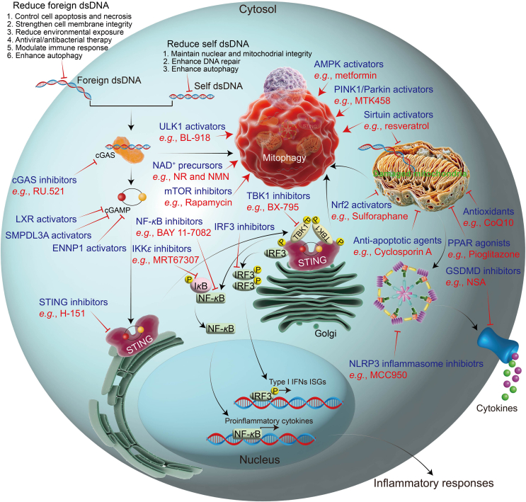
Mitophagy, essential for mitochondrial health, selectively degrades damaged mitochondria. It is intricately linked to the cGAS-STING pathway, which is crucial for innate immunity. This pathway responds to mitochondrial DNA and is associated with cellular stress response. Our review explores the molecular details and regulatory mechanisms of mitophagy and the cGAS-STING pathway. We critically evaluate the literature demonstrating how dysfunctional mitophagy leads to neuroinflammatory conditions, primarily through the accumulation of damaged mitochondria, which activates the cGAS-STING pathway. This activation prompts the production of pro-inflammatory cytokines, exacerbating neuroinflammation. This review emphasizes the interaction between mitophagy and the cGAS-STING pathways. Effective mitophagy may suppress the cGAS-STING pathway, offering protection against neuroinflammation. Conversely, impaired mitophagy may activate the cGAS-STING pathway, leading to chronic neuroinflammation. Additionally, we explored how this interaction influences neurodegenerative disorders, suggesting a common mechanism underlying these diseases. In conclusion, there is a need for additional targeted research to unravel the complexities of mitophagy-cGAS-STING interactions and their role in neurodegeneration. This review highlights potential therapies targeting these pathways, potentially leading to new treatments for neuroinflammatory and neurodegenerative conditions. This synthesis enhances our understanding of the cellular and molecular foundations of neuroinflammation and opens new therapeutic avenues for neurodegenerative disease research.

Keywords: Crosstalk; Innate immunity; Mitochondrial DNA; Mitophagy; Neurodegenerative diseases; Neuroinflammation; Therapeutic avenues; cGAS–STING.


Conflict of interest statement

The authors declare no conflicts of interest.

Figures

Image 1
Graphical abstract
Figure 1
Figure 1
This timeline depicts a selection of significant discoveries related to autophagy/mitophagy (light brown), cGAS–STING (light yellow), and their interactions (light green). Regarding the research on autophagy, Christian de Duve first coined the term ‘autophagy’ in 1963, leading to a series of crucial discoveries. These include the complex pathways of autophagy/mitophagy, ranging from simple yeast organisms to animals such as mice. In 2016, Yoshinori Ohsumi was rightfully awarded the Nobel Prize in Physiology or Medicine for his pioneering work in the field of autophagy, underscoring the importance of this area of research. In the study of cGAS–STING, notable discoveries include the identification of key proteins such as IFN, NF-κB, and IRF3; the recognition of STING as a crucial adaptor protein for intracellular DNA-induced IFN response; and the discovery of cGAS as a cytosolic DNA sensor activating STING. Over the past decade, there has been an increasing focus on the interplay between autophagy/mitophagy and cGAS–STING. This includes research on the regulatory effect of cGAS–STING on the induction of autophagy/mitophagy and the inhibitory impact of autophagy/mitophagy on the activation of cGAS–STING and subsequent immune responses.
Figure 2
Figure 2
The cellular functions of mitophagy and the c-GAS–STING pathway and their implications in various human diseases.
Figure 3
Figure 3
The molecular mechanisms of mitophagy. The diagram illustrates two classical mitophagy pathways: the ubiquitin-dependent pathway and the ubiquitin-independent pathway. In the ubiquitin-dependent pathway, when mitochondria are stressed, PINK1 is stabilized on the OMM and activated through autophosphorylation. It then phosphorylates Parkin and ubiquitin, enhancing Parkin's E3 ligase activity. This results in the ubiquitination of several OMM proteins, which results in the formation of polyubiquitin chains that PINK1 further phosphorylates, creating a self-amplifying loop. These chains attract adaptor proteins such as OPTN, nuclear dot protein 52 kDa (NDP52), NBR1, and p62, which bind to LC3, initiating autophagosome formation. The binding of OPTN to ubiquitin chains is further strengthened by TBK1-mediated phosphorylation. In the ubiquitin-independent pathway, under hypoxic conditions, proteins such as FUNDC1, BNIP3, and NIX play crucial roles in recruiting autophagosomes to mitochondria through their interaction with LC3. During mitophagy, Ambra1 is instrumental in relocating HUWE1 from the cytosol to the mitochondria, leading to the degradation of mitofusin 2, a necessary step for Ambra1-driven mitophagy. Additionally, the IκB kinase α phosphorylates S1014 on Ambra1, enabling its interaction with LC3. PHB2, an inner mitochondrial membrane protein, is key for directing mitochondria toward autophagic degradation. The externalization of cardiolipin (CL) to the OMM in response to mitochondrial damage acts as a signal for the selective autophagic removal of dysfunctional mitochondria, with CL interacting with LC3 in mammalian cortical neurons. Ceramide serves as a selective mitophagy receptor by directly binding to LC3, as does FKBP8, an OMM protein, promoting the degradation of damaged mitochondria through interactions with LC3.
Figure 4
Figure 4
DNA sensing and signaling in the cGAS–STING pathway. This pathway leads to the production of type I IFNs and inflammatory cytokines. Abnormal DNA in the cytoplasm, originating from either pathogen infection (bacteria, DNA viruses, or retroviruses) or cellular damage, activates this process. cGAS binds to double-stranded DNA (dsDNA) and becomes active, converting ATP and GTP into 2′,3′-cGAMP. This compound subsequently activates the adaptor protein STING. STING, upon binding to cGAMP, changes conformation and moves from the ER to the Golgi. In the Golgi, STING attracts TBK1, which phosphorylates the C-terminal tail of STING. This phosphorylated STING recruits IFN regulatory factor 3 (IRF3), which TBK1 also phosphorylates and activates. The activated IRF3 dimer travels to the nucleus, where it regulates type 1 IFN transcription and triggers type I IFN responses. Secreted IFN-β, through its receptor, activates TYK and JAK signals, influencing the STAT, MAPK, and PI3K pathways. This enhances IFN-stimulated gene transcription via a positive feedback loop. Concurrently, the cGAS–STING pathway activates NF-κB signaling, regulating inflammatory cytokine gene transcription. The NLRP3 inflammasome, which is triggered by increased ROS, is further upregulated by the STING protein. Additionally, STING initiates autophagy for degradation.
Figure 5
Figure 5
Crosstalk between the regulatory mechanism of autophagy/mitophagy and cGAS–STING. The cGAS–STING pathway is a crucial mechanism in the innate immune system and is primarily responsible for detecting cytoplasmic DNA, which is indicative of either pathogenic infection or cellular damage. When cGAS encounters this DNA, it becomes activated and synthesizes the messenger molecule cGAMP. This molecule then binds to STING, a protein on the ER, triggering its conformational change and relocation to the Golgi apparatus. STING activates a cascade of immune responses, primarily through the activation of TBK1 and subsequent phosphorylation of IRF3 and NF-κB activation, leading to the production of type I IFNs and inflammatory cytokines. Autophagy and mitophagy are sophisticated cellular processes for maintaining cellular homeostasis. Autophagy is activated by nutrient deprivation or other stressors, in turn engaging the ULK1 complex and the Beclin-1 complex, which are modulated by the balance of AKT and AMPK signaling. The ULK1 complex initiates autophagy, while the Beclin-1 complex nucleates the formation of autophagosomes. Mitophagy, a specialized form of autophagy that targets mitochondria, is primarily regulated by the PINK1/Parkin pathway. PINK1 accumulates on damaged mitochondria, where it recruits Parkin, which ubiquitinates mitochondrial proteins, signaling for autophagic degradation. Proteins such as p62 are involved in recognizing ubiquitinated proteins and facilitating mitophagy. Intriguingly, the cGAS–STING pathway intersects with these autophagic processes. cGAS and STING can undergo autophagic degradation mediated by p62, which recognizes ubiquitinated forms of these proteins. Beclin-1, an essential component of the autophagy initiation complex, can both inhibit and be activated by cGAS, indicating a complex regulatory interaction. Furthermore, ULK1 can inhibit the phosphorylation of STING, which is crucial for its activation, and attenuate the activity of cGAS. In this intricate network, TBK1 further enhances the ubiquitination of OPTN, another autophagy-related protein, adding another layer of regulation. This interplay between the autophagy/mitophagy pathway and the cGAS–STING pathway highlights the complexity of cellular responses to stress and infection, revealing potential therapeutic targets for diseases involving these pathways.
Figure 6
Figure 6
The role of autophagy/mitophagy and cGAS–STING crosstalk in microglial activation. In the advanced stages of neurodegenerative disease, resting M0 microglia can be activated to the M1 phenotypes under various stimuli. These include endogenous factors such as mtDNA, nuclear DNA, senescence, autophagy deficiency, and cell death, as well as exogenous factors such as microbial and foreign DNA. These factors are crucial for activating the cGAS–STING signaling pathway. In the early stages of neurodegenerative disease, resting M0 microglia can be polarized toward the M2 phenotype. This activation is achieved through the modulation of the type I IFN response, maintenance of cellular and protein homeostasis, clearance of damaged mitochondria, inhibition of the NLRP3 inflammasome, degradation of cGAS and STING, prevention of mtDNA release, control of cell death, and reduction of ROS. These processes are attributed to the induction of autophagy/mitophagy. When activated, M1 microglia release pro-inflammatory cytokines (such as TNF-α and IL-1β) and reactive species (including ROS and NO). These substances can induce neuronal damage and disrupt the integrity of the BBB. Conversely, M2 microglia release anti-inflammatory cytokines and growth factors that support neuronal survival and regeneration, and aid in the restoration and maintenance of BBB integrity.
Figure 7
Figure 7
Potential therapeutic targets and interventions focusing on the crosstalk between autophagy/mitophagy and the cGAS–STING pathway could be pivotal in mitigating the inflammatory responses associated with neurodegenerative diseases. By detecting dsDNA and subsequently activating the cGAS–STING pathway, this overview provides potential intervention sites. These include reducing foreign and self-dsDNA, targeting compounds that inhibit the cGAS–STING–TBK1 signaling pathway, targeting compounds that activate autophagy/mitophagy, targeting compounds that protect mitochondria, and targeting compounds that inhibit NLRP3 inflammasome-mediated pyroptosis.
Figure 8
Figure 8
The chemical structures of mitophagy activators are used in various neurological diseases.
Figure 9
Figure 9
The chemical structures of the identified cGAS, STING, TBK1, and IκB kinase ε (IKKε) inhibitors targeting the cGAS–STING–TBK1 signaling pathway.

Similar articles

Cited by

References

    1. Amor S., Puentes F., Baker D., van der Valk P. Inflammation in neurodegenerative diseases. Immunology. 2010;129:154–169. - PMC - PubMed
    1. Zhou X.G., Qiu W.Q., Yu L., Pan R., Teng J.F., Sang Z.P., et al. Targeting microglial autophagic degradation of the NLRP3 inflammasome for identification of thonningianin A in Alzheimer's disease. Inflamm Regen. 2022;42:25. - PMC - PubMed
    1. Brown G.C., Neher J.J. Microglial phagocytosis of live neurons. Nat Rev Neurosci. 2014;15:209–216. - PubMed
    1. Engelhardt B., Ransohoff R.M. Capture, crawl, cross: the T cell code to breach the blood–brain barriers. Trends Immunol. 2012;33:579–589. - PubMed
    1. He X., Wang X., Yang L., Yang Z., Yu W., Wang Y., et al. Intelligent lesion blood–brain barrier targeting nano-missiles for Alzheimer's disease treatment by anti-neuroinflammation and neuroprotection. Acta Pharm Sin B. 2022;12:1987–1999. - PMC - PubMed

LinkOut - more resources