. 2024 Aug 28:12:e17864.
doi: 10.7717/peerj.17864. eCollection 2024.

The role of DNA topoisomerase 1α (AtTOP1α) in regulating arabidopsis meiotic recombination and chromosome segregation

Affiliations

The role of DNA topoisomerase 1α (AtTOP1α) in regulating arabidopsis meiotic recombination and chromosome segregation

Ibrahim Eid Elesawi et al. PeerJ. .

Abstract

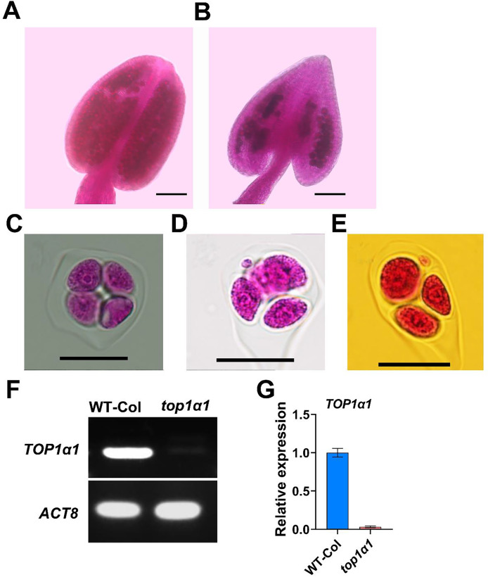
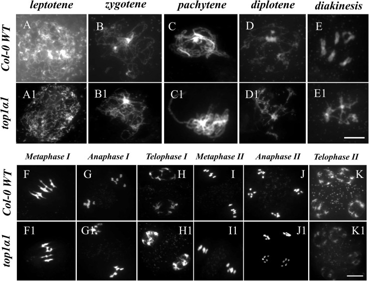
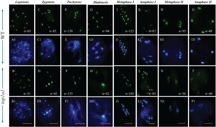
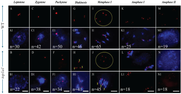
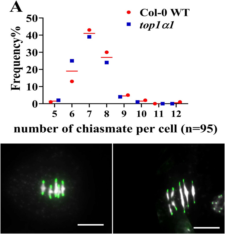
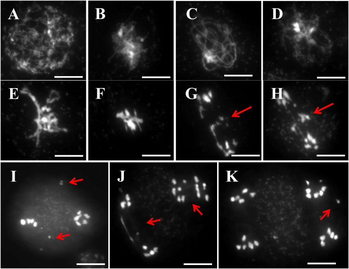
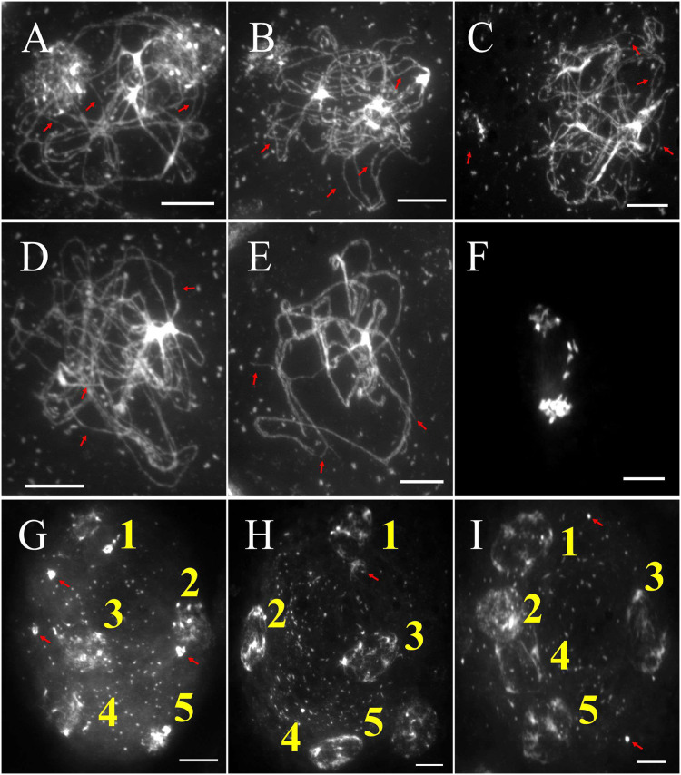
Meiosis is a critical process in sexual reproduction, and errors during this cell division can significantly impact fertility. Successful meiosis relies on the coordinated action of numerous genes involved in DNA replication, strand breaks, and subsequent rejoining. DNA topoisomerase enzymes play a vital role by regulating DNA topology, alleviating tension during replication and transcription. To elucidate the specific function of DNA topoisomerase 1α ( A t T O P 1 α ) in male reproductive development of Arabidopsis thaliana, we investigated meiotic cell division in Arabidopsis flower buds. Combining cytological and biochemical techniques, we aimed to reveal the novel contribution of A t T O P 1 α to meiosis. Our results demonstrate that the absence of A t T O P 1 α leads to aberrant chromatin behavior during meiotic division. Specifically, the top1α1 mutant displayed altered heterochromatin distribution and clustered centromere signals at early meiotic stages. Additionally, this mutant exhibited disruptions in the distribution of 45s rDNA signals and a reduced frequency of chiasma formation during metaphase I, a crucial stage for genetic exchange. Furthermore, the atm-2×top1α1 double mutant displayed even more severe meiotic defects, including incomplete synapsis, DNA fragmentation, and the presence of polyads. These observations collectively suggest that A t T O P 1 α plays a critical role in ensuring accurate meiotic progression, promoting homologous chromosome crossover formation, and potentially functioning in a shared DNA repair pathway with ATAXIA TELANGIECTASIA MUTATED (ATM) in Arabidopsis microspore mother cells.

Keywords: 45s rDNA; ATM; Centromere; DNA topoisomerase 1α; FISH; Meiosis.


Conflict of interest statement

Diaa Abd El-Moneim is an Academic Editor for PeerJ.

Figures

Figure 1
Figure 1. top1α1 plants exhibited reduced fertility and induced abnormal meiotic products.
The anther of the wild-type exhibited a density of pollen grains (A), while the anthers of the top1α1 mutant displayed a significantly lower abundance of pollen grains (B). Unlike the wild-type, top1α1, tetrads with four microspores contained polyads with small microspores, suggesting the occurrence of a meiotic abnormality. A total of 50 observations were conducted, revealing that 6% of the observed tetrads had abnormalities. A wild-type of tetrad with four microspores (C). top1α1 polyads with additional small microspores (D and E). AtTOP1α mRNA is disrupted across the T-DNA insertion sites in the top1α1 line (F and G). Scale bars = 50 µm (A and B) and 20 µm (C–E).
Figure 2
Figure 2. DAPI staining of meiotic chromosomes revealed abnormal chromosomal behavior in top1α1 mutants during prophase I.
Wild-type chromosome behavior is shown in (A)-(K), while (A1)-(K1) correspond to top1α1. Unlike the wild-type, top1α1 displayed abnormal chromosome features from zygotene to metaphase I, including the formation of multivalent links between chromosomes instead of normal bivalents and interlocks between homologous chromosomes. Chromosomes were stained with DAPI. Scale bar = 10 µm.
Figure 3
Figure 3. top1α1 induces unbalanced chromosome segregation. FISH analysis with a centromere probe.
(A–P) Wild-type (A, C, E, G, I, K, M, O) and top1α1 (B, D, F, H, J, L, N, P) meiotic cells at leptotene (A and B), zygotene (C and D), pachytene (E and F), diakinesis (G and H), metaphase I (I and J), anaphase I (K and L), metaphase II (M and N), and anaphase II (O and P) stages. FISH with a centromere probe revealed a similar number of signals in wild-type and top1α1 cells at leptotene (10 in wild-type and 8–10 in top1α1). In wild-type cells, the number of centromeric foci decreased from 10 at zygotene to 5 at pachytene. In contrast, top1α1 cells displayed fewer and larger foci at zygotene (4 foci) and pachytene (3 foci). While both wild-type and top1α1 diakinesis and metaphase I cells had the expected five pairs (10 signals) of centromeres, top1α1 cells frequently exhibited univalent and multivalent chromosomes. No significant differences were observed between WT and top1α1 cells from anaphase I to telophase II. Blue: DAPI staining of chromosomes; Green: centromere FISH signals. n = number of cells observed with the corresponding phenotype. Scale bar = 10 µm.
Figure 4
Figure 4. FISH with a 45S rDNA probe revealed the same number of signals in wild-type and top1α1 plantsat leptotene and zygotene.
At pachytene, the WT showed a single intense 45S rDNA signal, while top1α1 displayed two separate signals. At metaphase I, WT signals were located on two bivalent chromosomes (2–4), whereas top1α1 signals were on two bivalents and a univalent chromosome. Both WT and top1α1 cells displayed similar foci at diakinesis, anaphase I, and anaphase II. Blue indicates DAPI-stained chromosomes; red indicates 45S rDNA FISH signals. Scale bar = 10 µm.
Figure 5
Figure 5. Comparing the average number of chiasmata formations per one PMC in the wild-type and top1α1 at metaphase I.
Figure 6
Figure 6. The meiotic chromosomal behavior of atm (by DAPI).
(A) Leptotene, (B) zygotene, (C) pachytene. (D) At diplotene, the beginning of the emergence of interlocking and overlapping between non-homologous chromosomes. (E) At diakinesis, intertwined between the non-homologous chromosomes. (F) At metaphase I, atm formed a multivalent link between three or more chromosomes and univalent link. From anaphase I (G and H) through telophase II (K), bridges between chromosome groups and chromosome fragmentation are observed. Telophase II cells also exhibit unequal chromosome numbers. White indicates chromosomes stained with DAPI. Scale bar = 20 µm. Red arrows indicate incomplete migration of chromosomes.
Figure 7
Figure 7. The meiotic chromosomal behavior atm×top1α1 (by DAPI).
(A–C) Zygotene, (D and E) pachytene, (D) anaphase I, (G–I) telophase II. White indicates chromosomes stained with DAPI. Scale bar = 10 µm. (A–E) Red arrows indicate incomplete synapsis. (G–I) The red arrows indicate the presence of chromosome pieces that did not migrate to one of the daughter cells.

Similar articles

References

    1. Alavattam KG, Maezawa S, Andreassen PR, Namekawa SH. Meiotic sex chromosome inactivation and the XY body: a phase separation hypothesis. Cellular and Molecular Life Sciences. 2021;79(1):18. doi: 10.1007/s00018-021-04075-3. - DOI - PMC - PubMed
    1. Armstrong S. Plant Meiosis. Cham: Springer; 2013. Spreading and fluorescence in situ hybridization of male and female meiocyte chromosomes from Arabidopsis thaliana for cytogenetical analysis; pp. 3–11. - PubMed
    1. Chang S, Li C, Jiang Y, Long Y, Li Y, Yin J. Characteristics of the pollen morphology and viability of Bougainvillea (Nyctaginaceae) Scientia Horticulturae. 2021;277:109732. doi: 10.1016/j.scienta.2020.109732. - DOI
    1. Chang F, Zhang Z, Jin Y, Ma H. Flower Development. Cham: Springer; 2014. Cell biological analyses of anther morphogenesis and pollen viability in arabidopsis and rice; pp. 203–216. - PubMed
    1. Cui DL, Xu CX, Wang P, Gao TY, Wang B, Yu TY. Male gametogenesis in flowering plants. Frontiers in Sustainable Food Systems. 2024;7:1333544. doi: 10.3389/fsufs.2023.1333544. - DOI

MeSH terms

Substances

LinkOut - more resources