Randomized Controlled Trial
. 2024 Dec 1;9(12):1082-1092.
doi: 10.1001/jamacardio.2024.3200.

Lesion-Level Effects of LDL-C-Lowering Therapy in Patients With Acute Myocardial Infarction: A Post Hoc Analysis of the PACMAN-AMI Trial

Affiliations
Randomized Controlled Trial

Lesion-Level Effects of LDL-C-Lowering Therapy in Patients With Acute Myocardial Infarction: A Post Hoc Analysis of the PACMAN-AMI Trial

Flavio G Biccirè et al. JAMA Cardiol. .

Abstract

Importance: Previous studies investigated atherosclerotic changes induced by lipid-lowering therapy in extensive coronary segments irrespective of baseline disease burden (a vessel-level approach).

Objective: To investigate the effects of lipid-lowering therapy on coronary lesions with advanced atherosclerotic plaque features and presumably higher risk for future events.

Design, setting, and participants: The PACMAN-AMI randomized clinical trial (enrollment: May 2017 to October 2020; final follow-up: October 2021) randomized patients with acute myocardial infarction to receive alirocumab or placebo in addition to high-intensity statin therapy. In this post hoc lesion-level analysis, nonculprit lesions were identified as segments with plaque burden 40% or greater defined by intravascular ultrasound (IVUS). IVUS, near-infrared spectroscopy, and optical coherence tomography images at baseline and the 52-week follow-up were manually matched by readers blinded to treatment allocation. Data for this study were analyzed from October 2022 to November 2023.

Interventions: Alirocumab or placebo in addition to high-intensity statin therapy.

Main outcomes and measures: Lesion-level imaging outcome measures, including high-risk plaque characteristics and phenotypes.

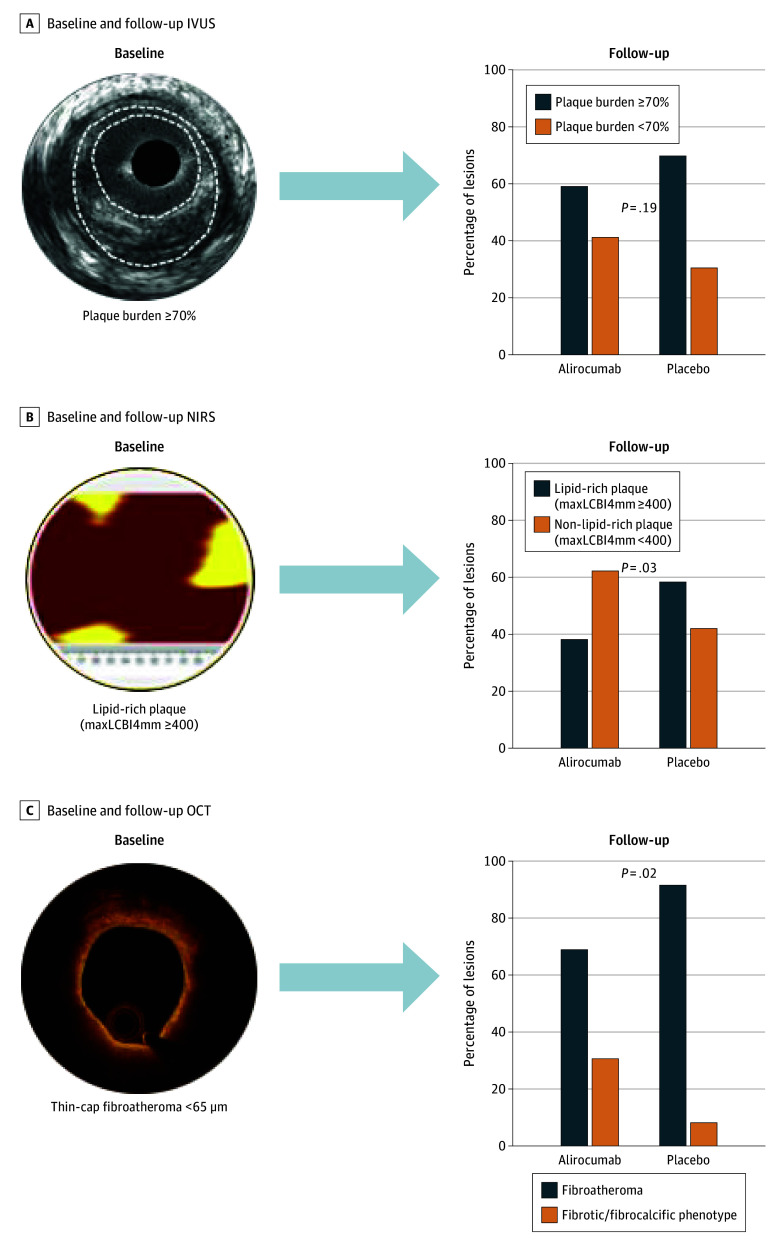
Results: Of the 245 patients in whom lesions were found, 118 were in the alirocumab group (mean [SD] age, 58.2 [10.0] years; 101 [85.6%] male and 17 [14.4%] female) and 127 in the placebo group (mean [SD] age, 57.7 [8.8] years; 104 [81.9%] male and 23 [18.1%] female). Overall, 591 lesions were included: 287 lesions (118 patients, 214 vessels) in the alirocumab group and 304 lesions (127 patients, 239 vessels) in the placebo group. Lesion-level mean change in percent atheroma volume (PAV) was -4.86% with alirocumab vs -2.78% with placebo (difference, -2.02; 95% CI, -3.00 to -1.05; P < .001). At the minimum lumen area (MLA) site, mean change in PAV was -10.14% with alirocumab vs -6.70% with placebo (difference, -3.36; 95% CI, -4.98 to -1.75; P < .001). MLA increased by 0.15 mm2 with alirocumab and decreased by 0.07 mm2 with placebo (difference, 0.21; 95% CI, 0.01 to 0.41; P = .04). Among 122 lipid-rich lesions, 34 of 55 (61.8%) in the alirocumab arm and 27 of 67 (41.8%) in the placebo arm showed a less lipid-rich plaque phenotype at follow-up (P = .03). Among 63 lesions with thin-cap fibroatheroma at baseline, 8 of 26 (30.8%) in the alirocumab arm and 3 of 37 (8.1%) in the placebo arm showed a fibrous/fibrocalcific plaque phenotype at follow-up (P = .02).

Conclusions and relevance: At the lesion level, very intensive lipid-lowering therapy induced substantially greater PAV regression than described in previous vessel-level analyses. Compared with statin therapy alone, alirocumab treatment was associated with greater enlargement of the lesion MLA and more frequent transition of presumably high-risk plaque phenotypes into more stable, less lipid-rich plaque phenotypes.

Trial registration: ClinicalTrials.gov Identifier: NCT03067844.


Conflict of interest statement

Conflict of Interest Disclosures: Dr Kakizaki received consulting fees from Infraredx; speaker fees from Abbott Medical, Boston Scientific, Philips, Orbusneich Medical, Eli Lilly, Mochida Pharmaceutical, Novartis, Kowa, Takeda Pharmaceuticals, Ono Pharmaceutical, Boehringer Ingelheim, Daiichi Sankyo, Mitsubishi Tanabe Pharma, and AstraZeneca; and manuscript writing fees from Orbusneich Medical and Philips outside the submitted work. Dr Koskinas reported receiving grants from Sanofi, Regeneron, and Infraredx during the conduct of the study and personal fees from Amgen and Daiichi Sankyo outside the submitted work. Dr Lønborg reported grants from Boston Scientific and speaker fees from Abbott and Boston Scientific outside the submitted work. Dr Spitzer reported institutional contracts/grants for which he receives no direct compensation from Abbott, Biosensors Europe, Boston Scientific, Cardiawave, Edwards Lifesciences, Medtronic, Mixin Medtech (Suzhou), Shanghai Microport Medical, NVT, Philips Healthcare, Pie Medical Imaging, Shanghai Shenqi Medical Technologies, and Siemens Healthcare and being a board member for Cardialysis, European Cardiovascular Research Institute, EU-MDR Cardiovascular Collaboratory, and Academic Research Consortium outside the submitted work. Dr Iglesias reported receiving grants to his institution from Biotronik, AstraZeneca, Abbott Vascular, Concept Medical, Philips Volcano, Terumo Corp, and Biosensors and personal fees from Biotronik, AstraZeneca, Abbott Vascular, Concept Medical, Biosensors, Philips Volcano, Terumo Corp, Bristol Myers Squibb/Pfizer, Cordis, Medtronic, Medalliance, ReCor Medical, and Novartis outside the submitted work. Dr Otsuka reported personal fees from Amgen, Nipro, and Kowa and nonfinancial support from Shimadzu outside the submitted work. Dr Stortecky reported receiving grants to the institution from Edwards Lifesciences, Medtronic, Boston Scientific, and Abbott and personal fees from Boston Scientific, Teleflex, and BTG outside the submitted work. Dr van Geuns reported grants from Amgen, Infraredx, AstraZeneca, and Sanofi and personal fees from Abbott outside the submitted work. Dr Spirk reported receiving personal fees from Sanofi-Aventis (Suisse) outside the submitted work. Dr Daemen reported receiving institutional grant/research support from AstraZeneca, Abbott Vascular, Boston Scientific, ACIST Medical, Medtronic, Microport, Pie Medical, and ReCor Medical and consultancy and speaker fees from Abbott Vascular, Abiomed, ACIST Medical, Boston Scientific, Cardialysis BV, CardiacBooster, Kaminari Medical, ReCor Medical, PulseCath, Pie Medical, Sanofi, Siemens Health Care, and Medtronic. Dr Mach reported grants to their institution from Sanofi during the conduct of the study. Dr Windecker reported receiving research and educational grants to the institution from Abbott, Abiomed, Amgen, AstraZeneca, Bayer, Bbraun, Biotronik, Boehringer Ingelheim, Boston Scientific, Bristol Myers Squibb, Cardinal Health, CardioValve, Cleery, Cordis Medical, Corflow Therapeutics, CSL Behring, Daiichi Sankyo, Edwards Lifesciences, Farapulse, Fumedica, GE Medical System, Gebro Pharma, Guerbet, Idorsia, Inari Medical, Infraredx, Janssen-Cilag, Johnson & Johnson, Medalliance, Medicure, Medtronic, Merck Sharp & Dohme, Miracor Medical, Neucomed, Novartis, Novo Nordisk, Organon, OrPha Suisse, Pharmintech, Pfizer, Philips AG, Polares, Regeneron, Sanofi-Aventis, Servier, Siemens Healthcare, Sinomed, SMT, Terumo, Vifor, V-Wave, and Zoll Medical outside the submitted work and serving as an unpaid advisory board member and/or unpaid member of the steering/executive group of trials funded by Abbott, Abiomed, Amgen, Astra Zeneca, Bayer, Boston Scientific, Biotronik, Bristol Myers Squibb, Edwards Lifesciences, Janssen, MedAlliance, Medtronic, Novartis, Polares, Recardio, Sinomed, Terumo, V-Wave, and Xeltis, but has not received personal payments by pharmaceutical companies or device manufacturers, and is a member of the steering/ executive committee group of several investigator-initiated trials that receive funding by industry without impact on his personal remuneration. Dr Engstrøm reported advisory board fees from Abbott and Novo Nordisk and speaker fees from Boston outside the submitted work. Dr Lang reported travel support from Medtronic during the conduct of the study and grants and/or personal fees from Janssen, AOP Orphan, MSD, and Neutrolis outside the submitted work. Dr Räber reported receiving grants from Sanofi, Regeneron, and Infraredx to Inselspital and speaker fees from Sanofi during the conduct of the study and grants from Abbott, Heartflow, Boston Scientific, and Biotronik to Inselspital and grants from Abbott, Amgen, AstraZeneca, Gentuity, Occlutech, Sanofi, Canon, and Medtronic for speaker and consultation fees outside the submitted work. No other disclosures were reported.

Figures

Figure 1.
Figure 1.. Incremental Atheroma Burden Reduction at the Vessel Level, Lesion Level, and Minimum Lumen Area (MLA) Site (n = 243 Patients)
Atheroma burden reduction is shown for both treatment groups at the vessel level (A), lesion level (B), and MLA site (C). Local regression plots (D-F) show the relationships between achieved low-density lipoprotein cholesterol (LDL-C) level and percent atheroma volume (PAV) regression at different levels (natural cubic spline using 4 knots). The solid line is the fitted line from a spline mixed-effect model; the shading indicates the 95% CIs. The x-axis is truncated at 110 mg/dL because of the small numbers of values that changed in PAV. To convert LDL-C to mmol/L, multiply by 0.0259.
Figure 2.
Figure 2.. Evolution of Different Plaque Phenotypes According to Optical Coherence Tomography Evaluation
TCFA indicates thin-cap fibroatheroma; ThCFA, thick-cap fibroatheroma.
Figure 3.
Figure 3.. Evolution of High-Risk Plaques as Defined by Different Intracoronary Imaging Modalities
Evolution of lesions with baseline high-risk plaque phenotypes (plaque burden ≥70%, maximum lipid core burden index ≥400, and thin cap fibroatheroma <65 μm) according to treatment group. IVUS indicates intravascular ultrasound; maxLCBI4mm, 4-mm maximum lipid core burden index; NIRS, near-infrared spectroscopy; OCT, optical coherence tomography.

Comment on

References

    1. Falk E, Shah PK, Fuster V. Coronary plaque disruption. Circulation. 1995;92(3):657-671. doi: 10.1161/01.CIR.92.3.657 - DOI - PubMed
    1. Erlinge D, Maehara A, Ben-Yehuda O, et al. ; PROSPECT II Investigators . Identification of vulnerable plaques and patients by intracoronary near-infrared spectroscopy and ultrasound (PROSPECT II): a prospective natural history study. Lancet. 2021;397(10278):985-995. doi: 10.1016/S0140-6736(21)00249-X - DOI - PubMed
    1. Waksman R, Di Mario C, Torguson R, et al. ; LRP Investigators . Identification of patients and plaques vulnerable to future coronary events with near-infrared spectroscopy intravascular ultrasound imaging: a prospective, cohort study. Lancet. 2019;394(10209):1629-1637. doi: 10.1016/S0140-6736(19)31794-5 - DOI - PubMed
    1. Sarraju A, Nissen SE. Atherosclerotic plaque stabilization and regression: a review of clinical evidence. Nat Rev Cardiol. 2024;21(7):487-497. doi: 10.1038/s41569-023-00979-8 - DOI - PubMed
    1. Nicholls SJ, Ballantyne CM, Barter PJ, et al. Effect of two intensive statin regimens on progression of coronary disease. N Engl J Med. 2011;365(22):2078-2087. doi: 10.1056/NEJMoa1110874 - DOI - PubMed

MeSH terms

Substances

Associated data

LinkOut - more resources