Review
. 2024 Oct 30;52(5):1995-2008.
doi: 10.1042/BST20230070.

How does CHD4 slide nucleosomes?

Affiliations
Review

How does CHD4 slide nucleosomes?

Xavier J Reid et al. Biochem Soc Trans. .

Abstract

Chromatin remodelling enzymes reposition nucleosomes throughout the genome to regulate the rate of transcription and other processes. These enzymes have been studied intensively since the 1990s, and yet the mechanism by which they operate has only very recently come into focus, following advances in cryoelectron microscopy and single-molecule biophysics. CHD4 is an essential and ubiquitous chromatin remodelling enzyme that until recently has received less attention than remodellers such as Snf2 and CHD1. Here we review what recent work in the field has taught us about how CHD4 reshapes the genome. Cryoelectron microscopy and single-molecule studies demonstrate that CHD4 shares a central remodelling mechanism with most other chromatin remodellers. At the same time, differences between CHD4 and other chromatin remodellers result from the actions of auxiliary domains that regulate remodeller activity by for example: (1) making differential interactions with nucleosomal epitopes such as the acidic patch and the N-terminal tail of histone H4, and (2) inducing the formation of distinct multi-protein remodelling complexes (e.g. NuRD vs ChAHP). Thus, although we have learned much about remodeller activity, there is still clearly much more waiting to be revealed.

Keywords: CHD4; chromatin; enzyme activity.


Conflict of interest statement

The authors declare that there are no competing interests associated with the manuscript.

Figures
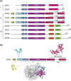
Figure 1.
Figure 1.. Domain architecture of human CHD-family remodellers.
(A) Domain schematics of CHD1–9. Subfamily I comprises CHD1–2, subfamily II comprises CHD3–5, and subfamily III comprises CHD6–9. HMG; high mobility group, PHD: plant homeodomain, CD: chromodomain, CTD: C-terminal domain, SANT: Swi3/Ada2/N-Cor/TFIIIB domain, SANT–SLIDE: Swi3/Ada2/N-Cor/TFIIIB-SANT-like ISWI domain, BRK: BRM and KIS domain. Dashed lines indicate sequence not drawn to scale. (B) Available structural information on CHD4. The structures of the N-terminal high mobility group (HMG)-like domain (yellow, PDB: 2N5N), the PHD1 domain (cyan, PDB: 2L5U), the PHD2 domain (cyan, PDB: 2L75), CHD4 (residues 446–1200) bound to the nucleosome (purple, PDB:6RYR), and the C-terminal SANT–SLIDE domain (maroon, PDB: 8D4Y) are indicated together with a schematic of CHD4 domain architecture.
Figure 2.
Figure 2.. Structure of Snf2 bound to the nucleosome and simple model of the remodelling mechanism based on structural snapshots.
(A) Cryo-EM structure of Snf2 (green) bound to the nucleosome (grey) (PDB: 5X0Y). (B) Chromatin remodelling mechanism derived from cryo-EM structures. The ATPase binds to the nucleosome in an open conformation in the absence of ATP and induces a 1-nt DNA distortion of the DNA tracking strand (pink, inset below). Binding of ATP (red) (red circle) results in a closed conformation of the ATPase and movement of the other DNA strand (guide strand, grey), with the overall effect of moving the DNA 1 bp in the indicated direction. ATP hydrolysis (to ADP, blue circle) resets the ATPase to an open conformation and again induces a 1-nt DNA distortion of the tracking strand. The translocated base pair is somehow temporarily absorbed into the nucleosome.
Figure 3.
Figure 3.. The mode of CHD4-DNA interaction is consistent with that observed for other remodeller complexes.
(A) Cryo-EM structure of CHD4 bound to the nucleosome (PDB: 6RYR). (B) Cryo-EM structure of CHD1 bound to the nucleosome (PDB: 5O9G). Binding of the CTD to DNA induces a significant opening of the nucleosome. (C) Overlay of the structures of a nucleosome bound to: SNF2 in the presence of ADP (dark green, 5Z3O), apo-CHD1 (orange, 7TN2), SNF2 in the presence of ADP.BeFx (light green, 5Z3U), CHD1 in the presence of ADP.BeFx (yellow, 5O9G) and CHD4 in the presence of AMP-PNP (purple, 6RYR). Structures are overlaid over the histone octamer. Positioning of the gating helix of ATPase lobe 2 reflects the nucleotide-bound state, even between different remodellers from different species. CHD4, SNF2, and CHD1 are observed in the closed conformation when bound to ATP mimics and the DNA path resembles that of an unbound nucleosome. Movement of the tracking strand is observed only for the apo or ADP-bound complexes in which SNF2 or CHD1 is in the open conformation (red, orange).
Figure 4.
Figure 4.. CHD4 activity is less reliant than other remodellers on the nucleosome acidic patch and the H4 arginine anchor motif.
Fold reduction in the rate of nucleosome sliding activity for seven remodelling enzymes/complexes following either: (1) mutation of the nucleosome acidic patch (blue bars) or (2) mutation of the arginine anchor motif in the N-terminal tail of histone H4 (orange bars).
Figure 5.
Figure 5.. Effect of histone PTMs and sequence changes on in vitro nucleosome remodelling rate.
Data are taken from Dann et al. [94] and are plotted as log(2) fold changes. The nucleosome sliding activity was measured for an unmodified nucleosome, together with 111 variants that include common histone PTMs, histone variants and several previously identified histone mutants. The mean and standard deviation are indicated for each of the eight remodellers tested and the dotted line indicates a two-fold increase in activity compared with unmodified nucleosome. Red data points indicate histone mutants.

References

    1. Petesch, S.J. and Lis, J.T. (2012) Overcoming the nucleosome barrier during transcript elongation. Trends Genet. 28, 285–294 10.1016/j.tig.2012.02.005 - DOI - PMC - PubMed
    1. Kornberg, R.D. (1974) Chromatin structure: a repeating unit of histones and DNA. Science 184, 868–871 10.1126/science.184.4139.868 - DOI - PubMed
    1. Luger, K., Mader, A.W., Richmond, R.K., Sargent, D.F. and Richmond, T.J. (1997) Crystal structure of the nucleosome core particle at 2.8 A resolution. Nature 389, 251–260 10.1038/38444 - DOI - PubMed
    1. Skene, P.J., Hernandez, A.E., Groudine, M. and Henikoff, S. (2014) The nucleosomal barrier to promoter escape by RNA polymerase II is overcome by the chromatin remodeler Chd1. Elife 3, e02042 10.7554/eLife.02042 - DOI - PMC - PubMed
    1. Quan, T.K. and Hartzog, G.A. (2010) Histone H3K4 and K36 methylation, Chd1 and Rpd3S oppose the functions of Saccharomyces cerevisiae Spt4-Spt5 in transcription. Genetics 184, 321–334 10.1534/genetics.109.111526 - DOI - PMC - PubMed

MeSH terms

LinkOut - more resources