. 2024 Sep;12(9):e70007.
doi: 10.1002/iid3.70007.

Impact of Microparticle Transarterial Chemoembolization (mTACE) on myeloid-derived suppressor cell subtypes in hepatocellular carcinoma: Clinical correlations and therapeutic implications

Affiliations

Impact of Microparticle Transarterial Chemoembolization (mTACE) on myeloid-derived suppressor cell subtypes in hepatocellular carcinoma: Clinical correlations and therapeutic implications

Yuanxun Yue et al. Immun Inflamm Dis. 2024 Sep.

Abstract

Background: Myeloid-derived suppressor cells (MDSCs) play a pivotal role in immunosuppression and tumor progression in hepatocellular carcinoma (HCC). While various treatments like surgical resection, ablation, and radiotherapy have been studied for their effects on circulating MDSC frequencies in HCC patients, the findings remain inconclusive. Transarterial Chemoembolization (TACE) stands as the standard care for unresectable HCC, with Microparticle TACE (mTACE) gaining prominence for its capacity to induce significant tumor necrosis. However, the immunological ramifications of such pathological outcomes are scarcely reported.

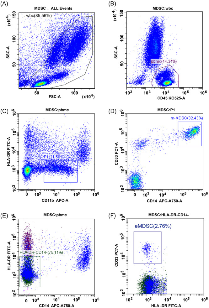
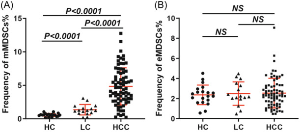
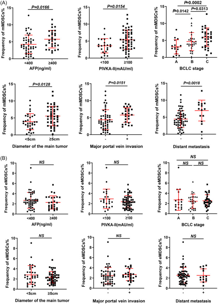
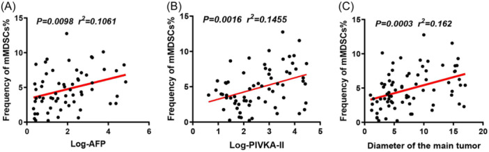
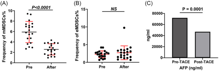
Methods and results: This study aims to elucidate the alterations in MDSC subtypes, specifically monocytic MDSCs (mMDSCs) and early-stage MDSCs (eMDSCs), post-mTACE and to investigate their clinical correlations in HCC patients. A cohort comprising 75 HCC patients, 16 liver cirrhosis patients, and 20 healthy controls (HC) was studied. Peripheral blood samples were collected and analyzed for MDSC subtypes. The study also explored the associations between MDSC frequencies and various clinical parameters in HCC patients. The frequency of mMDSCs was significantly elevated in the HCC group compared to liver cirrhosis and HC. Importantly, mMDSC levels were strongly correlated with aggressive clinical features of HCC, including tumor size, vascular invasion, and distant metastasis. Post-mTACE, a marked reduction in mMDSC frequencies was observed, while eMDSC levels remained stable.

Conclusions: Our findings underscore the critical role of mMDSCs in HCC pathogenesis and their potential as a therapeutic target. The study also highlights the efficacy of mTACE in modulating the immunosuppressive tumor microenvironment, thereby opening new avenues for combinatorial immunotherapeutic strategies in HCC management.

Keywords: Transarterial Chemoembolization (TACE); early stage MDSCs; gelatin sponge microparticles (GSMs); hepatocellular carcinoma (HCC); monocytic‐myeloid‐derived suppressor cells (mMDSCs).


Conflict of interest statement

The authors declare no conflict of interest.

Figures

Figure 1
Figure 1
Gating Strategy for Identifying MDSC Subsets in Peripheral Blood. The final frequencies of mMDSC and eMDSC in this patient were 6.97% (21.50% × 32.43% × 100) and 2.07% (75.11% × 2.76% × 100), respectively. (A) Fresh whole blood (WB) samples served as the substrate for the flow cytometric assay. Boxes indicate the cell populations selected for further analysis, with an initial exclusion of cell debris. (B) Live PBMC cells were selected using plots of CD45 versus SSC‐A. (C, D) Gating strategy for mMDSCs. The P1 box was selected by gating on cells positive for CD11b and negative or low‐negative for HLA‐DR antibodies. Subsequently, mMDSCs were selected by gating on CD14+ cells and CD33+ cells. (E, F) Gating strategy for eMDSCs. Based on the PBMC box (Figure 1B), cells were selected by gating on cells negative for CD14 and HLA‐DR antibodies. Subsequently, eMDSCs were selected by gating on CD33+ cells using plots of CD33 versus HLA‐DR. eMDSCs, early‐stage myeloid‐derived suppressor cells; PBMCs, peripheral blood mononuclear cells.
Figure 2
Figure 2
. Frequency of MDSCs in the Health Control (HC) Group, Liver Cirrhosis (LC) Group, and hepatocellular carcinoma (HCC) Group. A) Represents the frequency of mMDSCs. B) Represents the frequency of eMDSCs. eMDSCs, early‐stage myeloid‐derived suppressor cells; MDSCs, myeloid‐derived suppressor cells.
Figure 3
Figure 3
. The comparison of MDSCs frequencies between different tumor characteristics. (A) The frequency of mMDSCs in HCC patients with different clinical characteristics; (B) The frequency of eMDSCs in HCC patients with different clinical characteristics. eMDSCs, early‐stage myeloid‐derived suppressor cells; HCC, hepatocellular carcinoma; MDSCs, myeloid‐derived suppressor cells.
Figure 4
Figure 4
. Associations between mMDSCs Abundance and Clinical Parameters in HCC Patients. (A) Depicts the association between the abundance of mMDSCs and the serum level of AFP in HCC patients. The equation describing this relationship is: mMDSCs (%) = 0.7241 * log(AFP) (ng/mL) + 3.306. (B) Illustrates the association between the abundance of mMDSCs and the serum level of PIVKA‐II in HCC patients. The equation for this relationship is: mMDSCs (%) = 1.011 * log(PIVKA‐II) (mAU/ml) + 2.224. (C) Demonstrates the association between the abundance of mMDSCs and the major tumor size in HCC patients. The equation representing this correlation is: mMDSCs (%) = 0.2338 * diameter (cm) + 3.076. AFP, alpha‐fetoprotein; HCC, hepatocellular carcinoma; MDSCs, myeloid‐derived suppressor cells; PIVKA‐II, prothrombin induced by vitamin K absence‐II.
Figure 5
Figure 5
Alterations in Peripheral Blood MDSCs Frequencies Among HCC Patients in Response to mTACE Treatment. (A) Depicts the quantification of changes in mMDSCs (Monocytic MDSCs) frequencies before and after mTACE. (B) Depicts the quantification of changes in eMDSCs (Granulocytic MDSCs) frequencies before and after mTACE. (C) Changes in AFP Levels before and after mTACE. eMDSCs, early‐stage myeloid‐derived suppressor cells; MDSCs, myeloid‐derived suppressor cells; mTACE, Microparticle Transarterial Chemoembolization.
Figure 6
Figure 6
Response to mTACE Treatment in a 62‐Year‐Old Male Patient with a Massive HCC. The area indicated by the red arrow is where the tumor is located. (A) Enhanced MRI of the upper abdomen displaying a substantial mass with evident enhancement during the arterial phase, situated in the right lobe of the liver. The maximum diameter of the mass measures 4.1 cm. (B) Angiography depicting extensive tumor staining in the right lobe of the liver before mTACE intervention. (C) Post‐mTACE, the tumor's blood supply artery exhibits near‐complete occlusion. (D) Enhanced CT scan of the upper abdomen conducted 4 weeks following mTACE, illustrating significant tumor liquefaction and necrosis. eMDSCs, early‐stage myeloid‐derived suppressor cells; HCC, hepatocellular carcinoma; MDSCs, myeloid‐derived suppressor cells; mTACE, Microparticle Transarterial Chemoembolization.

Similar articles

Cited by

References

    1. Sung H, Ferlay J, Siegel RL, et al. Global cancer statistics 2020: GLOBOCAN estimates of incidence and mortality worldwide for 36 cancers in 185 countries. CA Cancer J Clin. 2021;71(3):209‐249. - PubMed
    1. Elwan N, Salem ML, Kobtan A, et al. High numbers of myeloid derived suppressor cells in peripheral blood and ascitic fluid of cirrhotic and HCC patients. Immunol Invest. 2018;47(2):169‐180. - PubMed
    1. Bhat DK, Olkhanud PB, Gangaplara A, et al. Early myeloid derived suppressor cells (eMDSCs) are associated with high donor myeloid chimerism following haploidentical HSCT for sickle cell disease. Front Immunol. 2021;12:757279. - PMC - PubMed
    1. Weber R, Fleming V, Hu X, et al. Myeloid‐derived suppressor cells hinder the anti‐cancer activity of immune checkpoint inhibitors. Front Immunol. 2018;9:1310. - PMC - PubMed
    1. Nan J, Xing YF, Hu B, et al. Endoplasmic reticulum stress induced LOX‐1+ CD 15+ polymorphonuclear myeloid‐derived suppressor cells in hepatocellular carcinoma. Immunology. 2018;154(1):144‐155. - PMC - PubMed

Publication types