

A Retrospective Study on the Clinical Characteristics and Computed Tomography, Biochemical, and Blood Parameters of Duodenal Papillary Diseases
- PMID: 39222361
- PMCID: PMC11369862
- DOI: 10.1177/10732748241278921
A Retrospective Study on the Clinical Characteristics and Computed Tomography, Biochemical, and Blood Parameters of Duodenal Papillary Diseases
Abstract
Objective: This study was conducted to investigate the imaging information, laboratory data, and clinical characteristics of duodenal papillary malignancies, aiming to contribute to the early diagnosis of these diseases.
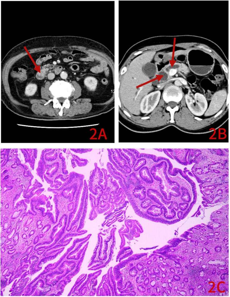
Methods: The clinical characteristics, laboratory data, and computed tomography (CT) findings of 17 patients with adenoma of the major duodenal papilla (the adenoma group) and 58 patients with cancer of the major duodenal papilla (the cancer group) were retrospectively analyzed. The measurement data were analyzed using t test and expressed as mean ± standard deviation. The counting data were analyzed using the χ2 test and expressed in n (%). Pearson correlation analysis was also conducted, and a scatter plot was drawn.
Results: There were significant differences in the diameter, shape, margin, and target sign of the major duodenal papilla, pancreatic duct diameter, common bile duct diameter, enhancement uniformity, fever, direct bilirubin, total bilirubin, carcinoembryonic antigen, carbohydrate antigen 19-9, and jaundice between the adenoma group and the cancer group (P < .01). The enhancement magnitude of the duodenal papilla was correlated with the lesion size, and the venous phase CT value of the enhanced scan was correlated with the duodenal papilla diameter (P < .05). Additionally, 12 patients in the cancer group suffered from malignant transformation of adenomas.
Conclusion: Firstly, CT is of high value in the diagnosis of duodenal papilla diseases. Secondly, the enhancement magnitude of the duodenal papilla is correlated with the lesion size. Thirdly, patients with duodenal papilla adenomas have a risk of progression into adenocarcinoma, thereby requiring close follow-up.
Keywords: adenoma; cancer; clinical characteristics; computed tomography; laboratory examination; major duodenal papilla.
Conflict of interest statement
Declaration of conflicting interestsThe author(s) declared no potential conflicts of interest with respect to the research, authorship, and/or publication of this article.
Figures


References
-
- Chen X, Wang J, Zhao J. Surgery. 9th ed. Beijing, China: People’s Medical Publishing House; 2018:224-225. doi:10.2018/468-473 - DOI
MeSH terms
LinkOut - more resources
Full Text Sources
Medical
