. 2024 Nov:49:102076.
doi: 10.1016/j.tranon.2024.102076. Epub 2024 Sep 1.

Effect of tumor-derived extracellular vesicle-shuttled lncRNA MALAT1 on proliferation, invasion and metastasis of triple-negative breast cancer by regulating macrophage M2 polarization via the POSTN/Hippo/YAP axis

Affiliations

Effect of tumor-derived extracellular vesicle-shuttled lncRNA MALAT1 on proliferation, invasion and metastasis of triple-negative breast cancer by regulating macrophage M2 polarization via the POSTN/Hippo/YAP axis

Xuedong Wang et al. Transl Oncol. 2024 Nov.

Abstract

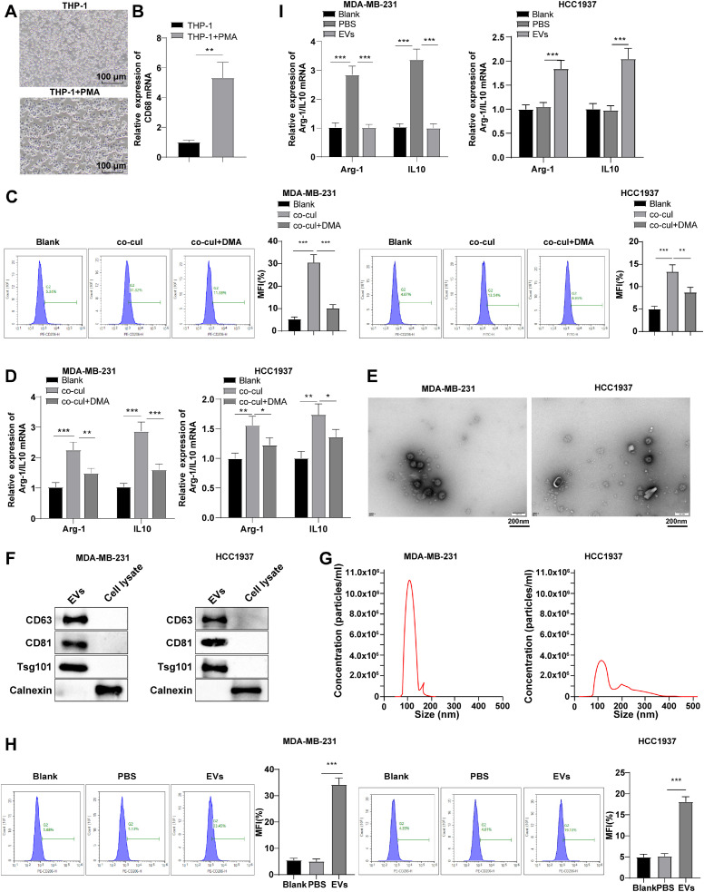
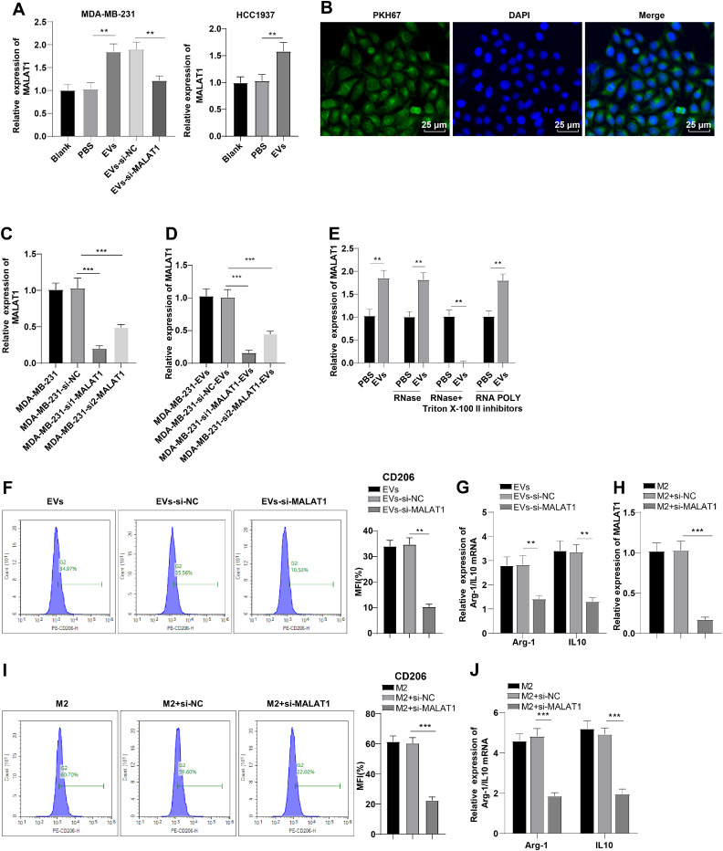
Objectives: Triple-negative breast cancer (TNBC) is the deadliest subtype of breast cancer (BC). Tumor-derived extracellular vesicles (EVs) trigger tumor progression by promoting M2 polarization. Some lncRNAs can be encapsulated into EVs for intercellular communication. Herein, we investigated the mechanism of TNBC-derived EV-shuttled lncRNA MALAT1 on macrophage polarization/tumorigenesis.

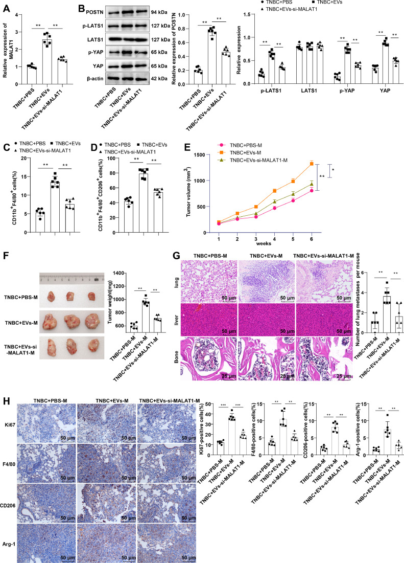
Methods: BC-associated targeted EV-derived lncRNAs were screened. Tumor tissues/tissues adjacent to cancer of TNBC patients, and blood samples of all subjects were collected. MALAT1/POSTN mRNA levels in tumor tissues/tissues adjacent to cancer, and MALAT1 expression in EVs and its correlation with TNBC patient overall survival were assessed by RT-qPCR/Kaplan-Meier survival analysis/log-rank test. TNBC patient M2 infiltration was detected by flow cytometry. MALAT1/POSTN levels in EVs/macrophages were regulated by transfection. Hippo/YAP activation was determined by Western blot. Nude mouse xenograft model was established and metastasis was detected by H&E staining.

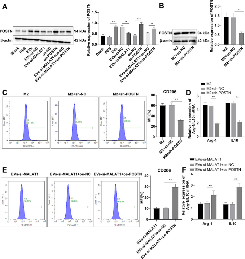
Results: MALAT1/POSTN were up-regulated and correlated with M2 infiltration/poor prognosis in TNBC patients. TNBC-derived EVs induced M2 polarization. MALAT1 was highly expressed in TNBC-derived EVs and could be transferred to macrophages via EVs to induce M2 polarization. POSTN overexpression diminished the inhibitory effect of MALAT1 knockdown on M2 markers. EVs activated the Hippo/YAP pathway in macrophages. The Hippo/YAP pathway inhibition abrogated the effect of POSTN overexpression on M2 marker expression. TNBC-EV-derived MALAT1 facilitated M2 polarization, and thus promoting occurrence and metastasis of TNBC in vitro and in vivo.

Conclusions: TNBC-EV-derived MALAT1 activated the Hippo/YAP axis by up-regulating POSTN, thereby inducing M2 polarization to promote TNBC occurrence and metastasis in vivo.

Keywords: Extracellular vesicles; Hippo/YAP; M2 macrophage; MALAT1; Triple negative breast cancer.


Conflict of interest statement

Declaration of competing interest The authors declare that they have no competing interests.

Figures

Fig. 1
Fig. 1
MALAT1 in serum EVs and POSTN expression in tumor tissues were up-regulated in TNBC patients and prominently linked with infiltration of M2 TAMs and poor prognosis. (A) 11 BC-associated target EV-derived lncRNAs were obtained by exoRBase 2.0, deepBase v3.0, RNADisease V4.0 (Score = 1), and LncRNADisease 2.0 (Score > 0.95) screening; (B) MALAT1 and (C) POSTN mRNA expression levels in tumors and tissues adjacent to cancer of TNBC patients were determined by RT-qPCR; Kaplan-Meier survival analysis and log-rank test were performed to determine the correlations between (D) MALAT1 and (E) POSTN mRNA in tumor tissues with OS; (F) EV morphology in serum of TNBC patients and healthy subjects was observed by TEM; (G) Western blot was implemented to measure the expression levels of CD63, CD81, TSG101 and Calnexin in serum EVs of TNBC patients and healthy subjects; (H) The concentration and size of EVs were analyze by NET; (I) RT-qPCR was carried out to determine the expression of MALAT1 in serum EVs of TNBC patients and healthy subjects; (J) Kaplan-Meier survival analysis and log-rank test were utilized to determine the correlation between serum EV-MALAT1 levels and OS in TNBC patients; (K) The percentages of TAMs (CD11b+F4/80+) and (L) M2 TAMs (CD11b+F4/80+CD206+) in tumors and tissues adjacent to cancer of TNBC patients were detected by flow cytometry; (M-N) Pearson was used to analyzed the correlations between MALAT1 level in serum EVs and POSTN mRNA level in tumor tissues with M2 macrophage infiltration level in TNBC patients. Data were expressed as mean ± standard deviation, and comparisons between 2 groups were made using paired/idependent t tests. ** p < 0.01.
Fig. 2
Fig. 2
TNBC-derived EVs induced macrophage M2 polarization. (A) Representative graphs of macrophages obtained from THP-1 cells treated with PMA for 24 h; (B) RT-qPCR to determine CD68 expression; (C/H) Flow cytometry to determine CD206 expression; (D/I) RT-qPCR to assess Arg-1 and IL10 expression patterns; (E) TEM to observe EV morphology; (F) Western blot to measure the expression levels of CD63, CD81, TSG101 and Calnexin in EVs; (G) NET to analyze the concentration and size of EVs. The cellular experiments were repeated three times. Data were expressed as the mean ± standard deviation. T test was conducted for the comparisons between 2 groups. One-way ANOVA was utilized for the comparisons of data among multiple groups. Tukey's multiple comparisons test was used for post-hoc analysis. ** P < 0.01, *** P < 0.001.
Fig. 3
Fig. 3
MALAT1 was highly expressed in TNBC-derived EVs and was transferred to macrophages via EVs to induce M2 polarization. (A/C/D/E/H) RT-qPCR to measure MALAT1 expression; (B) Representative immunofluorescence images showed that PKH67-labelled TNBC-derived EVs (green) were internalized by macrophages, and the nuclei were stained with 4′,6-diamidino-2-phenylindole (DAPI); (F/I) Flow cytometry to determine CD206 expression; (G/J) RT-qPCR to assess Arg-1 and IL10 expression levels. The cellular experiments were repeated three times. Data were expressed as mean ± standard deviation. One-way ANOVA was used to compare the data among multiple groups, and Tukey's multiple comparisons test was applied for post hoc analysis.** P < 0.01, *** P < 0.001.
Fig. 4
Fig. 4
TNBC-EV-derived MALAT1 induced macrophage M2 polarization by promoting POSTN expression. (A-B) Western blot to assess POSTN expression; (C/E) Flow cytometry to determine CD206 expression; (D/F) RT-qPCR to measure Arg-1 and IL10 expression patterns. The cellular experiments were repeated three times, and the data were expressed as mean ± standard deviation. One-way ANOVA was used to compare the data among multiple groups, and Tukey's multiple comparisons test was used for post-hoc analysis. ** P < 0.01.
Fig. 5
Fig. 5
POSTN induced macrophage M2 polarization by activating the Hippo-YAP pathway. (A-B) Western blot to assess the expression levels of the Hippo/YAP pathway-related proteins (p-LATS1, LATS1, p-YAP and YAP); (C) Flow cytometry to assess the expression of CD206; (D) RT-qPCR to determine the expression levels of Arg-1 and IL10. The cellular experiments were repeated three times, and the data were expressed as mean ± standard deviation. One-way ANOVA was utilized to compare the data among multiple groups, and Tukey's multiple comparisons test was used for post hoc analysis.* P < 0.05, ** P < 0.01.
Fig. 6
Fig. 6
M2 macrophages activated by TNBC-EV-derived MALAT1 faciltated the proliferation, migration and invasion of TNBC cells. MDA-MB-231 cells were collected after indirect co-culture with macrophages. (A) CCK-8 to detect changes of cell viability; (B) Transwell to detect changes of migration and invasion of cells. The cellular experiments were repeated three times. Data were expressed as mean ± standard deviation. One-way ANOVA was utilized to compare the data among multiple groups. Tukey's multiple comparisons test was applied for post-hoc analysis. ** P < 0.01.
Fig. 7
Fig. 7
TNBC-EV-derived MALAT1 facilitated the macrophage M2 polarization, thereby promoting the occurrence and metastasis of TNBC in vivo. (A) RT-qPCR to assess the expression of MALAT1 in tumor tissues; (B) Western blot to determine the expression patterns of POSTN and the Hippo/YAP pathway-related proteins (p-LATS1, LATS1, p-YAP and YAP); flow cytometry to assess the percentages of (C) TAMs (CD11b+ F4/80+) and (D) M2 TAMs (CD11b+F4/80+CD206+) in tumor tissues of mice; (E) The tumor volume and (F) weight of mice were recorded; (G) H&E staining of lungs, bones and livers. The blue arrow indicated metastatic tumor cells. n = 6. Data were expressed as mean ± standard deviation. One-way ANOVA was utilized to compare the data among multiple groups, and Tukey's multiple comparisons test was applied for post-hoc analysis. ** P < 0.01.

References

    1. DeSantis C.E., Fedewa S.A., Goding Sauer A., Kramer J.L., Smith R.A., et al. Breast cancer statistics, 2015: convergence of incidence rates between black and white women. CA Cancer J. Clin. 2016;66:31–42. doi: 10.3322/caac.21320. - DOI - PubMed
    1. Chodosh L.A. Breast cancer: current state and future promise. Breast. Cancer Res. 2011;13(113) doi: 10.1186/bcr3045. - DOI - PMC - PubMed
    1. Cronin K.A., Lake A.J., Scott S., Sherman R.L., Noone A.M., et al. Annual report to the nation on the status of cancer, part I: national cancer statistics. Cancer. 2018;124:2785–2800. doi: 10.1002/cncr.31551. - DOI - PMC - PubMed
    1. Wu H., Feng J., Zhong W., Zouxu X., Xiong Z., et al. Model for predicting immunotherapy based on M2 macrophage infiltration in TNBC. Front. Immunol. 2023;14 doi: 10.3389/fimmu.2023.1151800. - DOI - PMC - PubMed
    1. So J.Y., Ohm J., Lipkowitz S., Yang L. Triple negative breast cancer (TNBC): non-genetic tumor heterogeneity and immune microenvironment: emerging treatment options. Pharmacol. Ther. 2022;237 doi: 10.1016/j.pharmthera.2022.108253. - DOI - PMC - PubMed

LinkOut - more resources