. 2024 Sep;16(9):2221-2229.
doi: 10.1111/os.14217. Epub 2024 Sep 2.

The Efficacy of Modified Ilizarov Distraction-Tension Device on Limb Wound Difficult to Cover with Skin Flaps

Affiliations

The Efficacy of Modified Ilizarov Distraction-Tension Device on Limb Wound Difficult to Cover with Skin Flaps

Yueliang Zhu et al. Orthop Surg. 2024 Sep.

Abstract

Introduction: Closure of complex limb wounds poses challenges and requires innovative approaches. This research aimed to evaluate the effectiveness of a modified distraction-tension device using Ilizarov external fixation for wound closure in challenging cases.

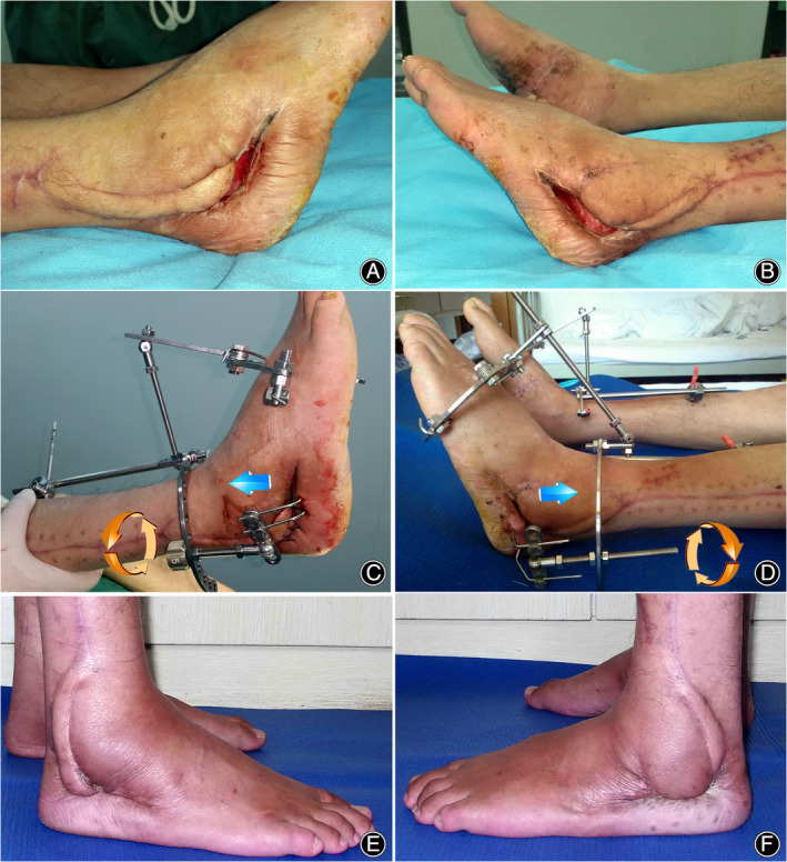
Methods: A retrospective analysis was conducted on 43 patients with extremity wounds that were difficult to cover with skin flaps between January 2019 and December 2022. Tension-relieving traction was applied using the Ilizarovexternal fixator apparatus, tailored to individual wound characteristics. Three types of wire-pin connections were used in this study. The distraction begins on the third postoperative day, with a speed of 0.5mm/d. Clinical wound healing scores were evaluated at 5 and 30 days postoperatively. Complications were documented following the Paley classification system.

Results: Traction using modified Ilizarovexternal fixation promoted a significant reduction in wound size. The mean traction period was 11.2 ± 7.3 days, and the mean healing duration was 17.0 ± 3.7 days. The clinical wound healing score improved from 3.7 ± 2.9 at 5 days to 1.7 ± 0.7 at 30 days postoperatively (p < 0.05). Complications were minimal, with no significant obstacles or sequelae observed. Direct closure healing was achieved in 21 cases, skin graft healing in 13 cases, and suture healing in 9 cases. No recurrences were reported. Using Paley's classified complications, there were 17 problems, 9 obstacles, and 0 sequelae.

Conclusion: The Ilizarov tension-relieving traction shows promise in facilitating wound closure that is challenging to manage with skin flaps. The modified three types of pin-skin connection configuration could satisfy various types of wound closure.

Keywords: Complex Limb Wounds; Ilizarov Tension‐Relieving Traction; Modified Ilizarov external fixation; Skin Flaps; Wound Closure Effectiveness.


Conflict of interest statement

All authors have no competing interests.

Figures

Figure 1
Figure 1
Modified Ilizarov distraction‐tension device for wound closure. The base usually involves two rings or half‐rings. The rings are located in the proximal and distal ends of the wound and are connected by threaded rods. The rings were fixed on the bone via half‐pins or K‐wires. Distraction‐tension devices are threaded rods through posts that were connected to the rings. Turning the nut (green arrows) on the three rods could produce distraction or compression forces (blue arrows) via the movements of the threaded rods. Wire–pin connection is of three types, discussed in the following. (1) Parallel pin–skin connection: The K‐wires are inset subcutaneously and parallelly to the wound length. This configuration is indicated for long wounds. (2) Hook–pin connections: The k‐wire ends were bent and hooked to the skin and subcutaneous tissue. The configuration is indicated for short and deep wounds with strong skins, such as the skin of the plantar foot. (3) Olive wire–skin connection. It consists of olive wires and washers of different sizes. Usually, a rubber penicillin vial or a gauze is inset between the washers and skin as a cushion. This configuration is very flexible and could be used in various wounds.
FIGURE 2
FIGURE 2
A male patient, 35 years old. Three months following the completion of tibial open bone defect reconstruction and bone grafting. The tension‐induced ossification zone in the anterior aspect of the proximal tibia exhibited signs of bone infection. After meticulous wound debridement and dressing changes, a residual bone and soft tissue defect measuring 5 cm× 3 cm persisted. The surrounding soft tissues presented with fibrotic scarring, posing challenges for both adjacent skin flap transfer and free flap coverage (attributable to the limited availability of suitable vascular anastomosis sites). To address the medial aspect of the defect, a 3.0 cm× 1.5 cm osteotomy was performed, and a gradual traction system employing an olive wire–skin connection was employed to progressively mobilize the bone and skin from the inner to the outer aspect (A). The traction pins were retained with additional pressure. Radiographic analysis before traction (B) delineated the osteotomy line represented by green dotted lines; yellow denotes the olive wires, and the arrows indicate the direction of traction. After three months, traction was discontinued, and the traction bone segment achieved osseous union with the tibial cortex on the outer aspect (C). Traction was administered gradually, at a rate of 0.5 mm/day, administered thrice daily, resulting in wound closure after one month (D). Subsequently, the external fixation was removed, and during the four‐month postoperative follow‐up, the skin exhibited complete healing without any signs of recurrence (E).
FIGURE 3
FIGURE 3
A male patient, 27 years old, presented with bilateral calcaneal comminuted fractures following a fall from a height. Open reduction and internal fixation with plates were performed, but the incision became infected. Bilateral fibular artery perforator propeller flaps were utilized for wound coverage; however, the wound became infected, leading to distal flap necrosis. The wound remained exposed (A,B). After one month of repeated debridement and dressing changes, a gradual traction device was implemented with a smaller wound size, using a hook–skin connection. Postoperatively, traction was gradually applied by rotating the nut three times a day, one side at a time, at a rate of 0.5 mm/day (C,D). After 10 days of traction, wound edges were approximated, and fixation was continued for an additional 15 days until wound closure was achieved in the right foot, leading to the removal of external fixation. The left side underwent sutured mattress closure. At the three‐month follow‐up, bilateral calcaneal bones exhibited deformity during the healing process, but the wounds healed without recurrence (E,F).
FIGURE 4
FIGURE 4
A female patient, 61 years old, presented with bilateral lower limb injuries resulting from the forceful impact of a mudslide. The distal tibia and fibula exhibited an open fracture, accompanied by extensive soft tissue degloving injuries and defects. Notably, both tendons and bones were exposed, as evidenced by the marked regions (A, highlighted in green). Following meticulous wound debridement on two occasions, a traction device was employed, utilizing an olive wire–skin connection system to facilitate bidirectional skin traction (B). The traction protocol was maintained at a consistent rate, with daily dressing changes. After three weeks, a reduction in wound size was observed, concomitant with the robust development of healthy granulation tissue. Notably, exuberant granulation tissue formation was evident on the previously exposed surface of the tibia (C), necessitating the implementation of a skin graft to achieve definitive wound closure. Subsequently, the patient underwent bilateral ankle joint fusion six months postoperatively. At the one‐year follow‐up, the patient demonstrated improved ambulatory function with the aid of an assistive device. However, it is important to note that a residual posterior tilt deformity of the right tibial plateau persisted (D).

References

    1. Teot L, Ohura N. Challenges and Management in Wound Care. Plast Reconstr Surg. 2021;147:9S–15S. 10.1097/PRS.0000000000007628 - DOI - PubMed
    1. Alga A, Haweizy R, Bashaireh K, Wong S, Lundgren KC, von Schreeb J, et al. Negative pressure wound therapy versus standard treatment in patients with acute conflict‐related extremity wounds: a pragmatic, multisite, randomised controlled trial. Lancet Glob Health. 2020;8:e423–e429. 10.1016/S2214-109X(19)30547-9 - DOI - PubMed
    1. Zhang YX, Hayakawa TJ, Levin LS, Hallock GG, Lazzeri D. The economy in autologous tissue transfer: part 1. The kiss flap technique. Plast Reconstr Surg. 2016;137:1018–1030. 10.1097/01.prs.0000479971.99309.21 - DOI - PubMed
    1. Rajput S, Kuruoglu D, Salinas CA, Sen I, Kalra M, Moran SL. Flap management of groin wounds following vascular procedures: a review of 270 flaps for vascular salvage. J Plast Reconstr Aesthet Surg. 2023;78:38–47. 10.1016/j.bjps.2023.01.028 - DOI - PubMed
    1. Jia Z, Liang G, Duan C, Zhang Z, Guo Y, Teng Y. Flow‐through Dorsoulnar perforator flap repair of soft‐tissue defect of the finger with segmental digital arterial defect. J Coll Physicians Surg Pak. 2022;32:685–687. 10.29271/jcpsp.2022.05.685 - DOI - PubMed

LinkOut - more resources