. 2024 Sep 2;14(1):20388.
doi: 10.1038/s41598-024-68510-8.

Induction of oxidative- and endoplasmic-reticulum-stress dependent apoptosis in pancreatic cancer cell lines by DDOST knockdown

Affiliations

Induction of oxidative- and endoplasmic-reticulum-stress dependent apoptosis in pancreatic cancer cell lines by DDOST knockdown

Richard Böhme et al. Sci Rep. .

Abstract

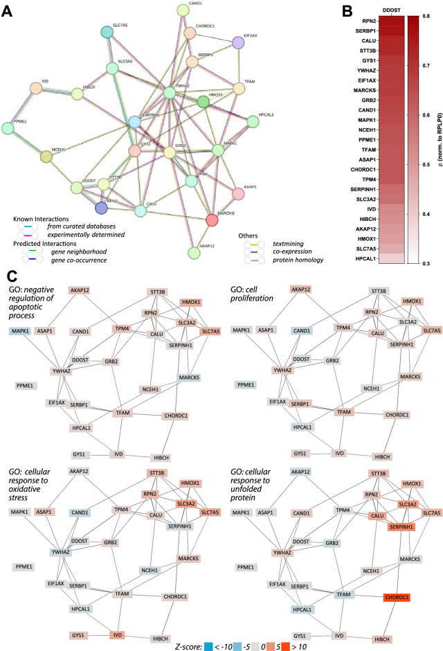
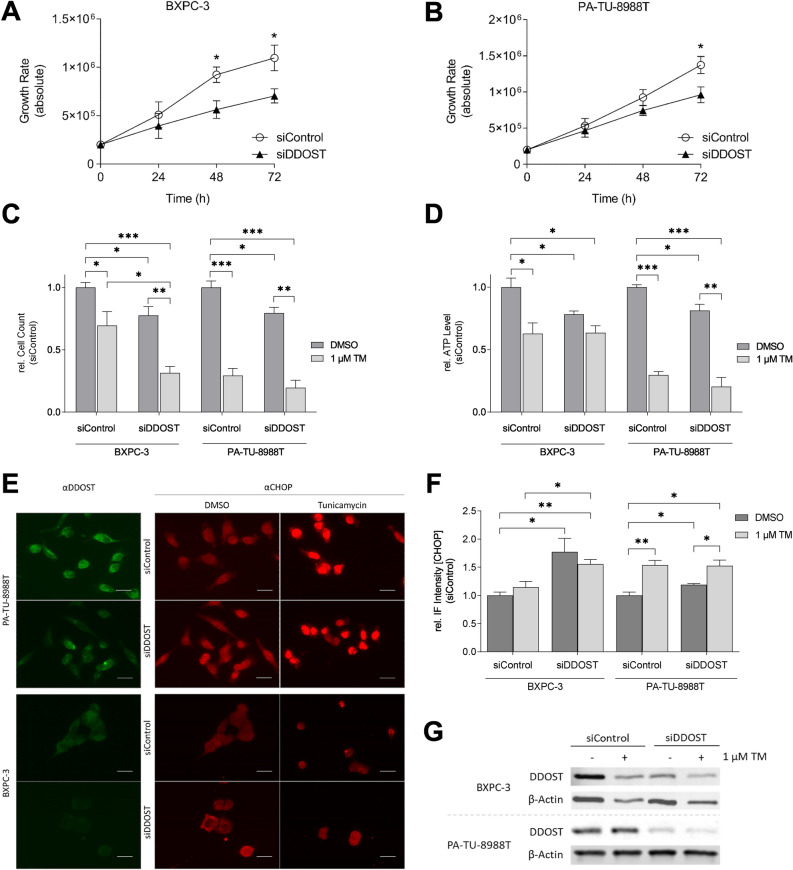
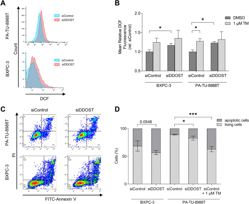
The dolichyl-diphosphooligosaccharide-protein glycosyltransferase non-catalytic subunit (DDOST) is a key component of the oligosaccharyltransferase complex catalyzing N-linked glycosylation in the endoplasmic reticulum lumen. DDOST is associated with several cancers and congenital disorders of glycosylation. However, its role in pancreatic cancer remains elusive, despite its enriched pancreatic expression. Using quantitative mass spectrometry, we identify 30 differentially expressed proteins and phosphopeptides (DEPs) after DDOST knockdown in the pancreatic ductal adenocarcinoma (PDAC) cell line PA-TU-8988T. We evaluated DDOST / DEP protein-protein interaction networks using STRING database, correlation of mRNA levels in pancreatic cancer TCGA data, and biological processes annotated to DEPs in Gene Ontology database. The inferred DDOST regulated phenotypes were experimentally verified in two PDAC cell lines, PA-TU-8988T and BXPC-3. We found decreased proliferation and cell viability after DDOST knockdown, whereas ER-stress, ROS-formation and apoptosis were increased. In conclusion, our results support an oncogenic role of DDOST in PDAC by intercepting cell stress events and thereby reducing apoptosis. As such, DDOST might be a potential biomarker and therapeutic target for PDAC.


Conflict of interest statement

The authors declare no competing interests.

Figures

Figure 1
Figure 1
Tissue Specificity Scores of DDOST protein expression in human body donors. If a gene has TS (tissue specificity) scores at least in one tissue ≥ 2.5, this gene is called tissue-enriched. Vertical lines indicate the threshold values of 2.5 and 4. Adapted from “A Quantitative Proteome Map of the Human Body” by Jiang et al., 2020.
Figure 2
Figure 2
Fold change (FC) of total proteins and phosphopeptides after DDOST KD in PA-TU-8988T cell line. (A) Western blot analysis of DDOST expression after DDOST KD. β-Actin was used as loading control. (B, C) Quantification of rel. RNA expression and rel. protein expression levels of DDOST after KD (**P < 0.01, ***P < 0.001; unpaired t-test). (D, F) Volcano plot of protein and phosphopeptide log2-FC. Highlighted proteins and phosphopeptides cutoff FDR < 0.05 (n = 5; ROTS-test). (E, G) Bar chart of protein and phosphopeptide log2-FC. Upregulated proteins and phosphopeptides in red, downregulated in blue.
Figure 3
Figure 3
Interactions and correlations of 26 proteins identified as DEPs. (A) PPI network consisting of 26 nodes and 67 edges from STRING database (PPI enrichment P = 2.83 × 10−4). (B) Spearman correlation analysis of mRNA levels (TCGA pancreatic adenocarcinoma) comparing DDOST with all identified DEPs (P < 1.00 × 10−4; n = 179). (C) Functional enrichment analysis as implemented in the GADO webserver (negative regulation of apoptotic process: P = 2.54 × 10−6; cell proliferation: P = 5.35 × 10−4; cellular response to oxidative stress: P = 1.11 × 10−3; cellular response to unfolded protein: P = 1.18 × 10−3).
Figure 4
Figure 4
Reduced proliferation and viability and induced ER stress after DDOST KD in PDAC cell lines. (A) Growth curve of BXPC-3 cells after DDOST KD. (B) Growth curve of PA-TU-8988T cells after DDOST KD. (C) Quantification of proliferation assay 72 h after DDOST KD and TM treatment. (D) Quantification of viability assay 72 h after DDOST KD and TM treatment. (E) Immunofluorescence images of DDOST and CHOP after DDOST KD and treatment with TM (scale bar = 50 µm). (F) Quantification of CHOP rel. IF intensity after DDOST KD and treatment with TM. (G) Western blot analysis of DDOST KD efficiency. β-Actin was used as loading control. (*P < 0.05, **P < 0.01, ***P < 0.001; unpaired t-test).
Figure 5
Figure 5
Induced ROS formation and Apoptosis after DDOST KD in PDAC cells. (A) Flow cytometry assay of ROS formation by DCF detection after DDOST KD. (B) Quantification of mean rel. DCF intensity after DDOST KD and treatment with TM. (C) Flow cytometry assay of FITC-Annexin V and PI staining after DDOST KD. (D) Quantification of FITC-Annexin V and PI positive cells after DDOST KD and treatment with TM. (*P < 0.05, ***P < 0.001; unpaired t-test). Western blot analysis of DDOST KD efficiency shown in Fig. 4G.

References

    1. Neoptolemos, J. P. et al. Therapeutic developments in pancreatic cancer: Current and future perspectives. Nat. Rev. Gastroenterol. Hepatol.15, 333–348 (2018). 10.1038/s41575-018-0005-x - DOI - PubMed
    1. Ferlay, J., Partensky, C. & Bray, F. More deaths from pancreatic cancer than breast cancer in the EU by 2017. Acta Oncol. Stockh. Swed.55, 1158–1160 (2016).10.1080/0284186X.2016.1197419 - DOI - PubMed
    1. Arnold, M. et al. Global burden of 5 major types of gastrointestinal cancer. Gastroenterology159, 335–349 (2020). 10.1053/j.gastro.2020.02.068 - DOI - PMC - PubMed
    1. Bause, E. Structural requirements of N-glycosylation of proteins. Studies with proline peptides as conformational probes. Biochem. J.209, 331–336 (1983). 10.1042/bj2090331 - DOI - PMC - PubMed
    1. Imperiali, B. & O’Connor, S. E. Effect of N-linked glycosylation on glycopeptide and glycoprotein structure. Curr. Opin. Chem. Biol.3, 643–649 (1999). 10.1016/S1367-5931(99)00021-6 - DOI - PubMed

MeSH terms

Substances