. 2024 Sep 2;14(1):20386.
doi: 10.1038/s41598-024-71416-0.

SGLT2 inhibition mitigates transition from acute kidney injury to chronic kidney disease by suppressing ferroptosis

Affiliations

SGLT2 inhibition mitigates transition from acute kidney injury to chronic kidney disease by suppressing ferroptosis

Yutaro Hirashima et al. Sci Rep. .

Abstract

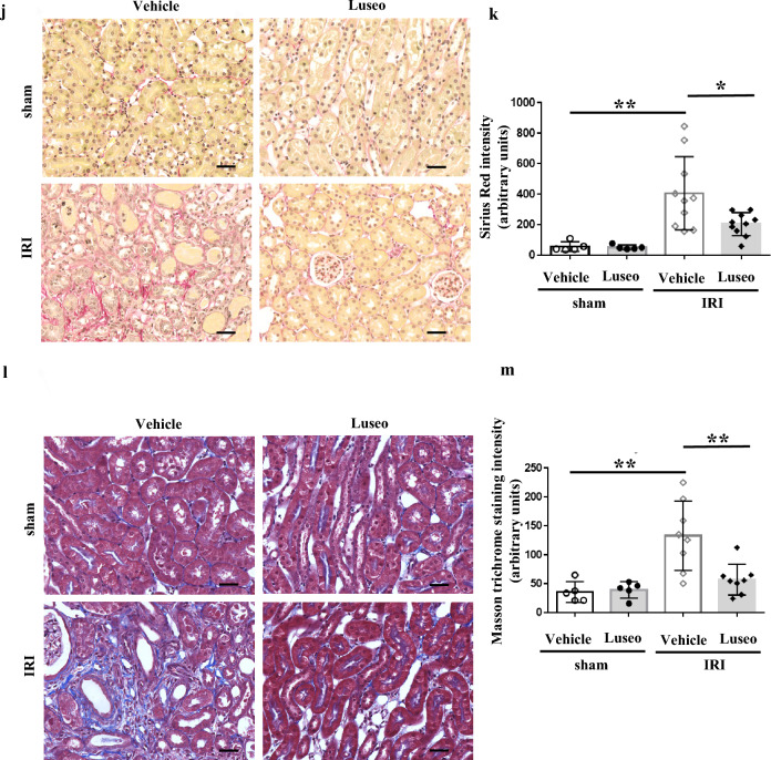
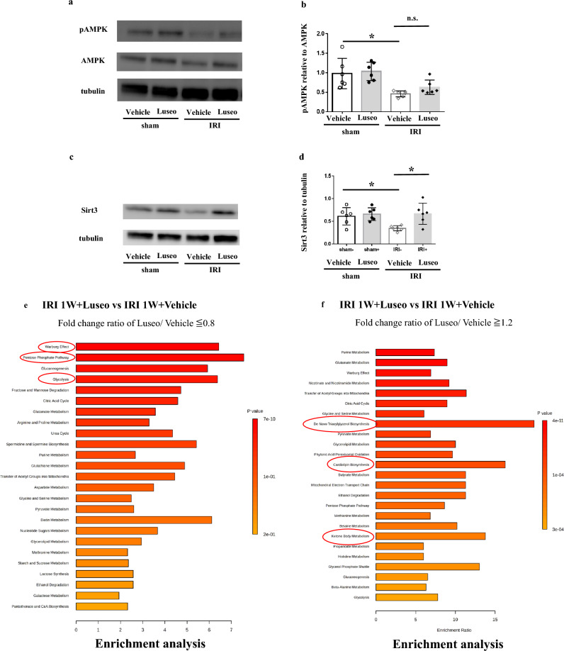
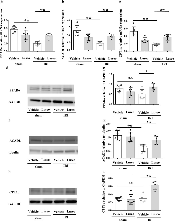
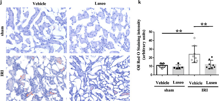
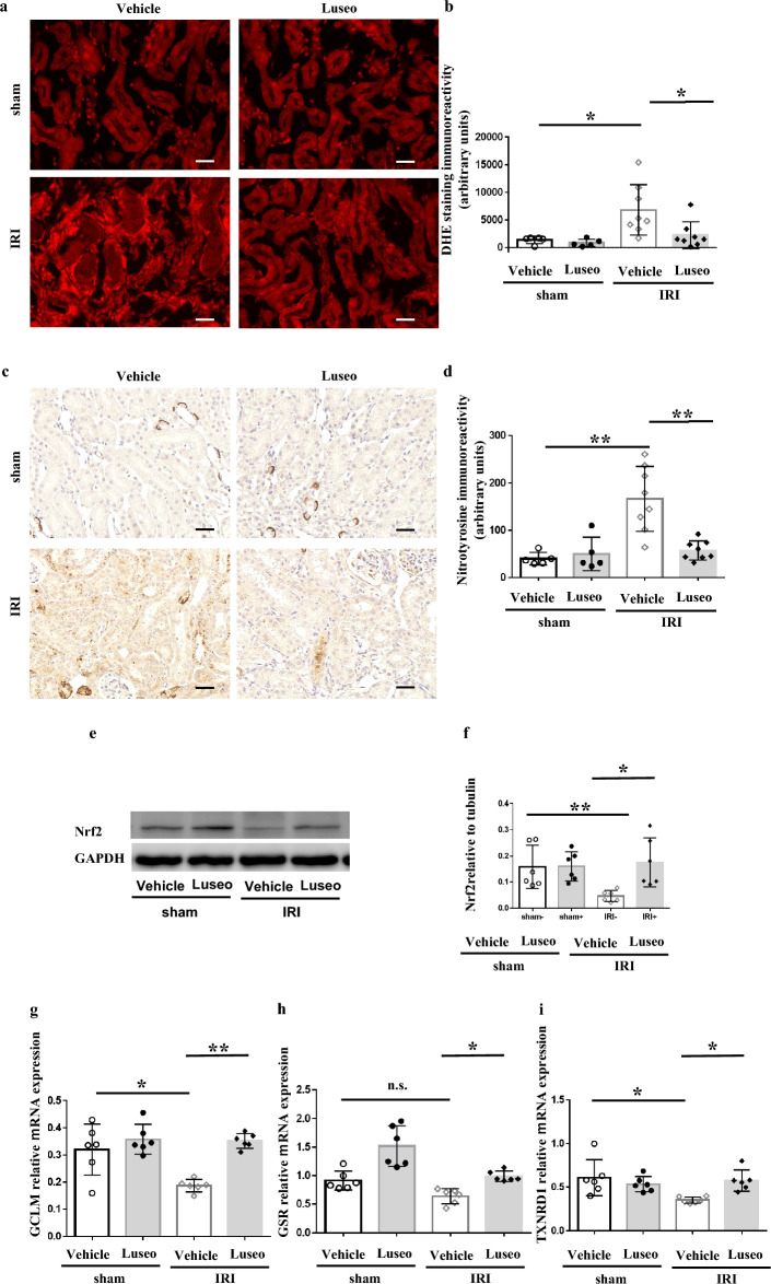
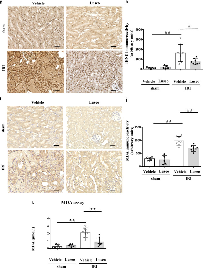
Sodium-glucose cotransporter 2 (SGLT2) inhibitors have been shown to be renoprotective in ischemia-reperfusion (I/R) injury, with several proposed mechanisms, though additional mechanisms likely exist. This study investigated the impact of luseogliflozin on kidney fibrosis at 48 h and 1 week post I/R injury in C57BL/6 mice. Luseogliflozin attenuated kidney dysfunction and the acute tubular necrosis score on day 2 post I/R injury, and subsequent fibrosis at 1 week, as determined by Sirius red staining. Metabolomics enrichment analysis of I/R-injured kidneys revealed suppression of the glycolytic system and activation of mitochondrial function under treatment with luseogliflozin. Western blotting showed increased nutrient deprivation signaling with elevated phosphorylated AMP-activated protein kinase and Sirtuin-3 in luseogliflozin-treated kidneys. Luseogliflozin-treated kidneys displayed increased protein levels of carnitine palmitoyl transferase 1α and decreased triglyceride deposition, as determined by oil red O staining, suggesting activated fatty acid oxidation. Luseogliflozin prevented the I/R injury-induced reduction in nuclear factor erythroid 2-related factor 2 activity. Western blotting revealed increased glutathione peroxidase 4 and decreased transferrin receptor protein 1 expression. Immunostaining showed reduced 4-hydroxynonenal and malondialdehyde levels, especially in renal tubules, indicating suppressed ferroptosis. Luseogliflozin may protect the kidney from I/R injury by inhibiting ferroptosis through oxidative stress reduction.

Keywords: Ferroptosis; Ischemia reperfusion injury; Luseogliflozin; Nrf2.


Conflict of interest statement

This work received the research grant by Taisho Pharmaceutical Co., the manufacturer of luseogliflozin. The funding entity did not participate in the study's design, data gathering and analysis, decision to publish, or manuscript preparation, with the sole exception of supplying pharmacokinetic data for luseogliflozin. All the authors declare no conflict of interest.

Figures

Fig. 1
Fig. 1
Luseogliflozin-mediated improvements in renal function and histology at 48 h post ischemia/reperfusion (I/R) injury in mice. (ac) Serum levels of blood urea nitrogen (BUN; a, and creatinine (Cr; b), and histological findings of kidney sections stained with hematoxylin and eosin (c) at 48 h post I/R injury. (d) Acute tubular necrosis (ATN) scores in the following groups: vehicle-treated + sham-operated (n = 5), luseogliflozin-treated + sham-operated (n = 5), vehicle-treated + I/R-injured (n = 10), and luseogliflozin-treated + I/R-injured (n = 10). (e and f) Histological findings and quantification of kidney sections immunostained for kidney injury molecule 1 (KIM-1) at 48 h post I/R injury; *P < 0.05, **P < 0.01, as determined by one-way analysis of variance. n.s., not significant.
Fig. 2
Fig. 2
Luseogliflozin-mediated improvement in renal interstitial fibrosis at 1 week post ischemia/reperfusion (I/R) injury in mice. (ac) Quantification of kidney mRNA levels of SM22 (a), COLA1 (b), and TGF (c) at 1 week post I/R injury. (d and e) Western blot analysis and quantification of αSMA expression in the kidney at 1 week post I/R injury in the following groups: vehicle-treated + sham-operated (n = 6), luseogliflozin-treated + sham-operated (n = 6), vehicle-treated + I/R-injured (n = 6), and luseogliflozin-treated + I/R-injured (n = 6). (f and g) Western blot analysis and quantification of collagen I expression in the kidney at 1 week post I/R injury in the following groups: vehicle-treated + sham-operated (n = 6), luseogliflozin-treated + sham-operated (n = 6), vehicle-treated + I/R-injured (n = 6), and luseogliflozin-treated + I/R-injured (n = 6). (h and i) Western blot analysis and quantification of fibronectin expression in the kidney at 1 week post I/R injury in the following groups: vehicle-treated + sham-operated (n = 6), luseogliflozin-treated + sham-operated (n = 6), vehicle-treated + I/R-injured (n = 6), and luseogliflozin-treated + I/R-injured (n = 6). (jm) Histological findings and quantification of kidney sections stained with Sirius red (j, k) and Masson’s trichrome staining (l, m) at 1 week post I/R injury in the following groups: vehicle-treated + sham-operated (n = 5), luseogliflozin-treated + sham-operated (n = 5), vehicle-treated + I/R-injured (n = 10), and luseogliflozin-treated + I/R-injured (n = 10) ; *P < 0.05, **P < 0.01, as determined by one-way analysis of variance. n.s., not significant.
Fig. 2
Fig. 2
Luseogliflozin-mediated improvement in renal interstitial fibrosis at 1 week post ischemia/reperfusion (I/R) injury in mice. (ac) Quantification of kidney mRNA levels of SM22 (a), COLA1 (b), and TGF (c) at 1 week post I/R injury. (d and e) Western blot analysis and quantification of αSMA expression in the kidney at 1 week post I/R injury in the following groups: vehicle-treated + sham-operated (n = 6), luseogliflozin-treated + sham-operated (n = 6), vehicle-treated + I/R-injured (n = 6), and luseogliflozin-treated + I/R-injured (n = 6). (f and g) Western blot analysis and quantification of collagen I expression in the kidney at 1 week post I/R injury in the following groups: vehicle-treated + sham-operated (n = 6), luseogliflozin-treated + sham-operated (n = 6), vehicle-treated + I/R-injured (n = 6), and luseogliflozin-treated + I/R-injured (n = 6). (h and i) Western blot analysis and quantification of fibronectin expression in the kidney at 1 week post I/R injury in the following groups: vehicle-treated + sham-operated (n = 6), luseogliflozin-treated + sham-operated (n = 6), vehicle-treated + I/R-injured (n = 6), and luseogliflozin-treated + I/R-injured (n = 6). (jm) Histological findings and quantification of kidney sections stained with Sirius red (j, k) and Masson’s trichrome staining (l, m) at 1 week post I/R injury in the following groups: vehicle-treated + sham-operated (n = 5), luseogliflozin-treated + sham-operated (n = 5), vehicle-treated + I/R-injured (n = 10), and luseogliflozin-treated + I/R-injured (n = 10) ; *P < 0.05, **P < 0.01, as determined by one-way analysis of variance. n.s., not significant.
Fig. 3
Fig. 3
Luseogliflozin-mediated enhancement of master regulators of starvation and improvement in mitochondrial function, as shown by kidney metabolomic analysis at 1 week post ischemia/reperfusion (I/R) injury. (ad) Western blot analysis and quantification of kidney expression of pAMPK (a, b) and Sirt3 (c, d) at 1 week post ischemia/reperfusion (I/R) injury in the following groups of mice: vehicle-treated + sham-operated (n = 6), luseogliflozin-treated + sham-operated (n = 6), vehicle-treated + I/R-injured (n = 6), and luseogliflozin-treated + I/R-injured (n=6). (e and f) Capillary electrophoresis combined with time-of-flight mass spectrometry (CE-TOFMS) and capillary electrophoresis combined with triple quadrupole mass spectrometry (QqQMS) measurements in cation and anion mode were used to generate metabolomics data, which were analyzed using MetaboAnalyst. Of the 116 metabolites identified, enrichment analysis was performed on those with a fold change of ≤ 0.8 (e) or ≥ 1.2 (f) in the kidneys of luseogliflozin-treated I/R-injured mice (n = 4) relative to those in vehicle-treated I/R-injured mice (n =4). *P < 0.05, **P < 0.01, as determined by one-way analysis of variance. n.s., not significant.
Fig. 4
Fig. 4
Luseogliflozin-mediated improvement in dysfunctional fatty acid oxidation at 1 week post ischemia/reperfusion (I/R) injury in mice. (ac) Quantification of kidney mRNA levels of PPARα (a), ACADL (b), and CPT1α (c) at 1 week post I/R injury. (dg) Western blot analysis and quantification of kidney protein expression of PPARα (d, e), ACADL (f, g), and CPT1α (h, i) at 1 week post I/R injury in the following groups: vehicle-treated + sham-operated (n = 6), luseogliflozin-treated + sham-operated (n = 6), vehicle-treated + I/R-injured (n = 6), and luseogliflozin-treated + I/R-injured (n = 6). (j and k) Histological findings and quantification Oil red O staining of kidney sections at 1 week post I/R injury in the following groups: vehicle-treated + sham-operated (n = 5), luseogliflozin-treated + sham-operated (n = 5), vehicle-treated + I/R-injured (n = 8), and luseogliflozin-treated + I/R-injured (n = 8); *P < 0.05, **P < 0.01, as determined by one-way analysis of variance. n.s., not significant.
Fig. 4
Fig. 4
Luseogliflozin-mediated improvement in dysfunctional fatty acid oxidation at 1 week post ischemia/reperfusion (I/R) injury in mice. (ac) Quantification of kidney mRNA levels of PPARα (a), ACADL (b), and CPT1α (c) at 1 week post I/R injury. (dg) Western blot analysis and quantification of kidney protein expression of PPARα (d, e), ACADL (f, g), and CPT1α (h, i) at 1 week post I/R injury in the following groups: vehicle-treated + sham-operated (n = 6), luseogliflozin-treated + sham-operated (n = 6), vehicle-treated + I/R-injured (n = 6), and luseogliflozin-treated + I/R-injured (n = 6). (j and k) Histological findings and quantification Oil red O staining of kidney sections at 1 week post I/R injury in the following groups: vehicle-treated + sham-operated (n = 5), luseogliflozin-treated + sham-operated (n = 5), vehicle-treated + I/R-injured (n = 8), and luseogliflozin-treated + I/R-injured (n = 8); *P < 0.05, **P < 0.01, as determined by one-way analysis of variance. n.s., not significant.
Fig. 5
Fig. 5
Luseogliflozin-mediated improvement in mitochondrial function. (ad) Western blot analysis and quantification of kidney expression of PGC1α (a, b) and TOM20 (c, d) at 1 week post ischemia/reperfusion (I/R) injury in the following groups: vehicle-treated + sham-operated (n = 6), luseogliflozin-treated + sham-operated (n = 6), vehicle-treated + I/R-injured (n = 6), and luseogliflozin-treated + I/R-injured (n = 6). (e and f) Histological findings and quantification of immunostaining for TOM20 in kidney sections at 1 week post I/R injury in the following groups: vehicle-treated + sham-operated (n = 5), luseogliflozin-treated + sham-operated (n = 5), vehicle-treated + I/R-injured (n = 8), and luseogliflozin-treated + I/R-injured (n = 8); *P < 0.05, **P < 0.01, as determined by one-way analysis of variance. n.s., not significant.
Fig. 6
Fig. 6
Luseogliflozin-mediated amelioration of oxidative stress and enhancement of Nrf2, the master regulator of anti-oxidative responses. (a and b) Histological findings and quantification of reactive oxygen species in the kidney with dihydroethidium (DHE) staining at 1 week post ischemia/reperfusion (I/R) injury. (c and d) Histological findings and quantification of immunostaining for nitrotyrosine in the kidney at 1 week post I/R injury in the following groups: vehicle-treated + sham-operated (n = 5), luseogliflozin-treated + sham-operated (n = 5), vehicle-treated + I/R-injured (nv8), and luseogliflozin-treated + I/R-injured (n = 8). (e and f) Western blot analysis and quantification of kidney expression of Nrf2 at 1 week post I/R injury in the following groups: vehicle-treated + sham-operated (n = 6), luseogliflozin-treated + sham-operated (n = 6), vehicle-treated + I/R-injured (n = 6), and luseogliflozin-treated + I/R-injured (n = 6). (gi) Quantification of mRNA levels of GCLM (g), GSR (h), and TXNRD1 (i) in the kidney at 1 week post I/R injury; *P < 0.05, **P < 0.01, as determined by one-way analysis of variance. n.s., not significant.
Fig. 7
Fig. 7
Luseogliflozin-mediated inhibition of ferroptosis and amelioration of ischemia/reperfusion (I/R) injury. (a and b) Histological findings and positive area of TUNEL assay staining of the kidney at 1 week post I/R injury in the following groups: vehicle-treated + sham-operated (n = 10), luseogliflozin-treated + sham-operated (n = 10), vehicle-treated + I/R-injured (n = 5), and luseogliflozin-treated + I/R-injured (n = 5). (cf) Western blot analysis and quantification of kidney expression of Gpx4 (c, d) and TfR1 (e, f) at 1 week post I/R injury in the following groups: vehicle-treated + sham-operated (n = 6), luseogliflozin-treated + sham-operated (n = 6), vehicle-treated + I/R-injured (n = 6), and luseogliflozin-treated + I/R-injured (n = 6). (g-j) Histological findings and quantification of kidney immunostaining for 4-hydroxynonenal (4-HNE; g, h) and malondialdehyde (MDA; i, j) at 1 week post I/R injury in the following groups: vehicle-treated + sham-operated (n = 8), luseogliflozin-treated + sham-operated (n = 8), vehicle-treated + I/R-injured (n = 5), and luseogliflozin-treated + I/R-injured (n = 5). (k) MDA concentrations in the kidney at 1 week post I/R injury in the following groups: vehicle-treated + sham-operated (n = 8), luseogliflozin-treated + sham-operated (n = 8), vehicle-treated + I/R-injured (n = 5), and luseogliflozin-treated + I/R-injured (n = 5); * P < 0.05, ** P < 0.01, as determined by one-way analysis of variance. n.s., not significant.
Fig. 7
Fig. 7
Luseogliflozin-mediated inhibition of ferroptosis and amelioration of ischemia/reperfusion (I/R) injury. (a and b) Histological findings and positive area of TUNEL assay staining of the kidney at 1 week post I/R injury in the following groups: vehicle-treated + sham-operated (n = 10), luseogliflozin-treated + sham-operated (n = 10), vehicle-treated + I/R-injured (n = 5), and luseogliflozin-treated + I/R-injured (n = 5). (cf) Western blot analysis and quantification of kidney expression of Gpx4 (c, d) and TfR1 (e, f) at 1 week post I/R injury in the following groups: vehicle-treated + sham-operated (n = 6), luseogliflozin-treated + sham-operated (n = 6), vehicle-treated + I/R-injured (n = 6), and luseogliflozin-treated + I/R-injured (n = 6). (g-j) Histological findings and quantification of kidney immunostaining for 4-hydroxynonenal (4-HNE; g, h) and malondialdehyde (MDA; i, j) at 1 week post I/R injury in the following groups: vehicle-treated + sham-operated (n = 8), luseogliflozin-treated + sham-operated (n = 8), vehicle-treated + I/R-injured (n = 5), and luseogliflozin-treated + I/R-injured (n = 5). (k) MDA concentrations in the kidney at 1 week post I/R injury in the following groups: vehicle-treated + sham-operated (n = 8), luseogliflozin-treated + sham-operated (n = 8), vehicle-treated + I/R-injured (n = 5), and luseogliflozin-treated + I/R-injured (n = 5); * P < 0.05, ** P < 0.01, as determined by one-way analysis of variance. n.s., not significant.
Fig. 8
Fig. 8
Schematic diagram of the therapeutic effect of luseogliflozin, showing how it effectively reduces I/R injury and prevents fibrosis in mice. Under normal conditions, I/R injury reduces tubular mitochondrial function and increases oxidative stress. In addition, tubular metabolism shifts from fatty acid oxidation to glycolysis, resulting in accumulation of polyunsaturated fatty acids (PUFAs) and leading to lipid peroxidation and tubular ferroptosis. Next, macrophage infiltration and inflammatory cytokine production occur, leading to tubular fibrosis and the transition from acute kidney injury (AKI) to chronic kidney disease (CKD). Sodium-glucose cotransporter 2 (SGLT2) inhibitors increase the starvation regulator Sirtuin-3 (Sirt3) and also show a tendency to increase pAMPK. Mitochondrial function is improved, oxidative stress is lowered, and PUFA accumulation is prevented by inhibiting the tubular metabolic shift from fatty acid oxidation to the glycolytic system. Consequently, ferroptosis and development of the AKI to CKD transition are prevented.

References

    1. Kellum, J. A. et al. Acute kidney injury. Nat. Rev. Dis. Prim.7(1), 52 (2021). 10.1038/s41572-021-00284-z - DOI - PubMed
    1. Peerapornratana, S., Manrique-Caballero, C. L., Gomez, H. & Kellum, J. A. Acute kidney injury from sepsis: Current concepts, epidemiology, pathophysiology, prevention and treatment. Kidney Int.96(5), 1083–1099 (2019). 10.1016/j.kint.2019.05.026 - DOI - PMC - PubMed
    1. Carroll, W. R. & Esclamado, R. M. Ischemia/reperfusion injury in microvascular surgery. Head Neck22(7), 700–713 (2000). 10.1002/1097-0347(200010)22:7<700::AID-HED10>3.0.CO;2-H - DOI - PubMed
    1. Kosieradzki, M. & Rowinski, W. Ischemia/reperfusion injury in kidney transplantation: Mechanisms and prevention. Transplant. Proc.40(10), 3279–3288 (2008). 10.1016/j.transproceed.2008.10.004 - DOI - PubMed
    1. Wanner, C. et al. Empagliflozin and progression of kidney disease in type 2 diabetes. N. Engl. J. Med.375(4), 323–334 (2016). 10.1056/NEJMoa1515920 - DOI - PubMed

MeSH terms

Substances