. 2024 Oct;6(10):1886-1896.
doi: 10.1038/s42255-024-01125-5. Epub 2024 Sep 2.

Mitochondria transfer-based therapies reduce the morbidity and mortality of Leigh syndrome

Affiliations

Mitochondria transfer-based therapies reduce the morbidity and mortality of Leigh syndrome

Ritsuko Nakai et al. Nat Metab. 2024 Oct.

Abstract

Mitochondria transfer is a recently described phenomenon in which donor cells deliver mitochondria to acceptor cells1-3. One possible consequence of mitochondria transfer is energetic support of neighbouring cells; for example, exogenous healthy mitochondria can rescue cell-intrinsic defects in mitochondrial metabolism in cultured ρ0 cells or Ndufs4-/- peritoneal macrophages4-7. Exposing haematopoietic stem cells to purified mitochondria before autologous haematopoietic stem cell transplantation allowed for treatment of anaemia in patients with large-scale mitochondrial DNA mutations8,9, and mitochondria transplantation was shown to minimize ischaemic damage to the heart10-12, brain13-15 and limbs16. However, the therapeutic potential of using mitochondria transfer-based therapies to treat inherited mitochondrial diseases is unclear. Here we demonstrate improved morbidity and mortality of the Ndufs4-/- mouse model of Leigh syndrome (LS) in multiple treatment paradigms associated with mitochondria transfer. Transplantation of bone marrow from wild-type mice, which is associated with release of haematopoietic cell-derived extracellular mitochondria into circulation and transfer of mitochondria to host cells in multiple organs, ameliorates LS in mice. Furthermore, administering isolated mitochondria from wild-type mice extends lifespan, improves neurological function and increases energy expenditure of Ndufs4-/- mice, whereas mitochondria from Ndufs4-/- mice did not improve neurological function. Finally, we demonstrate that cross-species administration of human mitochondria to Ndufs4-/- mice also improves LS. These data suggest that mitochondria transfer-related approaches can be harnessed to treat mitochondrial diseases, such as LS.


Figures

Extended Data Fig. 1.
Extended Data Fig. 1.. Additional metabolic cage parameters from NDUFS4-deficient mice treated with wildtype or NDUFS4-deficient bone marrow transplantation.
(a) Distance travelled and (b) average respiratory exchange ratio (RER) over 3 hr after a 1 hr air equilibration period. Data are expressed as mean +/− SEM. All data points are unique biological replicates. n=5 KO, n=17 WT. Data accompanies Fig. 1.
Extended Data Fig. 2.
Extended Data Fig. 2.. Engraftment of CD45.1+ cells in blood, spleen, and liver in mice transplanted with wildtype or mtD2 bone marrow.
(a) Representative gating for identification of CD45.1+ CD45.2 donor immune cells and CD45.1 CD45.2+ radioresistant host cells. (b) Chimerism expressed as a percentage of the ratio of CD45.1+ cells to the sum of CD45.1+ and CD45.2+ cells. (c) Representative gating in the blood to exclude red blood cells (RBCs) and platelets from particles less than 2 μm in diameter for identification of extracellular mtD2+ mitochondria. Data are expressed as mean +/− SEM. All data points are unique biological replicates. For b, n=3 WT, n=5 mtD2 (except n=4 for spleen). Data accompanies Fig. 2.
Extended Data Fig. 3.
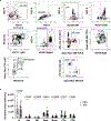
Extended Data Fig. 3.. Bone marrow transplantation leads to transfer of mtD2+ mitochondria to host cells in the blood, spleen, and liver.
(a) Representative gating for identification of CD45.1 CD45.2+ radioresistant host B cells, T cells, neutrophils, and monocytes as well as CD45.1 CD45.2 host epithelial, endothelial, and stromal cells in the spleen. (b) Representative gating to identify the proportions of host B cells, T cells, neutrophils, monocytes, epithelial, endothelial, and stromal cells that received mtD2+ mitochondria in the spleen. (c) Proportions of B cells (B), T cells (T), neutrophils (neut), monocytes (mono), epithelial cells (epi), endothelial cells (endo), and stromal cells that received mtD2+ mitochondria in the liver and (d) peripheral blood. For c-d, closed circles are WT and open squares are mtD2. Data are expressed as mean +/− SEM. All data points are unique biological replicates. For b-d, n=3 WT, n=5 mtD2 (except n=4 for spleen). For b-d, 2-way ANOVA with Sidak post-hoc test. ***P<0.001, ****P<0.0001. Data accompanies Fig. 2.
Extended Data Fig. 4.
Extended Data Fig. 4.. Mitochondria isolates from the liver are enriched in mtD2+ events and produce more yield than bone marrow mitochondria isolates.
(a) Flow cytometric identification of the proportion of mtD2+ mitochondria. Pre-gated on CD41 CD45 TER-119 events less than 2 μm in diameter. (b) Mitochondrial yield from mouse liver or bone marrow (BM). Data are expressed as mean +/− SEM. All data points are unique biological replicates. For a-b, n=4 biological replicates/group. For b, Student’s t-test (two-sided). *P<0.05 Data accompanies Fig. 3.
Extended Data Fig. 5.
Extended Data Fig. 5.. Additional metabolic cage parameters from NDUFS4-deficient mice treated with mitochondria or PBS.
(a) Body weight, and (b) rectal core body temperature of 7-week-old KO mice treated with PBS or 100μg mitochondria 1–2 times per week. (c) Average energy expenditure (d) distance travelled, and (e) average respiratory exchange ratio (RER) over 3 hr after a 1 hr air equilibration period. Data are expressed as mean +/− SEM. All data points are unique biological replicates. For a, n=11 PBS, n=12 WT. For b, n=11/group. For c-e, n=15/group. Variation in n is due to mouse mortality. For c, Student’s t-test (two-sided). *P<0.05. Data accompanies Fig. 3.
Extended Data Fig. 6.
Extended Data Fig. 6.. Administration of wildtype mitochondria to NDUFS4-deficient mice does not alter immune cell composition in the blood.
(a) Representative gating to identify immune cells in the peripheral blood of Ndufs4—/— (KO) mice. (b) Overall number of B cells (B), eosinophils (eos), natural killer cells (NK), neutrophils (neut), monocytes (mono), CD4 T cells, and CD8 T cells per mL of peripheral blood from KO mice treated weekly with PBS (closed circle) or 100μg mitochondria (open square) for 5 weeks. Data are expressed as mean +/− SEM. All data points are unique biological replicates. For b, n=14 PBS, n=18 Mito. For b, 2-way ANOVA with Sidak post-hoc test. Data accompanies Fig. 3.
Extended Data Fig. 7.
Extended Data Fig. 7.. Administration of mitochondria to NDUFS4-deficient mice does not alter immune cell composition in the blood or tissues.
(a) Overall number of immune cells in peripheral blood, (b) spleen, (c) bone marrow, and (d) liver of Ndufs4—/— mice treated weekly with 100μg KO or WT mitochondria for 7 weeks. For a-d, closed circles are KO mito and open squares are WT mito. Data are expressed as mean +/− SEM. All data points are unique biological replicates. For a, n=3 KO Mito, n=4 WT Mito. For b and d, n=4/group. For c, n=4 KO Mito, n=3 WT Mito. For a-d, 2-way ANOVA with Sidak post-hoc test. Data accompanies Fig. 3.
Extended Data Fig. 8.
Extended Data Fig. 8.. Administration of mitochondria to NDUFS4-deficient mice does not alter expression of inflammatory cytokines.
(a) Inflammatory cytokine expression in the spleen (b) peritoneal exudate cells (PECS), and (c) liver of Ndufs4—/— mice treated weekly with 100μg KO or WT mitochondria for 7 weeks. For a-c, closed circles are KO mito and open squares are WT mito. Data are expressed as mean +/− SEM. All data points are unique biological replicates. For a-c, n=4 biological replicates/group. For a-c, 2-way ANOVA with Sidak post-hoc test. Data accompanies Fig. 3.
Extended Data Fig. 9.
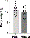
Extended Data Fig. 9.. Administration of MRC-Q does not alter body weight in NDUFS4-deficient mice.
Body weight of Ndufs4—/— mice treated with PBS or 50 μg MRC-Q once per week from 3- to 7-weeks old. Data are expressed as mean +/− SEM. All data points are unique biological replicates. n=12 PBS, n=14 MRC-Q. Data accompanies Fig. 4.
Figure 1.
Figure 1.. Wildtype bone marrow transplantation improves the morbidity and mortality of Leigh Syndrome in NDUFS4-deficient mice.
(a) Experimental design. Ndufs4—/— (KO) mice were lethally irradiated at 3–5-weeks and transplanted with wildtype (WT) or KO bone marrow cells. (b) Survival expressed as postnatal age in days. Solid black line is WT to KO, dashed blue line is KO to KO. (c) Body weight, (d) rectal core body temperature, (e) time until falling off a rotarod, (f) rotarod rotations per minute (rpm) at the time of falling (starting at rpm=4), and (g) four-limb grip strength at age 7-weeks. (h) Energy expenditure shown per interval. Closed black circles are WT to KO, open blue squares are KO to KO (i) Average energy expenditure. Data are expressed as mean +/− SEM. All data points are unique biological replicates, and all statistical tests are two-sided. For b, n=29 WT to KO, n=11 KO to KO. For c-i, n=5 KO to WT, n=17 WT to WT. For b, Mantel-Cox log-rank test. For e-f, i, Student’s t-tests. For h, two-way ANOVA with repeated measures. *P<0.05, **P<0.01.
Figure 2.
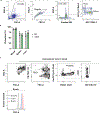
Figure 2.. Engrafted hematopoietic cells release mitochondria into the blood and transfer mitochondria to NDUFS4-deficient cells in vivo.
(a) Experimental design. Ndufs4—/— (KO) mice were lethally irradiated and transplanted with bone marrow cells from CD45.1 WT or CD45.1 mtD2 mitochondria reporter mice. (b) Flow cytometric identification of CD45.1+ CD45.2 donor immune cells and CD45.1 CD45.2+ radioresistant host immune cells. Histograms show mtD2 signal in the host and donor cells within each group. (c) Flow cytometry plots of extracellular mtD2+ mitochondria in peripheral blood. Pre-gated on CD41 CD45 TER-119 events less than 2 μm in diameter. (d) Numbers of mtD2+ extracellular mitochondria (ex-mito) per mL of peripheral blood. (e) Proportions of singlet, live, CD45.1 CD45.2+ radioresistant host immune cells and (f) CD45.1 CD45.2 host non-immune cells that received mtD2 signal from donor cells in the indicated tissues. (g) Proportions of host B cells (B), T cells (T), neutrophils (Neut), monocytes (Mono), epithelial cells (Epi), endothelial cells (Endo) and stromal cells that received mtD2 signal from donor cells in the spleen. For d-g, closed circles are WT, and open squares are mtD2. Data are expressed as mean +/− SEM. All data points are unique biological replicates, and all statistical tests are two-sided. For c-d n=7/group. For e-g, n=3 WT, n=5 mtD2 (except n=4 for spleen). For c-d, Mann-Whitney test. For e-g, two-way ANOVA with Sidak post-hoc test. **P<0.01, ***P<0.001, ****P<0.0001.
Figure 3.
Figure 3.. Administration of exogenous mouse mitochondria reduces the morbidity and mortality of Leigh Syndrome.
(a) Experimental design for a-e. Survival expressed as postnatal age in days of Ndufs4—/— (KO) mice treated 1–2 times per week with PBS (dashed blue line) or 100μg WT mitochondria (Mito, solid black line). (b) Pictures of mice, (c) time until falling off a stationary rotarod, (d) four-limb grip strength, and (e) energy expenditure at 6–8 weeks of age. (f) Basal oxygen consumption rate (OCR) of isolated KO or WT mitochondria. (g) Time until falling off a stationary rotarod by KO mice were treated once a week with 100 μg KO or WT mitochondria for 4 weeks. (h) Tissue biodistribution of NZB mtDNA in Ndufs4—/—(KO) mice treated once a week with 100 μg NZB mitochondria for 4 weeks. Background amplification was corrected for with KO mice treated with 100 μg C57BL6/J mitochondria for 4 weeks. Samples were normalized to β-actin as a nuclear DNA control. Data are expressed as mean +/− SEM. All data points are unique biological replicates, and all statistical tests are two-sided.For c, n=15 PBS, n=19 Mito. For d, n=11 PBS, n=12 mito. For e, n=15/group. For d-e, variation in n is due to mortality kinetics. For f, n=6/group. For g, n=9 KO mitochondria, n=8 WT mitochondria. For h, n=6/tissue. For a, Mantel-Cox log-rank test. For c-d, f-g, Student’s t-tests. For e, Mann-Whitney test. *P<0.05.
Figure 4.
Figure 4.. Administering a human mitochondria isolate called MRC-Q improves the morbidity and mortality of Leigh Syndrome.
(a) Production of ATP in the presence of DMSO or oligomycin (Omy) by mitochondria isolated from HeLa cells by a commercial kit (H-mito) or mitochondria organelle complex Q (MRC-Q). (b) Outer mitochondrial membrane (OMM) and (c) inner mitochondrial membrane (IMM) integrity. (d) Tissue biodistribution of MRC-Q mtDNA in Ndufs4—/— (KO) mice treated with 50 μg MRC-Q once per week for 4 weeks. Background amplification was corrected for with KO mice treated with PBS for 4 weeks. Samples were normalized to β-actin as a nuclear DNA control. (e) Survival expressed as postnatal age in days. MRC-Q, solid black line. PBS, dashed blue line. (f) Pictures of mice, (g) time until falling off a hang-wire at age 7-weeks-old, and (h) righting reflex delay when mice were placed on their backs. Data are expressed as mean +/− SEM. All data points are unique biological replicates, and all statistical testing is two-sided. For a and d, n=7/group. For c, n=6/group. For d, n=3/tissue. For g, n=10 PBS, n=14 MRC-Q. For h, n=10/group. For a, two-way ANOVA. For b-c and g-h, Student’s t-tests. For e, Mantel-Cox log-rank test. *P<0.05, ****P<0.0001.

References

    1. Al Amir Dache Z. & Thierry AR Mitochondria-derived cell-to-cell communication. Cell Reports 42, 112728, doi: 10.1016/j.celrep.2023.112728 (2023). - DOI - PubMed
    1. Zhu M. et al. Mitochondria Released by Apoptotic Cell Death Initiate Innate Immune Responses. ImmunoHorizons 2, 384–397, doi: 10.4049/immunohorizons.1800063 (2018). - DOI - PMC - PubMed
    1. Borcherding N. & Brestoff JR The power and potential of mitochondria transfer. Nature 623, 283–291, doi: 10.1038/s41586-023-06537-z (2023). - DOI - PMC - PubMed
    1. Borcherding N. et al. Dietary lipids inhibit mitochondria transfer to macrophages to divert adipocyte-derived mitochondria into blood. Cell Metabolism 34, 1499–1513.e1498 (2022). - PMC - PubMed
    1. Spees JL, Olson SD, Whitney MJ & Prockop DJ Mitochondrial transfer between cells can rescue aerobic respiration. Proceedings of the National Academy of Sciences 103, 1283–1288, doi: 10.1073/pnas.0510511103 (2006). - DOI - PMC - PubMed

Substances

LinkOut - more resources