Review
. 2024 Sep;26(9):1420-1433.
doi: 10.1038/s41556-024-01491-y. Epub 2024 Sep 2.

Innate immune sensing of cell death in disease and therapeutics

Affiliations
Review

Innate immune sensing of cell death in disease and therapeutics

Si Ming Man et al. Nat Cell Biol. 2024 Sep.

Abstract

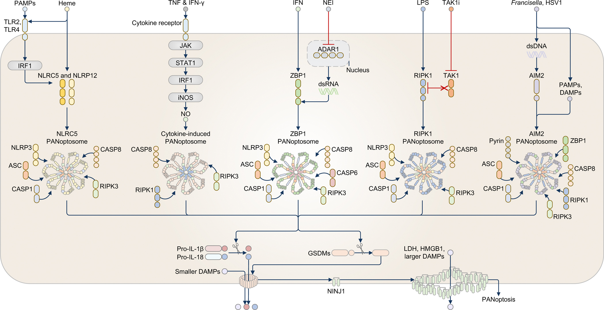
Innate immunity, cell death and inflammation underpin many aspects of health and disease. Upon sensing pathogens, pathogen-associated molecular patterns or damage-associated molecular patterns, the innate immune system activates lytic, inflammatory cell death, such as pyroptosis and PANoptosis. These genetically defined, regulated cell death pathways not only contribute to the host defence against infectious disease, but also promote pathological manifestations leading to cancer and inflammatory diseases. Our understanding of the underlying mechanisms has grown rapidly in recent years. However, how dying cells, cell corpses and their liberated cytokines, chemokines and inflammatory signalling molecules are further sensed by innate immune cells, and their contribution to further amplify inflammation, trigger antigen presentation and activate adaptive immunity, is less clear. Here, we discuss how pattern-recognition and PANoptosome sensors in innate immune cells recognize and respond to cell-death signatures. We also highlight molecular targets of the innate immune response for potential therapeutic development.


Conflict of interest statement

Competing financial interests

T-D.K. and S.M.M. have no interests to declare.

Figures

Fig. 1.
Fig. 1.. Dying or dead cells release PAMPs, DAMPs, and cytokines that contribute to innate immunity and disease pathogenesis.
Following infection or sterile injury such as trauma, dying or dead cells release a plethora of pathogen-associated molecular patterns (PAMPs), damage-associated molecular patterns (DAMPs), and cytokines that activate pattern-recognition receptors (PRRs) and other receptors such as cytokine receptors in responding immune cells. These cells respond by liberating more PAMPs (in the case of infection), DAMPs, or cytokines that contribute to the manifestation, or in some cases attenuation, of localized and systemic diseases. A subset of cytokines called chemokines induce the recruitment of additional immune cells to the site of injury, amplifying the inflammatory response. Collectively, PAMPs, DAMPs, and cytokines fuel the inflammation circuitry, resulting in the progression and perpetuation of inflammatory diseases.
Fig. 2.
Fig. 2.. Necrotic cells release DAMPs that activate Toll-like receptors and inflammasomes.
a, TLR2 and TLR4 share the immune sensing of many DAMPs. TLR2 can additionally recognize nucleosomes and versican, whereas TLR4 can additionally recognize DAMPs such as oxidized low-density lipoprotein-immune complexes (OxLDL-IC). TLR3 can sense RNA from necrotic cells, whereas TLR7 might sense single-stranded RNA (ssRNA) and TLR9 might sense single-stranded DNA (ssDNA). TLR9 can also sense DNA complexed with the antimicrobial peptide LL37 or the nuclear protein HMGB1, or DNA-containing immune complexes, and DNA from dying or dead hepatocytes or chemotherapy-killed cancer cells, mitochondrial DNA (mtDNA), and neutrophil-derived DNA. b, Priming via TLRs (also known as Step 1) induces transcription of the genes encoding NLRP3 and pro-IL-1β. Necrotic cells release DAMPs which trigger several common events that activate NLRP3 (also known as Step 2). The most widely accepted mechanism is the efflux of potassium ions (K+) from within the cell. In many cases, NLRP3 interacts with the kinase NEK7 in order to assemble an inflammasome complex. AIM2 binds directly to and is activated by double-stranded DNA, which is readily released by dying or dead cells. Both inflammasome sensor proteins form a separate inflammasome complex of 0.5 to 1 micron in diameter comprised of the inflammasome adaptor protein ASC and the cysteine protease caspase-1. Caspase-1 is activated to induce the proteolytic cleavage of pro-IL-1β and pro-IL-18, and the pyroptosis-inducing protein gasdermin D (GSDMD). The bioactive N-terminal fragment of GSDMD assembles into pores on the plasma membrane, through which these pores mediate the secretion of bioactive IL-1β and IL-18 and hundreds of other smaller DAMPs, including IL-1α and galectin-1 that can escape through the diameter of the GSDMD pores. The GSDMD pores eventually drive ninjurin-1 (NINJ1) activation and oligomerization, leading to physical plasma membrane rupture, allowing the release of lactate dehydrogenase (LDH), HMGB1, and other larger DAMPs. Sufficient tears on the plasma membrane eventually lead to pyroptosis. NLRP3 inflammasome specks are released by pyroptotic cells to further perpetuate inflammation in the extracellular environment. These specks are also phagocytosed by neighbouring macrophages to induce a second round of NLRP3 activation.
Fig. 3.
Fig. 3.. PAMPs, DAMPs, and cytokines activate PANoptosomes.
Heme in combination with PAMPs or TNF (not shown) triggers the activation of the NLRC5-PANoptosome, which also contains NLRP12. PAMPs induce the activation of the transcription factor IRF1 to upregulate the NLRC5 and NLRP12 proteins. Heme then induces PANoptosome formation by recruiting NLRP3, ASC, caspase-1 (CASP1), caspase-8 (CASP8), and RIPK3, leading to PANoptosis. The cytokine combination of TNF and interferon (IFN)-γ triggers a second PANoptosome. TNF and IFN-γ upregulate the production of nitric oxide (NO) via JAK, STAT1, IRF1, and inducible nitric oxide synthase (iNOS), triggering a cytokine-induced PANoptosome requiring CASP8, RIPK1, and RIPK3 to drive PANoptosis. The ZBP1-PANoptosome functions in sensing cytokines and DAMPs, as well as infections. In response to the combination of IFN and a nuclear export inhibitor (NEI), IFN upregulates the expression of ZBP1 and ADAR1, and NEI restricts ADAR1 to the nucleus, allowing ZBP1 to freely sense accumulated dsRNA and assemble a PANoptosome. The ZBP1-PANoptosome contains NLRP3, ASC, CASP1, CASP8, caspase-6 (CASP6), and RIPK3. The ZBP1-PANoptosome can also be activated by Influenza A virus infection (not shown). The RIPK1-PANoptosome senses the combination of the PAMP LPS and DAMPs in the form of TAK1 inhibition (TAK1i). TAK1i unleashes RIPK1, allowing LPS or other PAMPs or DAMPs to induce the assembly of the RIPK1-PANoptosome comprised of RIPK1, NLRP3, ASC, CASP1, CASP8, and RIPK3. The RIPK1-PANoptosome can also be activated by Yersinia infection (not shown). In response to PAMPs and DAMPs, such as those released during Francisella or herpes simplex virus 1 (HSV1) infection (not shown), dsDNA derived from these pathogens serves as a PAMP to activate the AIM2-PANoptosome. Other PAMPs carried by these pathogens and possibly DAMPs resulting from these infection events are likely required to drive the assembly of the AIM2-PANoptosome because transfection of dsDNA alone cannot recapitulate this response. The AIM2-PANoptosome comprises AIM2, Pyrin, ZBP1, ASC, CASP1, CASP8, RIPK1, and RIPK3. PANoptosomes activate or induce the proteolytic cleavage of many substrates, including gasdermin proteins, cytokines, and caspases, resulting in inflammatory lytic cell death and the secretion of cytokines and DAMPs. The symbols in the legend denote the colors and shapes used to depict each molecule; the number of shapes in a molecule reflects the general domain structure and Is not intended to suggest the number of molecules present.
Fig. 4.
Fig. 4.. Necrotic cells release DAMPs that activate C-type lectin receptors.
MINCLE binds to spliceosome-associated protein 130 (SAP130) and associates with the signalling receptor FcRγ and signals via SYK and CARD9 to drive the production of TNF and chemokines, inducing neutrophil recruitment. MINCLE activation by SAP130 also promote pro-tumorigenic immunosuppression in pancreatic cancer. MINCLE also recognizes β-glucosylceramide and activates CARD9, leading to cytokine and chemokine production, increased co-stimulation, and activation of adaptive immunity. Clec1a binds to the histidine-rich glycoprotein, mediating phagocytosis of bacteria. Clec1a also binds to TRIM21 and inhibits cross-presentation, promoting tumour growth. Clec2d is a sensor of DNA-free histones (not shown) and DNA-bound histones. Clec2d delivers DNA to TLR9 within the endosome to trigger cytokine production, enhancing liver injury and lethality. Clec9a binds to actin filaments and F-actin, activating SYK and promoting the delivery of the necrotic cell debris to the recycling endosome and cross presentation to CD8+ T cells. Further, Clec9a activates the tyrosine phosphatase SHP-1 and inhibits the production of the chemokine CXCL2, thereby preventing neutrophil recruitment, necrotizing pancreatitis, and pathology during infection with Candida albicans. Clec12a senses monosodium urate crystals from dead cells and inhibits SYK-dependent cytokine and chemokine production, reducing neutrophil influx. Clec12a also mediates the production of type I interferons (IFNs) by enhancing RIG-I signalling. Dectin-1 binds to N-glycan structures found on the cell-surface of live tumour cells and presumably also dead tumour cells, and via the membrane-associated protein MS4A4A, activates IRF5 and induces the cell-surface expression of IRF3-dependent NK-activating molecule (INAM) on dendritic cells. INAM mediates enhanced contact between dendritic cells and NK cells, promoting NK-cell anti-tumour immunity. Dectin-1 also senses galectin-9 found on the cell-surface of tumour cells and promotes immunosuppression and the growth of pancreatic ductal adenocarcinoma.
Fig. 5.
Fig. 5.. Necrotic cells release DAMPs that activate the cytosolic DNA-sensing and RNA-sensing pathways.
In phagocytic cells, cGAS recognizes DNA from dying or dead cells. DNA that results from DNase II deficiency (not shown), apoptotic cells, erythroid precursor cells or mitochondria accumulates within the cytoplasm of phagocytes. cGAS catalyzes cGAMP production for STING activation to trigger the production of inflammatory cytokines via NF-κB and type I interferons (IFNs) via IRF3/7, resulting in embryonic lethality and inflammatory arthritis. DNA from dying tumour cells and tumour-derived mitochondrial DNA taken up by dendritic cells is also sensed by cGAS, leading to IFN-β production and cross-presentation to CD8+ T cells, inducing anti-tumour immunity. Due to a loss of the mitochondrial RNA helicase SUV3 and polynucleotide phosphorylase PNPase (not shown), mitochondrial double-stranded RNA (dsRNA) from inside the cell or from dying or dead cells accumulates and escapes into the cytoplasm to activate MDA5, and via MAVS, triggers the production of inflammatory cytokines and type I IFNs. RIG-I can sense cytoplasmic RNA (not shown), but there is currently no evidence suggesting that RIG-I can sense RNA directly from dead cells. Defects in adenosine-to-inosine RNA editing induced by deficiencies or mutations in ADAR1 also lead to accumulation of endogenous RNA that is sensed by MDA5.

References

    1. Brennan MA & Cookson BT Salmonella induces macrophage death by caspase-1-dependent necrosis. Molecular microbiology 38, 31–40 (2000). - PubMed
    1. Kayagaki N et al. Non-canonical inflammasome activation targets caspase-11. Nature 479, 117–121 (2011). - PubMed
    1. Kayagaki N et al. Caspase-11 cleaves gasdermin D for non-canonical inflammasome signalling. Nature 526, 666–671 (2015). - PubMed
    1. Shi J et al. Cleavage of GSDMD by inflammatory caspases determines pyroptotic cell death. Nature 526, 660–665 (2015). - PubMed
    1. Christgen S et al. Identification of the PANoptosome: A Molecular Platform Triggering Pyroptosis, Apoptosis, and Necroptosis (PANoptosis). Front Cell Infect Microbiol 10, 237 (2020). - PMC - PubMed

Substances

LinkOut - more resources