

Quantitative susceptibility and T1 mapping of knee articular cartilage at 3T
- PMID: 39224132
- PMCID: PMC11367491
- DOI: 10.1016/j.ocarto.2024.100509
Quantitative susceptibility and T1 mapping of knee articular cartilage at 3T
Abstract
T1 and Quantitative Susceptibility Mapping (QSM) are evolving as substrates for quantifying the progressive nature of knee osteoarthritis.
Objective: To evaluate the effects of spin lock time combinations on depth-dependent T1 estimation, in adjunct to QSM, and characterize the degree of shared variance in QSM and T1 for the quantitative measurement of articular cartilage.
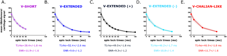
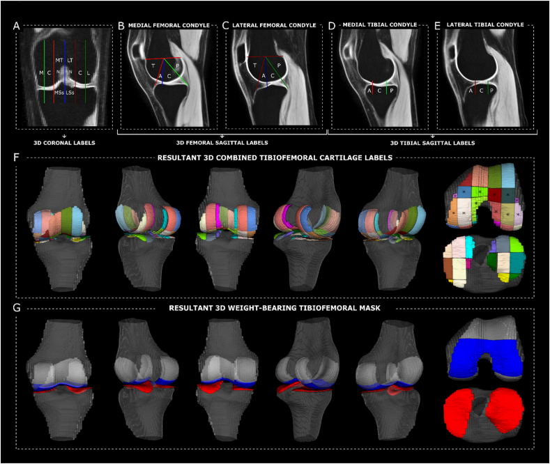
Design: Twenty healthy participants (10 M/10F, 22.2 ± 3.4 years) underwent bilateral knee MRI using T1 MAPPS sequences with varying TSLs ([0-120] ms), along with a 3D spoiled gradient echo for QSM. Five total TSL combinations were used for T1 computation, and direct depth-based comparison. Depth-wide variance was assessed in comparison to QSM as a basis to assess for depth-specific variation in T1 computations across healthy cartilage.
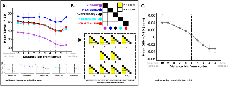
Results: Longer T1 relaxation times were observed for TSL combinations with higher spin lock times. Depth-specific differences were documented for both QSM and T1 , with most change found at ∼60% depth of the cartilage, relative to the surface. Direct squared linear correlation revealed that most T1 TSL combinations can explain over 30% of the variability in QSM, suggesting inherent shared sensitivity to cartilage microstructure.
Conclusions: T1 mapping is subjective to the spin lock time combinations used for computation of relaxation times. When paired with QSM, both similarities and differences in signal sensitivity may be complementary to capture depth-wide changes in articular cartilage.
Keywords: Arthritis; Articular cartilage; Magnetic resonance imaging; Microstructural integrity; QSM; T1ρ.
© 2024 The Authors.
Conflict of interest statement
Dr. Myer consults with commercial entities to support commercialization strategies and applications to the US Food and Drug Administration but has no direct financial interest in the products. Dr. Myer's institution receives current and ongoing grant funding from National Institutes of Health/NIAMS Grants U01AR067997, R01 AR070474, R01AR055563, R01AR076153, R01 AR077248 and industry sponsored research funding related to injury prevention and sport performance to his institution. Dr. Myer receives author royalties from Human Kinetics and Wolters Kluwer. Dr. Myer is an inventor of biofeedback technologies (Patent No: US11350854B2, Augmented and Virtual reality for Sport Performance and Injury Prevention Application, Approval Date: July 06, 2022, Software Copyrighted) designed to enhance rehabilitation and prevent injuries and receives licensing royalties. Dr. Myer and Dr. Diekfuss receive inventor-related royalties resultant from biofeedback technologies (Include Health: LIC1907082014-0706). Dr. Diekfuss also receives author royalties from Kendall Hunt Publishing Company. Dr. Mandava is an employee of GE HealthCare.
Figures


References
-
- Saarakkala S., et al. Depth-wise progression of osteoarthritis in human articular cartilage: investigation of composition, structure and biomechanics. Osteoarthritis Cartilage. 2010;18(1):73–81. - PubMed
-
- Link T.M., et al. Establishing compositional MRI of cartilage as a biomarker for clinical practice. Osteoarthritis Cartilage. 2018;26(9):1137–1139. - PubMed
-
- Duvvuri U., et al. T(1rho) relaxation can assess longitudinal proteoglycan loss from articular cartilage in vitro. Osteoarthritis Cartilage. 2002;10(11):838–844. - PubMed
LinkOut - more resources
Full Text Sources
