. 2024 Aug 29:17:5741-5762.
doi: 10.2147/JIR.S473665. eCollection 2024.

Ferulic Acid Methyl Ester Attenuates Cerebral Ischemia-Reperfusion Injury in Rats by Modulating PI3K/HIF-1α/VEGF Signaling Pathway

Affiliations

Ferulic Acid Methyl Ester Attenuates Cerebral Ischemia-Reperfusion Injury in Rats by Modulating PI3K/HIF-1α/VEGF Signaling Pathway

Peijie Zhou et al. J Inflamm Res. .

Abstract

Background: Cerebral ischaemia-reperfusion injury (CIRI) could worsen the inflammatory response and oxidative stress in brain tissue. According to previous studies, ferulic acid methyl ester (FAME), as the extract with the strongest comprehensive activity in the traditional Chinese medicine Huang Hua oil dot herb, has significant anti-oxidative stress and neuroprotective functions, and can effectively alleviate CIRI, but its mechanism of action is still unclear.

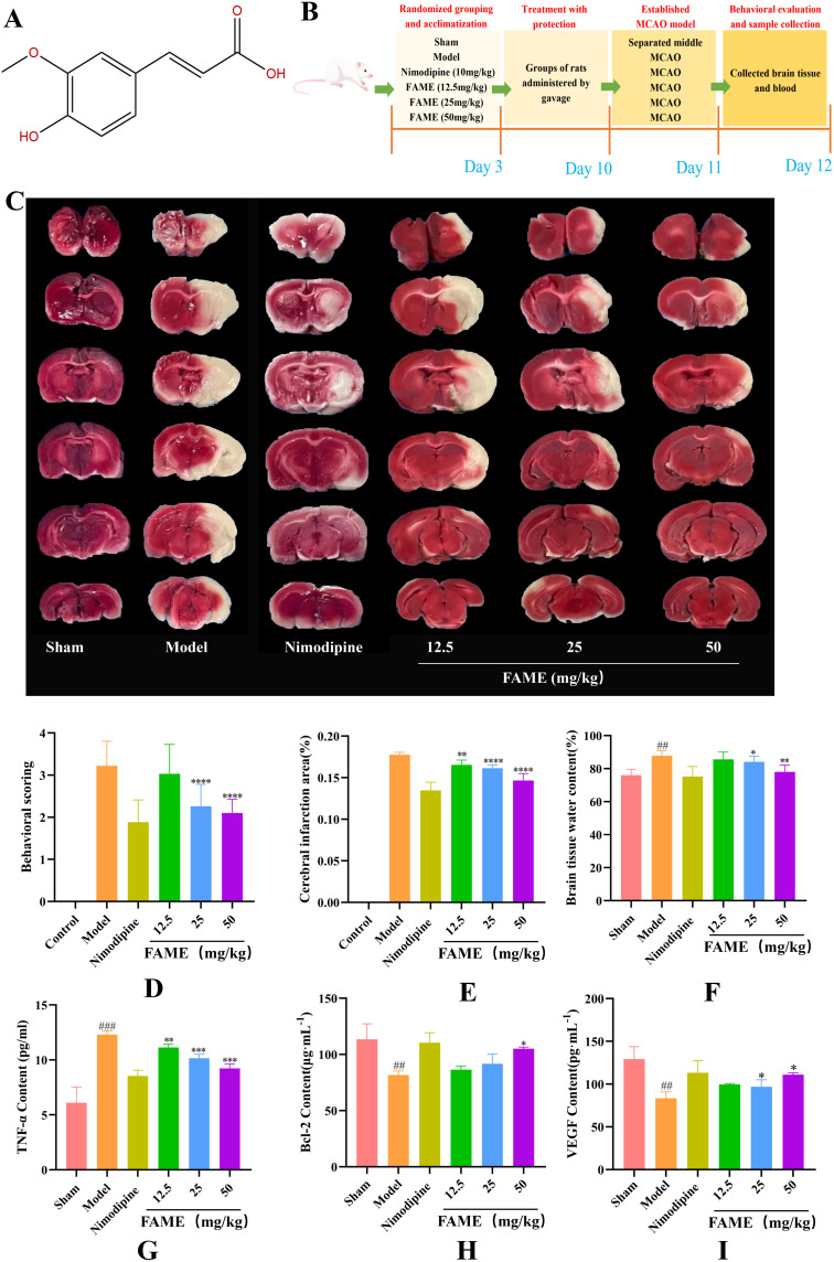
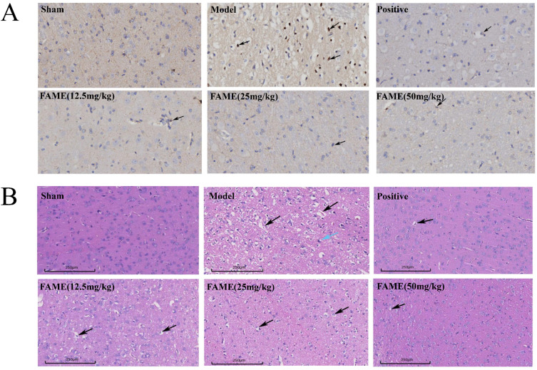
Methods: Firstly, the pharmacological effects of FAME were investigated by in vitro oxidative stress and inflammatory experiments. Secondly, evaluate the therapeutic effects of FAME in the treatment of CIRI by brain histopathological staining and cerebral infarct area by replicating the in vivo MACO model. Thirdly, RNA-Seq and network pharmacology were utilized to predict the possible targets and mechanisms of FAME for CIRI at the molecular level. Finally, the expression of key target proteins, as well as the key regulatory relationships were verified by molecular docking visualization, Western Blotting and immunohistochemistry.

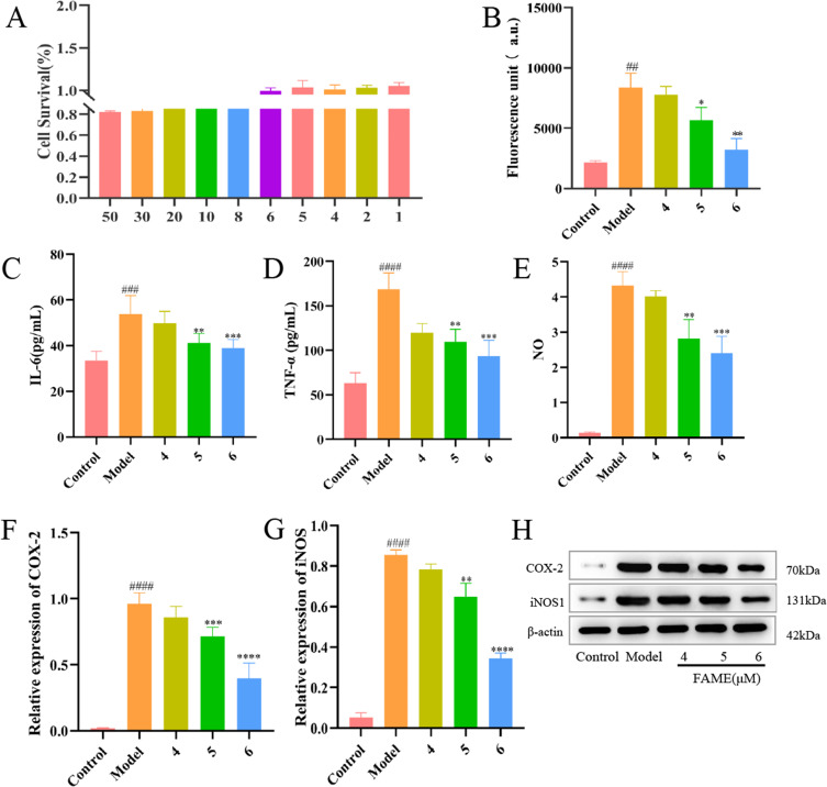
Results: The results of in vitro experiments concluded that FAME could significantly reduce the content of TNF-α, IL-1β and ROS, inhibiting COX-2 and iNOS protein expression in cells(p<0.01). FAME was demonstrated to have anti-oxidative stress and anti-inflammatory effects. The results of in vivo experiments showed that after the administration of FAME, the area of cerebral infarction in rats with CIRI was reduced, the content of Bcl-2 and VEGF was increased(p<0.05). Network pharmacology and RNA-Seq showed that the alleviation of CIRI by FAME may be through PI3K-AKT and HIF-1 signaling pathway. Enhanced expression of HIF-1α, VEGF, p-PI3K, p-AKT proteins in the brain tissues of rats in the FAME group was verified by molecular docking and Western Blotting.

Conclusion: FAME possesses significant anti-inflammatory and anti-oxidative stress activities and alleviates CIRI through the PI3K/HIF-1α/VEGF signaling pathway.

Keywords: RNA-Seq; WGCNA; cerebral ischemia-reperfusion injury; ferulic acid methyl ester; molecular docking techniques; network pharmacology.


Conflict of interest statement

The authors report no conflicts of interest in this work.

Figures

None
Graphical abstract
Figure 1
Figure 1
Results of in vitro antioxidant activity of FAME. (A) Cell survival rate at different concentrations. (B) Results of intracellular ROS content determination. (C) Results of intracellular IL-6 content determination. (D) Results of intracellular TNF-α content determination. (E) Results of intracellular NO content determination. (F) Intracellular COX-2 protein expression. (G) Intracellular iNOS protein expression. (H) Western blotting of intracellular COX-2 and iNOS proteins.
Figure 2
Figure 2
Therapeutic effects of FAME in CIRI rats. (A) Structural formula of ferulic acid methyl ester. (B) Therapeutic course of ferulic acid methyl ester in the treatment of cerebral ischemia-reperfusion injury. (C) TTC staining of rat brain tissue in each group (n=10). (D) Neurological deficit score of rats in each group (n=10). (E) Brain infarct area in each group (n=10). (F) The water content of brain tissues in each group (n=10). (G) levels of TNF-α in serum (n=10). (H) levels of Bcl-2 in serum (n=10). (I) levels of VEGF in serum (n=10).
Figure 3
Figure 3
Brain histopathologic staining results. (A) Apoptosis of rat TUNEL stained brain cells in each group 40.00x. (B) HE staining results of rat brain tissue for each group 40.00x.
Figure 4
Figure 4
Constructed component disease interaction maps using network pharmacology approach. (A) Intersectional gene target map of Ferulic acid methyl ester and farcical ischemia-reperfusion injury. (B) PPI network map. (C) Cytoscape network map. (D) PPI Degree Value Ranking.
Figure 5
Figure 5
Network pharmacology enrichment to predict the possible mechanism of action of Ferulic acid methyl ester in the treatment of cerebral ischemia-reperfusion injury. (A) Top 20 pathway analysis based on the targets regulated by MA in CIRI. (B) Biological processes predicted by network pharmacology. (C) Cellular component predicted by network pharmacology. (D) Molecular function predicted by network pharmacology.
Figure 6
Figure 6
RNA-Seq enrichment results of FAME for CIRI. (A) Sample aggregation distribution map. (B) Heatmap of genes differentially expressed. Difference comparison volcano map (C). Difference comparison volcano map. (D) Differential gene analysis veeny plots. (E) Results of differential gene GO enrichment based on RNA-Seq. (F) Results of differential gene KEGG enrichment based on RNA-Seq (G). GSEA analysis of the HIF-1 signaling pathway.
Figure 7
Figure 7
WGCNA results of FAME for CIRI. (A) The soft threshold was set at 8, where the blue horizontal line indicates the correlation coefficient 0.8 and the red horizontal line indicates the correlation coefficient 0.9. (B) module hierarchical clustering plot (C). Module trait clustering plot (D). Modules and inter-module correlation. (E) Samples and Modules correlation heat map. (F) trait association correlation map. (G) Gene enrichment heat map for each module (H). MM-GS Correlation Plot of Cerebral Infarct disease manifestation and Module Gene Association Analysis. (I) Gene regulatory network map between PIK3R1, STAT3 and HIF1Α. (J) Network diagram of HIF-1Α upstream and downstream regulatory relationships.
Figure 8
Figure 8
Molecular docking validation of Ferulic acid methyl ester. (A) The docking diagrams of protein PIK3R1 with FAME component (left) and Wortmannin positive drug (right). (B) The docking diagrams of protein STAT3 with FAME component(left) and Colforsin positive drug(right). (C) The docking diagrams of protein MTOR docking plot with FAME component(left) and Oleandrin positive drug(right). (D) The docking plot of protein TLR4 with FAME component(left) and Papain positive drug (right). (E) The docking plot of PIK3R1 with standard ligand 1LT (left) and docking plot of STAT3 with standard ligand ATP (right). (F) The docking plot of MTOR with standard ligand X6K (left) and docking plot of TLR4 with standard ligand DAO (right).
Figure 9
Figure 9
Immunohistochemical results of relevant factors in rat brain tissue after MACO model establishment.
Figure 10
Figure 10
Western blot analysis of the expression levels of the relevant proteins in the brain tissue of each group of rats. (A) Western blotting of key target proteins in brain tissues of various groups of rats. (B) VEGF protein expression in brain tissue of rats in each group. (C) HIF-1α protein expression in brain tissue of rats in each group. (D) P-PI3K/PI3K protein expression in brain tissue of rats in each group. (E) P-AKT/AKT protein expression in brain tissue of rats in each group.
Figure 11
Figure 11
Mechanism of ferulic acid methyl ester as a novel target modulator of HIF-1α to alleviate cerebral ischemia-reperfusion injury.

References

    1. Zhao Y, Zhang X, Chen X, Wei Y, Li Y, Liu N. Neuronal injuries in cerebral infarction and ischemic stroke: from mechanisms to treatment (Review). Int J Mol Med. 2022;49(4):49. doi:10.3892/ijmm.2022.5104 - DOI - PMC - PubMed
    1. Ajoolabady A, Wang S, Kroemer G, et al. Targeting autophagy in ischemic stroke: from molecular mechanisms to clinical therapeutics. Pharmacol Ther. 2021;225:107848. doi:10.1016/j.pharmthera.2021.107848 - DOI - PMC - PubMed
    1. Xu S, Lu J, Shao A, Zhang JH, Zhang J. Glial Cells: role of the Immune Response in Ischemic Stroke. Front Immunol. 2020a;11:294. doi:10.3389/fimmu.2020.00294 - DOI - PMC - PubMed
    1. Yang JL, Mukda S, Chen SD. Diverse roles of mitochondria in ischemic stroke. Redox Biol. 2018;16:263–275. doi:10.1016/j.redox.2018.03.002 - DOI - PMC - PubMed
    1. Jurcau A, Ardelean IA. Molecular pathophysiological mechanisms of ischemia/reperfusion injuries after recanalization therapy for acute ischemic stroke. J Integr Neurosci. 2021;20:727–744. - PubMed

LinkOut - more resources