. 2024 Sep 27;135(8):856-872.
doi: 10.1161/CIRCRESAHA.124.325023. Epub 2024 Sep 3.

Monocytes Reprogrammed by 4-PBA Potently Contribute to the Resolution of Inflammation and Atherosclerosis

Affiliations

Monocytes Reprogrammed by 4-PBA Potently Contribute to the Resolution of Inflammation and Atherosclerosis

Shuo Geng et al. Circ Res. .

Abstract

Background: Chronic inflammation initiated by inflammatory monocytes underlies the pathogenesis of atherosclerosis. However, approaches that can effectively resolve chronic low-grade inflammation targeting monocytes are not readily available. The small chemical compound 4-phenylbutyric acid (4-PBA) exhibits broad anti-inflammatory effects in reducing atherosclerosis. Selective delivery of 4-PBA reprogrammed monocytes may hold novel potential in providing targeted and precision therapeutics for the treatment of atherosclerosis.

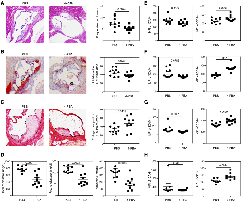
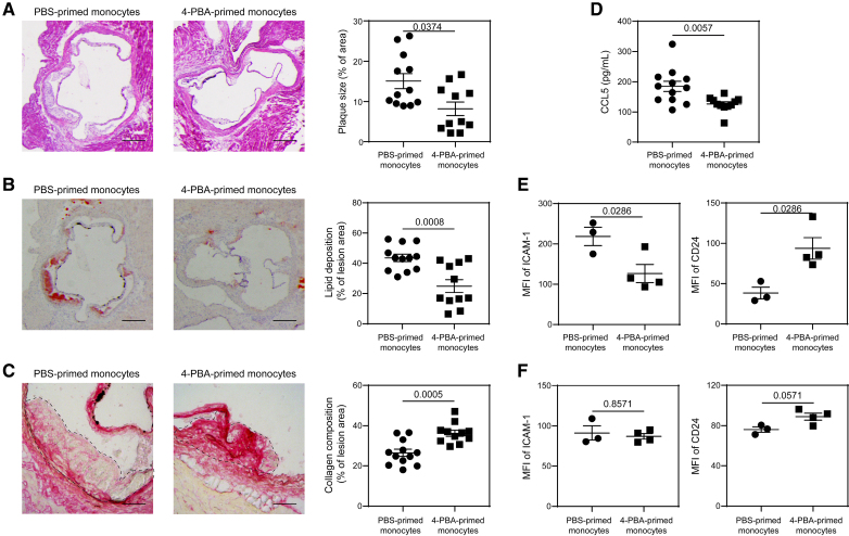
Methods: Systems analyses integrating single-cell RNA sequencing and complementary immunologic approaches characterized key resolving characteristics as well as defining markers of reprogrammed monocytes trained by 4-PBA. Molecular mechanisms responsible for monocyte reprogramming were assessed by integrated biochemical and genetic approaches. The intercellular propagation of homeostasis resolution was evaluated by coculture assays with donor monocytes trained by 4-PBA and recipient naive monocytes. The in vivo effects of monocyte resolution and atherosclerosis prevention by 4-PBA were assessed with the high-fat diet-fed ApoE-/- mouse model with IP 4-PBA administration. Furthermore, the selective efficacy of 4-PBA-trained monocytes was examined by IV transfusion of ex vivo trained monocytes by 4-PBA into recipient high-fat diet-fed ApoE-/- mice.

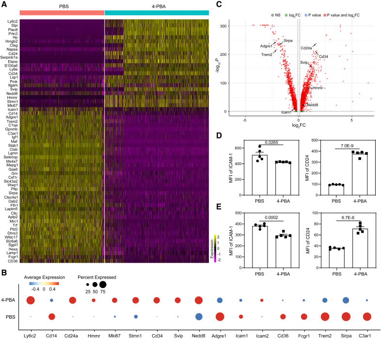
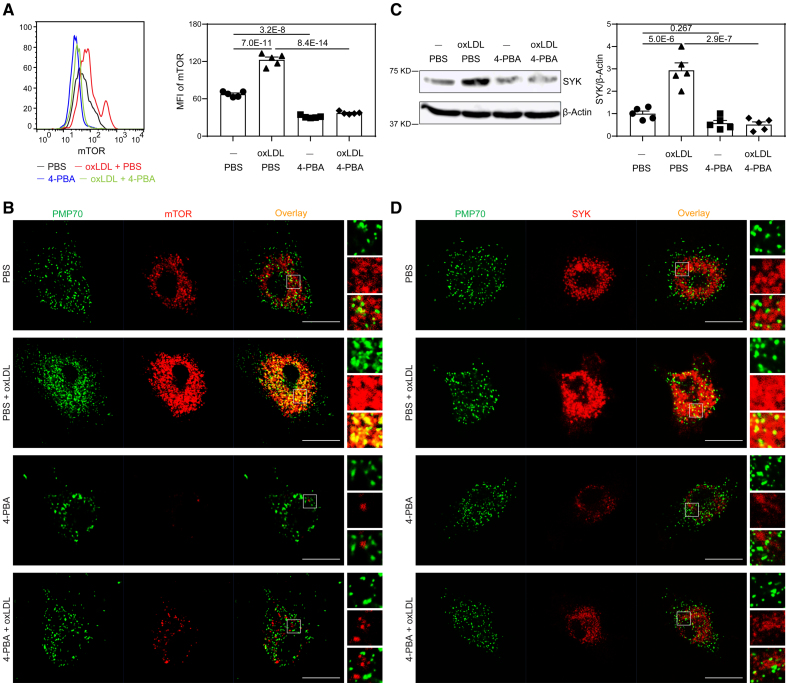
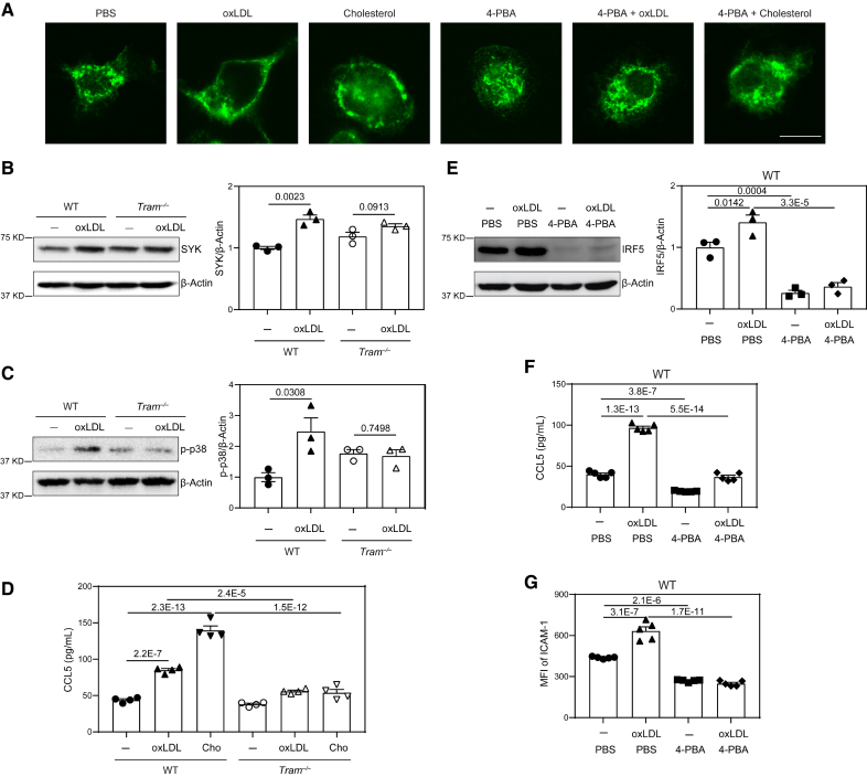
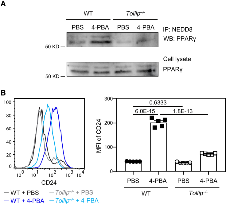
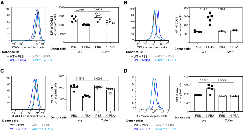
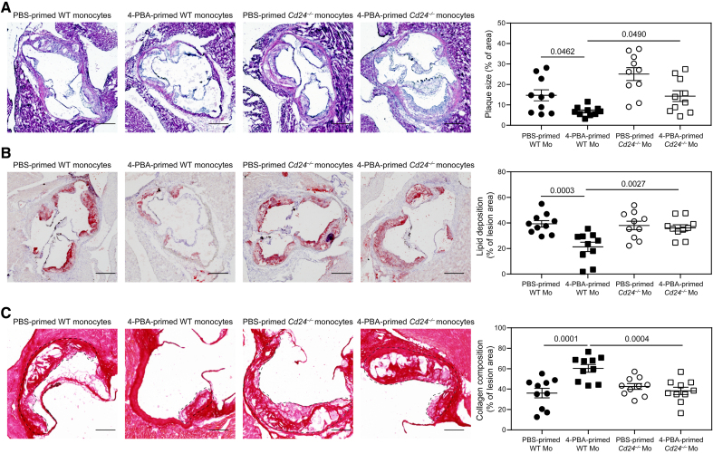
Results: In this study, we found that monocytes can be potently reprogrammed by 4-PBA into an immune-resolving state characterized by reduced adhesion and enhanced expression of anti-inflammatory mediator CD24. Mechanistically, 4-PBA reduced the expression of ICAM-1 (intercellular adhesion molecule 1) via reducing peroxisome stress and attenuating SYK (spleen tyrosine kinase)-mTOR (mammalian target of rapamycin) signaling. Concurrently, 4-PBA enhanced the expression of resolving mediator CD24 through promoting PPARγ (peroxisome proliferator-activated receptor γ) neddylation mediated by TOLLIP (toll-interacting protein). 4-PBA-trained monocytes can effectively propagate anti-inflammation activity to neighboring monocytes through CD24. Our data further demonstrated that 4-PBA-trained monocytes effectively reduce atherosclerosis pathogenesis when administered in vivo.

Conclusions: Our study describes a robust and effective approach to generate resolving monocytes, characterizes novel mechanisms for targeted monocyte reprogramming, and offers a precision therapeutics for atherosclerosis based on delivering reprogrammed resolving monocytes.

Keywords: atherosclerosis; immunity, innate; inflammation; monocytes; therapeutics.


Conflict of interest statement

None.

Figures

Figure 1.
Figure 1.
Treatment with 4-phenylbutyric acid (4-PBA) induces anti-inflammatory resolving characteristics of monocytes. A through D, Bone marrow–derived monocytes (BMMs) from wild-type (WT) C57 BL/6 mice were cultured in vitro with macrophage colony-stimulating factor (M-CSF; 10 ng/mL) in the presence 4-PBA (1 mmol/L) or PBS for 5 days. Single-cell RNA sequencing (scRNAseq) was performed, and data sets were processed to compare naive murine monocytes with monocytes trained by 4-PBA. A, Heatmaps demonstrating representative genes differentially expressed in different clusters of monocytes challenged with 4-PBA. B, Dot plot comparison of representative genes differentially expressed between PBS- vs 4-PBA–trained monocytes. C, Volcano plot showing differentially expressed genes between PBS- vs 4-PBA–trained monocytes. D, Surface expression of ICAM-1 (intercellular adhesion molecule 1) and CD24 on CD11b+ monocytes cultured was analyzed by flow cytometry. E, Peripheral blood mononuclear cells (PBMCs) isolated from healthy human individuals were cultured in vitro with M-CSF (100 ng/mL) in the presence 4-PBA (1 mmol/L) or PBS for 2 days. Surface expression of ICAM-1 and CD24 on total monocytes (including CD14hi, CD16hi, and intermediate monocytes) was analyzed by flow cytometry. Data in D and E (n=5 for each group, biological replicates) were analyzed with Student t test. Error bars represent means±SEM.
Figure 2.
Figure 2.
Administration of 4-phenylbutyric acid (4-PBA) alleviates atherosclerotic pathogenesis. Male ApoE−/− mice were fed with high-fat diet (HFD) for 4 weeks and intraperitoneally injected with 4-PBA (5 mg/kg body weight) or PBS every 3 days for additional 4 weeks. A, Representative images of H&E-stained atherosclerotic lesions and quantification of plaque size demonstrated as the percentage of lesion area within aortic root area. Scale bars, 300 µm. B, Representative images of oil red O–stained atherosclerotic plaques and quantification of lipid deposition within lesion area. Scale bars, 300 µm. C, Representative images of Picrosirius red–stained atherosclerotic plaques and quantification of collagen content within lesion area. Scale bars, 100 µm. D, Detection of total cholesterol, free cholesterol, and triglyceride levels in the plasma. E through H. Surface expressions of ICAM-1 (intercellular adhesion molecule 1) and CD24 on CD11b+ Ly6G Ly6Chi monocytes in the peripheral blood (E), bone marrow (BM; F), spleen (G), and aorta (H) were examined by flow cytometry. Data in A through E were analyzed using Student t test, and data in F through H were analyzed using Mann-Whitney U test (n=10 for each group in A through G; n=8 for each group in H; biological replicates). Error bars represent means±SEM.
Figure 3.
Figure 3.
4-phenylbutyric acid (4-PBA) inhibits mTOR (mammalian target of rapamycin) signaling and restores peroxisome homeostasis in monocytes. Bone marrow–derived monocytes (BMMs) from wild-type (WT) C57 BL/6 mice were cultured in vitro with M-CSF (macrophage colony-stimulating factor; 10 ng/mL) in the presence of oxLDL (oxidized low-density lipoprotein; 10 µg/mL), 4-PBA (1 mmol/L), or PBS for 5 days. A, Representative histogram and quantification of mTOR level in CD11b+ Ly6Chi monocytes as determined by flow cytometry. B, Monocytes were stained with anti-PMP70 and anti-mTOR antibodies, and the localization of PMP70+ peroxisomes and mTOR was examined by confocal microcopy. Scale bars, 10 µm. C, Protein level of SYK in BMMs was examined by Western blotting, and SYK expression was quantified after normalizing to β-Actin expression. D, Monocytes were stained with anti-PMP70 and anti-SYK antibodies, and the localization of peroxisomes and SYK was examined by confocal microcopy. Scale bars, 10 µm. Data in A and C were analyzed using 2-way ANOVA followed by Šídák post hoc test (n=5 for each group; biological replicates). Error bars represent means±SEM.
Figure 4.
Figure 4.
TRAM (Trif-related adapter molecule) serves as a general membrane stress sensor mediating the inflammatory effects of lipids on monocytes. A, Bone marrow–derived monocytes (BMMs) from wild-type (WT) C57 BL/6 mice were cultured in vitro with M-CSF (macrophage colony-stimulating factor; 10 ng/mL) in the presence of oxLDL (oxidized low-density lipoprotein; 10 µg/mL), cholesterol (10 µg/mL), 4-phenylbutyric acid (4-PBA; 1 mmol/L) or PBS for 5 days. The cells were stained with anti-TRAM antibody, and cellular distribution of TRAM was examined by confocal microcopy. Scale bars, 10 µm. B and C, BMMs from WT C57 BL/6 mice and Tram−/− mice were cultured with M-CSF (10 ng/mL) in the presence of oxLDL (10 µg/mL) or PBS for 5 days. Protein level of SYK (B) and phosphorylation of p-38 (C) were examined by Western blotting and quantified after normalizing to β-Actin expression. D, BMMs from WT C57 BL/6 mice and Tram−/− mice were cultured with M-CSF (10 ng/mL) in the presence of oxLDL (10 µg/mL), cholesterol (10 µg/mL) or PBS for 5 days. Production of chemokine ligand 5 (CCL5) was determined by ELISA. E through G, BMMs from WT C57 BL/6 mice were cultured with M-CSF (10 ng/mL) in the presence of oxLDL (10 µg/mL), 4-PBA (1 mmol/L), or PBS for 5 days. Protein level of IRF5 was examined by Western blotting and quantified after normalizing to β-Actin expression (E). Production of CCL5 was determined by ELISA (F), and surface expression of ICAM-1 on CD11b+ monocytes was determined by flow cytometry (G). Data in B and C were analyzed using Student t test, and data in D through G were analyzed using 2-way ANOVA followed by Šídák post hoc test (n=3 for each group in B, C, and E; n=4 for each group in D; n=5 for each group in F and G; biological replicates. Error bars represent means±SEM.
Figure 5.
Figure 5.
4-phenylbutyric acid (4-PBA) promotes the expression of CD24 through enhanced PPARγ (peroxisome proliferator-activated receptor γ) activation in a Tollip-dependent manner. Bone marrow–derived monocytes (BMMs) from wild-type (WT) C57 BL/6 mice and Tollip−/− mice were cultured in vitro with M-CSF (macrophage colony-stimulating factor; 10 ng/mL) in the presence of 4-PBA (1 mmol/L) or PBS for 5 days. A, Cell lysate was isolated and subjected to immunoprecipitation with anti-NEDD8 antibodies conjugated to resin. The association of PPARγ with NEDD8 was examined by Western blotting. PPARγ in cell lysate was also examined by Western blotting. B, Surface expression of CD24 on CD11b+ Ly6G Ly6Chi monocytes was examined by flow cytometry. Mean Fluorescence Intensity (MFI) of CD24 was quantified. Data in B were analyzed using 2-way ANOVA followed by Šídák post hoc test (n=5 for each group; biological replicates). Error bars represent means±SEM.
Figure 6.
Figure 6.
4-phenylbutyric acid (4-PBA) trained monocytes propagate resolving nature to neighboring monocytes through CD24 and Tollip. A and B, Bone marrow–derived monocytes (BMMs) from wild-type (WT) C57 BL/6 mice and Cd24−/− mice, which are both CD45.2+, were cultured in vitro with M-CSF (macrophage colony-stimulating factor; 10 ng/mL) in the presence of 4-PBA (1 mmol/L) or PBS for 5 days. Recipient BMMs prepared from B6 SJL mice, which are CD45.1+, were treated with oxLDL (oxidized low-density lipoprotein; 10 µg/mL) for 3 days and then cocultured in vitro with CD45.2+ donor cells for 2 days. Surface expressions of ICAM-1 (intercellular adhesion molecule 1; A) and CD24 (B) on CD45.1+ recipient BMMs were examined by flow cytometry. C and D, BMMs from WT C57 BL/6 mice and Tollip−/− mice, which are both CD45.2+, were cultured with M-CSF (10 ng/mL) in the presence of 4-PBA (1 mmol/L) or PBS for 5 days. Recipient BMMs prepared from B6 SJL mice, which are CD45.1+, were treated with oxLDL (10 µg/mL) for 3 days and then cocultured with CD45.2+ donor cells in vitro for 2 days. Surface expression of ICAM-1 (C) and CD24 (D) on CD45.1+ recipient BMMs was examined by flow cytometry. Data were analyzed using 2-way ANOVA followed by Šídák post hoc test (n=5 for each group; biological replicates). Error bars represent means±SEM.
Figure 7.
Figure 7.
Adoptive transfer of monocytes polarized by 4-phenylbutyric acid (4-PBA) alleviates atherosclerosis. A through D, Male ApoE−/− mice, serving as recipients, were fed with high-fat diet (HFD) for 4 weeks. Bone marrow–derived monocytes (BMMs) from ApoE−/− mice were treated with PBS or 4-PBA (1 mmol/L) for 5 days. PBS- or 4-PBA-polarized monocytes (3×10⁶ cells per mouse) were then adoptively transferred by intravenous injection to HFD-fed ApoE−/− mice once a week for 4 weeks. Tissues were harvested 1 week after the last monocyte transfer. A, Representative images of H&E-stained atherosclerotic lesions and quantification of plaque size demonstrated as the percentage of lesion area within aortic root area. Scale bars, 300 µm. B, Representative images of oil red O–stained atherosclerotic plaques and quantification of lipid deposition within lesion area. Scale bars, 300 µm. C, Representative images of Picrosirius red–stained atherosclerotic plaques and quantification of collagen content within lesion area. Scale bars, 100 µm. D, Detection of chemokine ligand 5 (CCL5) level in the plasma by ELISA. E and F, PBS- or 4-PBA-polarized monocytes were labeled with CFSE immediately before adoptive transfer, and tissues were harvested 1 week after the last monocyte transfer. Surface expressions of ICAM-1 and CD24 on host CFSE CD11b+ Ly6G Ly6Chi monocytes in the aorta (E) and bone marrow (BM; F) were determined by flow cytometry. Data in A, E, and F were analyzed using Mann-Whitney U test, and data in B through D were analyzed using Student t test (n=12 for PBS-trained monocytes group and n=11 for 4-PBA–trained monocytes group in A through D; n=3 for PBS-trained monocytes group and n=4 for 4-PBA-trained monocytes group in E and F; biological replicates). Error bars represent means±SEM.
Figure 8.
Figure 8.
CD24 mediates the resolving efficacy of 4-phenylbutyric acid (4-PBA)-trained monocytes in atherosclerotic mice. A through C, Male wild-type (WT) C57 BL/6 mice, serving as recipients, were intravenously injected with a single dose of AAV8-mPCSK9-D377Y (5×10¹¹ vector genomes per mouse) and fed with high-fat diet (HFD) for 4 weeks. Bone marrow–derived monocytes (BMMs) from WT C57 BL/6 and Cd24−/− mice were treated with PBS or 4-PBA (1 mmol/L) for 5 days. PBS- or 4-PBA-polarized monocytes (3×10⁶ cells per mouse) were then adoptively transferred by intravenous injection to HFD-fed recipient mice once a week for 4 weeks. Tissues were harvested 1 week after the last monocyte transfer. A, Representative images of H&E-stained atherosclerotic lesions and quantification of plaque size demonstrated as the percentage of lesion area within aortic root area. Scale bars, 300 µm. B, Representative images of Oil Red O–stained atherosclerotic plaques and quantification of lipid deposition within lesion area. Scale bars, 300 µm. C, Representative images of Picrosirius red–stained atherosclerotic plaques and quantification of collagen content within lesion area. Scale bars, 100 µm. Data were analyzed using 2-way ANOVA followed by Šídák post hoc test (n=10 for each group; biological replicates). Error bars represent means±SEM.

Update of

References

    1. Engelen SE, Robinson AJB, Zurke YX, Monaco C. Therapeutic strategies targeting inflammation and immunity in atherosclerosis: how to proceed? Nat Rev Cardiol. 2022;19:522–542. doi: 10.1038/s41569-021-00668-4 - PMC - PubMed
    1. Soehnlein O, Libby P. Targeting inflammation in atherosclerosis - from experimental insights to the clinic. Nat Rev Drug Discov. 2021;20:589–610. doi: 10.1038/s41573-021-00198-1 - PMC - PubMed
    1. Hetherington I, Totary-Jain H. Anti-atherosclerotic therapies: milestones, challenges, and emerging innovations. Mol Ther. 2022;30:3106–3117. doi: 10.1016/j.ymthe.2022.08.024 - PMC - PubMed
    1. Woollard KJ, Geissmann F. Monocytes in atherosclerosis: subsets and functions. Nat Rev Cardiol. 2010;7:77–86. doi: 10.1038/nrcardio.2009.228 - PMC - PubMed
    1. Ghattas A, Griffiths HR, Devitt A, Lip GY, Shantsila E. Monocytes in coronary artery disease and atherosclerosis: where are we now? J Am Coll Cardiol. 2013;62:1541–1551. doi: 10.1016/j.jacc.2013.07.043 - PubMed

MeSH terms