. 2024;19(24):1977-1993.
doi: 10.1080/17435889.2024.2382076. Epub 2024 Sep 3.

Novel Tubeimoside I liposomal drug delivery system in combination with gemcitabine for the treatment of pancreatic cancer

Affiliations

Novel Tubeimoside I liposomal drug delivery system in combination with gemcitabine for the treatment of pancreatic cancer

Shuhui Li et al. Nanomedicine (Lond). 2024.

Abstract

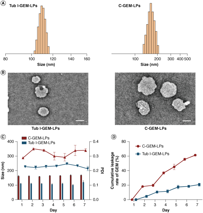
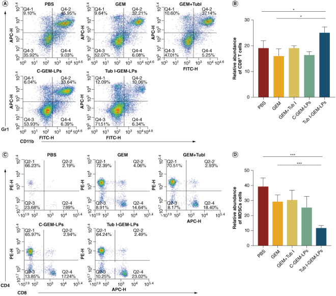
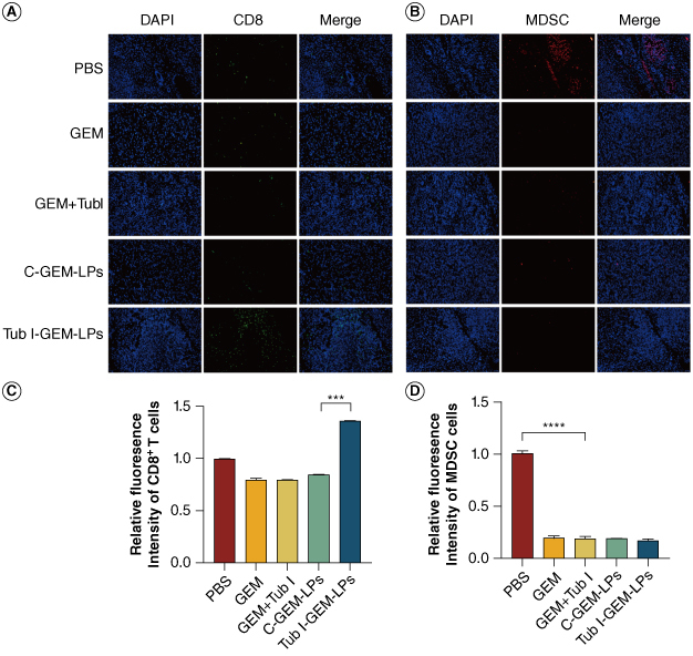
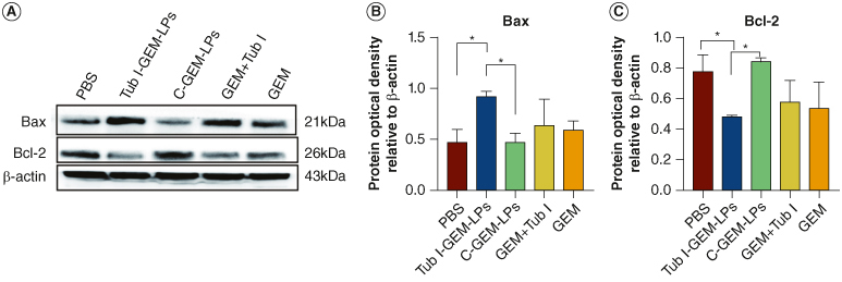
Aim: To evaluate the anti-pancreatic cancer effect of novel Tubeimoside I multifunctional liposomes combined with gemcitabine.Methods: Liposomes were prepared through the thin film hydration method, with evaluations conducted on parameters including encapsulation efficiency (EE%), particle size, polydispersity index (PDI), zeta potential (ZP), storage stability, and release over a 7-day period. The cellular uptake rate, therapeutic efficacy in vitro and in vivo and the role of immune microenvironment modulation were evaluated.Results: The novel Tubeimoside I multifunctional liposomal exhibited good stability, significant anti-cancer activity, and immune microenvironment remodeling effects. Furthermore, it showed a safety profile.Conclusion: This study underscores the potential of Novel Tubeimoside I multifunctional liposomal as a promising treatment option for pancreatic cancer.

Keywords: F18-FDG micro-PET/CT imaging; gemcitabine; liposomes; pancreatic cancer; tubular alfalfa glycosides I; tumor microenvironment.

Plain language summary

[Box: see text].


Conflict of interest statement

The authors have no competing interests or relevant affiliations with any organization or entity with the subject matter or materials discussed in the manuscript. This includes employment, consultancies, honoraria, stock ownership or options, expert testimony, grants or patents received or pending, or royalties.

Figures

Figure 1.
Figure 1.
The strategic creation of a versatile Tub I-GEM-LPs for targeting tumors includes replacing cholesterol with Tubeimoside I in Tub I-GEM-LPs. This switch acts as both a barrier enhancer and chemotherapy supplement, offering the liposome a variety of advantages. These advantages encompass improved absorption by malignant cells, restructuring of the tumor environment and heightened ability to eradicate tumor cells. By incorporating Tubeimoside I into the liposome, we have created a versatile therapeutic delivery system with the potential to improve the effectiveness of cancer treatments.
Figure 2.
Figure 2.
Characterization of Tubeimoside I liposomes. (A) Size distribution of Tub I-LPs and C-LPs was examined with nanoparticle sizer. (B) Transmission electron microscope images of Tubeimoside I liposomes (Tub I-LPs) and cholesterol liposome (C-LPs); scale bar = 100 nm. (C) Stability of two kinds liposome formulations kept in 4°C conditions monitored by nanoparticle sizer. (n = 3; mean ± SD).(D) Drug leakage profles of GEM loaded C-LPs and Tub I-LPs (n = 3; mean ± SD).
Figure 3.
Figure 3.
The liposome internalization and cellular uptake in Panc 02 and MIA PaCa 02 cells was investigated. Qualitative analysis (A & B) and quantitative analysis (C & D) of the uptake of carboxyfluorescein Coumarin 6-loaded C-LPs and Tub I-LPs in Panc 02 and MIA PaCa 02 cells was conducted. The cells were treated with 500 ng/ml Coumarin 6-loaded liposomes and incubated at 37°C for 4 h (n = 3; mean ± SD).
Figure 4.
Figure 4.
In vitro anticancer effect of Tub I-GEM-LPs. (A) The cytotoxic effect of various groups on Panc02 cells was evaluated. (n = 5, mean ± SD). (B) The study also examined the cytotoxicity of different groups on MIA PaCa cells (n = 5, mean ± SD). (C) Representative scatter plots of Annexin V/PI analysis were provided for Panc02 cells after drug treatments. (D) The percentage of cells with early, late, and complete apoptosis in Panc 02 cells (n = 3; mean ± SD). (E) Similar analysis was conducted for MIA PaCa 02 cells (n = 3; mean ± SD). *p < 0.05; **p < 0.01 and ***p < 0.001.
Figure 5.
Figure 5.
(A) Tumor weights of excised tumors n = 5; mean ± SD). (B) Tumor volume of excised tumors (n = 5; mean ± SD). (C) Tumors were removed from C57 mice with Panc 02 tumors the day following the final injection (n = 5; mean ± SD). (D) micro-PET/CT visualization of tumor-bearing rats after treatment. (E) Mean SUV was calculated for both groups of hormonal rats. (n = 3; mean ± SD) (F) Hematoxylin and eosin stained sections of the heart, liver, lungs and kidneys were examined in representative samples (100 μm scale bar). Statistical significance was observed (p < 0.001).
Figure 6.
Figure 6.
While employed as a chemotherapy adjuvant, Tub I assisted in reversing the immunosuppressive TME. (A) Flow cytometry analysis detected the presence of CD4+ and CD8+ T cells through the use of CD3 FITC, CD8 APC and CD4 PE markers in spleen tissues. (B) The histograms analysis of CD8+ T cells. (C) Flow cytometry gating analysis of CD11b+/Gr1 + MDSC cells in tumors. (D) The histograms analysis of CD11b+/Gr1 + MDSC cells in spleen tissues p < 0.05, **p < 0.01 and ***p < 0.001, (n = 5; mean ± SD).
Figure 7.
Figure 7.
(A & B) Immunofluorescence was employed to uncover the quantity of MDSC and CD8+ cells in tumors of mice bearing the Panc 02 tumor. Scale bar: 100 μm. (C & D) Immunofluorescence detection of CD8+ and MDSC cell numbers histogram analysis. *p < 0.05; **p < 0.01 and ***p < 0.001, (n = 5; mean ± SD).
Figure 8.
Figure 8.
(A) The Western blotting analysis determined the protein expression of Bax and Bcl-2, with β-actin serving as the loading control. (B & C) Band densities of the proteins were quantified using the Image J software. *p < 0.05; **p < 0.01; ***p < 0.001; mean ± S.D; n = 5.

Similar articles

References

    1. Khalaf N, El-Serag HB, Abrams HR, et al. Burden of pancreatic cancer: from epidemiology to practice. Clin Gastroenterol Hepatol. 2021;19(5):876–884. doi: 10.1016/j.cgh.2020.02.054 - DOI - PMC - PubMed
    1. Park W, Chawla A, O'Reilly EM. Pancreatic cancer: a review. JAMA. 2021;326(20):2081. doi: 10.1001/jama.2021.19984 - DOI - PMC - PubMed
    1. Zhang J, Wang Y, Wang L, et al. Pancreatic ductal adenocarcinoma chemoresistance: from metabolism reprogramming to novel treatment. Chin Med J (Engl). 2024; 137(4):408–420. doi: 10.1097/CM9.0000000000002758 - DOI - PMC - PubMed
    1. Gu M, Liu Y, Xin P, et al. Fundamental insights and molecular interactions in pancreatic cancer: pathways to therapeutic approaches. Cancer Lett. 2024;588:216738. doi: 10.1016/j.canlet.2024.216738 - DOI - PubMed
    1. Moosavian SA, Bianconi V, Pirro M, et al. Challenges and pitfalls in the development of liposomal delivery systems for cancer therapy. Semin Cancer Biol. 2021:69:337–348. doi: 10.1016/j.semcancer.2019.09.025 - DOI - PubMed

Publication types

MeSH terms

LinkOut - more resources