. 2024 Oct 22;98(10):e0011924.
doi: 10.1128/jvi.00119-24. Epub 2024 Sep 3.

Neu5Gc binding loss of subtype H7 influenza A virus facilitates adaptation to gallinaceous poultry following transmission from waterbirds

Affiliations

Neu5Gc binding loss of subtype H7 influenza A virus facilitates adaptation to gallinaceous poultry following transmission from waterbirds

Minhui Guan et al. J Virol. .

Abstract

Between 2013 and 2018, the novel A/Anhui/1/2013 (AH/13)-lineage H7N9 virus caused at least five waves of outbreaks in humans, totaling 1,567 confirmed human cases in China. Surveillance data indicated a disproportionate distribution of poultry infected with this AH/13-lineage virus, and laboratory experiments demonstrated that this virus can efficiently spread among chickens but not among Pekin ducks. The underlying mechanism of this selective transmission remains unclear. In this study, we demonstrated the absence of Neu5Gc expression in chickens across all respiratory and gastrointestinal tissues. However, Neu5Gc expression varied among different duck species and even within the tissues of the same species. The AH/13-lineage viruses exclusively bind to acetylneuraminic acid (Neu5Ac), in contrast to wild waterbird H7 viruses that bind both Neu5Ac and N-glycolylneuraminic acid (Neu5Gc). The level of Neu5Gc expression influences H7 virus replication and facilitates adaptive mutations in these viruses. In summary, our findings highlight the critical role of Neu5Gc in affecting the host range and interspecies transmission dynamics of H7 viruses among avian species.IMPORTANCEMigratory waterfowl, gulls, and shorebirds are natural reservoirs for influenza A viruses (IAVs) that can occasionally spill over to domestic poultry, and ultimately humans. This study showed wild-type H7 IAVs from waterbirds initially bind to glycan receptors terminated with N-acetylneuraminic acid (Neu5Ac) or N-glycolylneuraminic acid (Neu5Gc). However, after enzootic transmission in chickens, the viruses exclusively bind to Neu5Ac. The absence of Neu5Gc expression in gallinaceous poultry, particularly chickens, exerts selective pressure, shaping IAV populations, and promoting the acquisition of adaptive amino acid substitutions in the hemagglutinin protein. This results in the loss of Neu5Gc binding and an increase in virus transmissibility in gallinaceous poultry, particularly chickens. Consequently, the transmission capability of these poultry-adapted H7 IAVs in wild water birds decreases. Timely intervention, such as stamping out, may help reduce virus adaptation to domestic chicken populations and lower the risk of enzootic outbreaks, including those caused by IAVs exhibiting high pathogenicity.

Keywords: H7; H7N9; N-glycolylneuraminic acid; acetylneuraminic acid; duck; influenza A virus; receptor binding; spread; transmission; virus-host interactions.


Conflict of interest statement

The authors declare no conflict of interest.

Figures

Fig 1
Fig 1
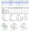
Receptor binding profile of H7 influenza A viruses. (a) N-glycan microarray binding profiles of five H7 viruses isolated from wild waterbirds in Eurasia and North America. (b) N-glycan microarray binding profiles of eight AH/13-lineage H7N9 viruses were collected during the first five waves of outbreaks in humans from 2013 to 2017 in China. (c) Quantitative analyses of virus glycan binding avidity using biolayer interferometry for two representative H7 viruses, rgCk/WX13 and rgMuSn/RI08 (Table 1). We categorized 75 glycans on the microarray based on the linkage and terminal glycan sequence into α2,3-linked Neu5Ac, α2,3-linked Neu5Gc, α2,6-linked Neu5Ac, α2,8-linked Neu5Ac, α2,6-linked Neu5Gc, and non-sialic acid glycans. The glycan sequences are detailed in Table S5. In the plot showing microarray data, the mean relative fluorescent units ± the standard deviations (vertical bars) are shown on the y-axis, and the x-axis represents the glycan number corresponding to the array. A microarray contains six replicates of each glycan. Biolayer interferometry analyses were performed using an Octet RED instrument (Pall FortéBio, Fremont, CA, USA) (see Materials and Methods), and binding curves were fitted using the saturation binding method in GraphPad Prism 8 (https://www.graphpad.com/scientific-software/prism/). We quantified and compared the 50% relative sugar loading concentration (RSL0.5) at half the fractional saturation (f = 0.5) of the virus against glycan analogs to determine the binding avidity. A higher RSL0.5 indicates a lower binding avidity. The BLI was performed with at least eight concentrations of glycan loadings. In the structures of biotinylated glycan analogs used in BLI, Neu5Acα2-3Galβ1-4GlcNAcβ (3’SLN), Neu5Acα2-6Galβ1-4GlcNAcβ) (6’SLN), Neu5Acα2-3Galβ1-4(Fucα1-3)GlcNAcβ (sLeX), Neu5Gcα2-3Galβ1-4GlcNAcβ (3’GLN), and Neu5Gcα2-3Galβ1-4(Fucα1-3)GlcNAcβ (GLeX), the following glycan symbols were used: light blue diamond indicated Neu5Gc, purple diamond indicated Neu5Ac, yellow circle represents galactose, blue square represents GlcNAc, and red triangle represents fucose.
Fig 2
Fig 2
Multiple individual amino acid substitutions facilitate the acquisition of virus binding avidity to Neu5Gc for H7 IAVs. (a) Sequence alignment of the RBS of H7 IAVs, including three groups of H7 viruses with distinct binding patterns to glycans terminated with Neu5Ac and Neu5Gc: equine H7N7 viruses bound exclusively to Neu5Gc, wild waterbird viruses bound to both Neu5Ac and Neu5Gc, and AH/13-lineage H7N9 viruses bound exclusively to Neu5Ac. (b) Amino acid diversity at the residues close to or within the hemagglutinin RBS of AH/13-lineage H7N9 viruses isolated from domestic poultry in China, as well as H7 viruses from dabbling ducks in Eurasia and North America (see additional details in Table 2). (c) Quantitative analyses of virus glycan binding avidity using biolayer interferometry for Ck/WX13, Ck/WX13-A121N, and Ck/WX13-V179I. Please refer to the legend of Fig. 1 and Online Methods for the details of biolayer interferometry analyses. The BLI was performed with at least 8 concentrations of glycan loadings. (d) The crystal structure of the HA protein from A/Anhui/1/2013 (H7N9), is identical to that of Ck/WX13. (e) Structural model of wild-type H7 in complex with Neu5Ac (green) versus Neu5Gc (magenta). HA residues less than 3 Å away from the modeled receptor are shown in sticks. (f) Structural model of the V179I H7 mutant in complex with Neu5Ac (green) versus Neu5Gc (magenta).
Fig 3
Fig 3
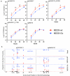
Neu5Gc affects virus replication of AH/13-lineage H7N9 viruses and drives adaptive mutations in the HA protein. (a) Growth kinetics of H7 influenza A viruses and mutants in MDCK-wt and MDCK-Gc cells. All viruses had HA genes from H7 viruses and the other seven from PR8, and three mutants were generated using the HA gene of Ck/WX13 as a template. Supernatants were collected at 12, 24, 48, and 72 hours post-infection and titrated by 50% tissue culture infection dose (TCID50) in MDCK CCL-34 cells. Two-way repeated measures analysis of variance were used to compare time-course growth data of H7 viruses among different cells. Statistical comparisons were shown as follows: not significantly different from n.s. (P > 0.05); P ≤ 0.05 as *; P < 0.01 as **; P < 0.001 as ***; and P < 0.0001 as ****. (b) HA amino acid polymorphisms were detected in the seed viruses and the viruses from the fifth passage in MDCK-wt and MDCK-Gc cells. The viruses rgCk/WX13 and rgMall/NJ10, which have a HA gene from rgCk/WX13 and rgMall/NJ10, respectively, and other seven genes from PR8 were passaged five times in MDCK-wt and MDCK-Gc cells. The two most abundant non-synonymous mutations in the HA protein were plotted to visualize adaptive amino acid substitutions caused by cell passages. The location of the RBS in the HA protein was marked as a black bar. The predominant variant is highlighted in blue and the minor variant in red.
Fig 4
Fig 4
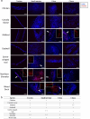
Distribution Neu5Gc glycan in the tissues of chicken, wild dabbling ducks, and Canada goose (B. canadensis). (a) The trachea, small intestine (duodenum/jejunum), colon, and cloaca of chicken (Gallus gallus), Canada goose, mallard (A. platyrhynchos), gadwall (M. strepera), green-winged teal (A. carolinensis), northern shoveler (S. clypeata), and wood duck (A. sponsa). Glycan terminated with Neu5Gc (red) was detected by immunofluorescence assay with an anti-Neu5Gc polyclonal antibody. Nuclei were stained with DAPI (blue). The white arrows indicated positive staining of Neu5Gc, the areas of which have been enlarged at the side of each image. The scale bar at the bottom of each image was 100 µm. (b) The abundance of Neu5Gc expression. We categorized the glycan receptor abundance: none or limited staining (−) without stained cells, moderate and sporadic staining (+) with <30% of stained cells, and strong staining (++) with ≥30% of the stained cells.
Fig 5
Fig 5
A transmission model illustrating the mechanisms of H7 IAV transmission and evolution between wild waterbirds and domestic poultry. An H7 IAV capable of binding to sialic acid receptors containing either Neu5Gc or Neu5Ac can be transmitted among wild waterbirds possessing these receptors, and can also transit between wild and domestic waterbirds expressed with the same receptors. This virus may then spill over into domestic poultry species (or another wild bird species) that express only Neu5Ac. Subsequent to this, the virus could acquire adaptive amino acid substitutions in the HA protein, leading it to lose its Neu5Gc binding ability and exclusively bind to Neu5Ac. Consequently, the transmission capability of these adapted viruses in waterbirds decreases. The figure was created by using BioRender.

Update of

References

    1. Olsen B, Munster VJ, Wallensten A, Waldenström J, Osterhaus ADME, Fouchier RAM. 2006. Global patterns of influenza A virus in wild birds. Science 312:384–388. doi:10.1126/science.1122438 - DOI - PubMed
    1. Long JS, Mistry B, Haslam SM, Barclay WS. 2019. Host and viral determinants of influenza A virus species specificity. Nat Rev Microbiol 17:67–81. doi:10.1038/s41579-018-0115-z - DOI - PubMed
    1. Jiang W, Hou G, Li J, Peng C, Wang S, Liu S, Zhuang Q, Chen J, Liu H. 2019. Prevalence of H7N9 subtype avian influenza viruses in poultry in China, 2013-2018. Transbound Emerg Dis 66:1758–1761. doi:10.1111/tbed.13183 - DOI - PubMed
    1. Xiang N, Li X, Ren R, Wang D, Zhou S, Greene CM, Song Y, Zhou L, Yang L, Davis CT, et al. . 2016. Assessing change in avian influenza A(H7N9) virus infections during the fourth epidemic - China, September 2015-August 2016. MMWR Morb Mortal Wkly Rep 65:1390–1394. doi:10.15585/mmwr.mm6549a2 - DOI - PubMed
    1. Chen Y, Liang W, Yang S, Wu N, Gao H, Sheng J, Yao H, Wo J, Fang Q, Cui D, et al. . 2013. Human infections with the emerging avian influenza A H7N9 virus from wet market poultry: clinical analysis and characterisation of viral genome. Lancet 381:1916–1925. doi:10.1016/S0140-6736(13)60903-4 - DOI - PMC - PubMed

MeSH terms

Substances

LinkOut - more resources