. 2024 Sep 1;4(9):2525-2538.
doi: 10.1158/2767-9764.CRC-24-0144.

MYC is Sufficient to Generate Mid-Life High-Grade Serous Ovarian and Uterine Serous Carcinomas in a p53-R270H Mouse Model

Affiliations

MYC is Sufficient to Generate Mid-Life High-Grade Serous Ovarian and Uterine Serous Carcinomas in a p53-R270H Mouse Model

Alexandra Blackman et al. Cancer Res Commun. .

Abstract

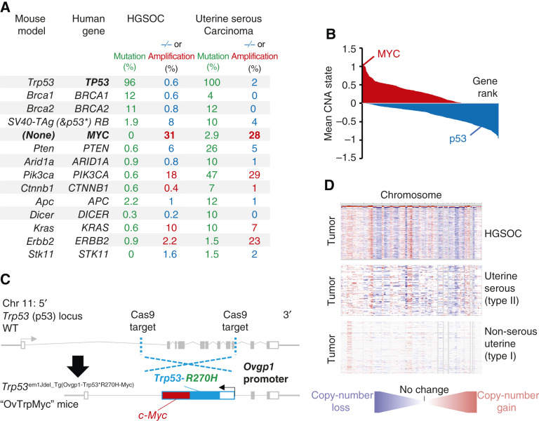
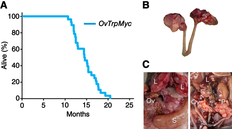
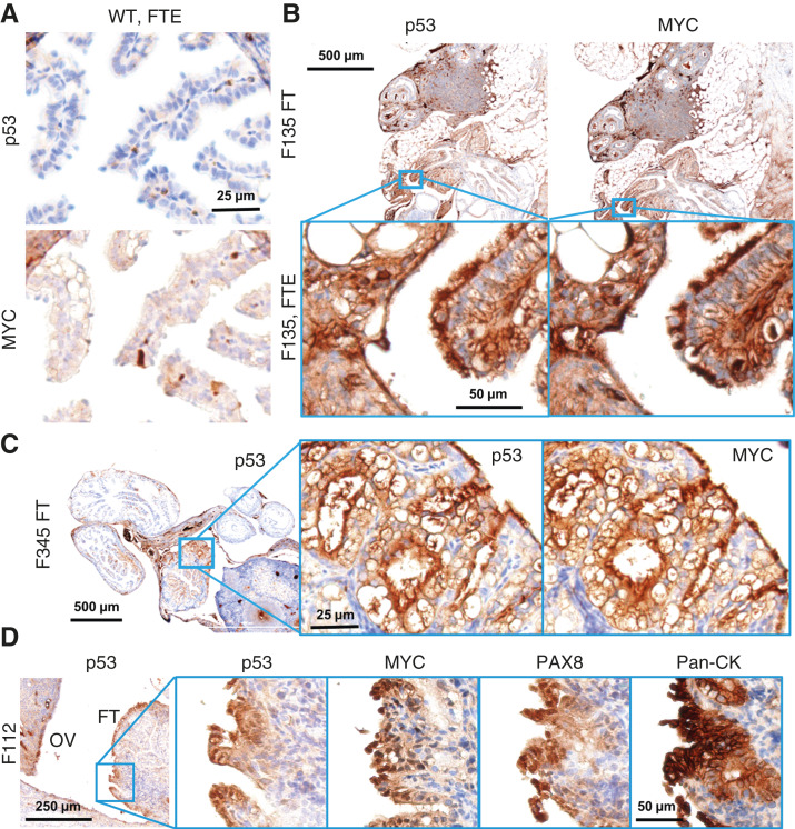
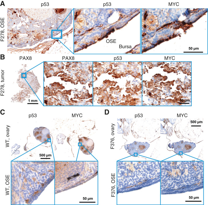
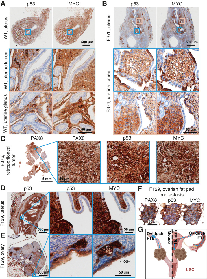
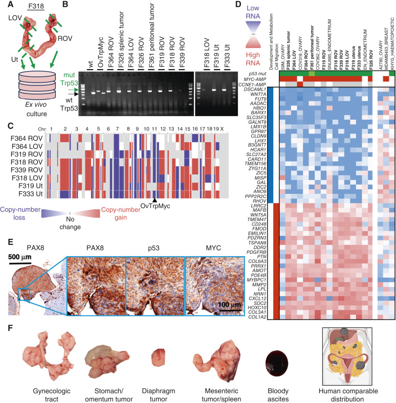
Genetically engineered mouse models (GEMM) have fundamentally changed how ovarian cancer etiology, early detection, and treatment are understood. MYC, an oncogene, is amongst the most amplified genes in high-grade serous ovarian cancer (HGSOC), but it has not previously been utilized to drive HGSOC GEMMs. We coupled Myc and dominant-negative mutant p53-R270H with a fallopian tube epithelium (FTE)-specific promoter Ovgp1 to generate a new GEMM of HGSOC. Female mice developed lethal cancer at an average of 14.5 months. Histopathologic examination of mice revealed HGSOC characteristics, including nuclear p53 and nuclear MYC in clusters of cells within the FTE and ovarian surface epithelium. Unexpectedly, nuclear p53 and MYC clustered cell expression was also identified in the uterine luminal epithelium, possibly from intraepithelial metastasis from the FTE. Extracted tumor cells exhibited strong loss of heterozygosity at the p53 locus, leaving the mutant allele. Copy-number alterations in these cancer cells were prevalent, disrupting a large fraction of genes. Transcriptome profiles most closely matched human HGSOC and serous endometrial cancer. Taken together, these results demonstrate that the Myc and Trp53-R270H transgenes were able to recapitulate many phenotypic hallmarks of HGSOC through the utilization of strictly human-mimetic genetic hallmarks of HGSOC. This new mouse model enables further exploration of ovarian cancer pathogenesis, particularly in the 50% of HGSOC which lack homology-directed repair mutations. Histologic and transcriptomic findings are consistent with the hypothesis that uterine serous cancer may originate from the FTE.

Significance: Mouse models using transgenes which generate spontaneous cancers are essential tools to examine the etiology of human diseases. Here, the first Myc-driven spontaneous model is described as a valid HGSOC model. Surprisingly, aspects of uterine serous carcinoma were also observed in this model.


Conflict of interest statement

G. Fullbright reports grants from the NIH/National Institute of General Medical Sciences during the conduct of the study. R.L. Carpenter reports a patent to 18/418,110 pending. D.T. Long reports grants from the NIH/National Institute of General Medical Sciences during the conduct of the study. J.R. Delaney reports grants from the NIH and Rivkin Foundation during the conduct of the study. No disclosures were reported by the other authors.

Figures

Figure 1
Figure 1
Rationale and design of the transgenic Ovgp1-Trp53-R270H-Myc mouse model. A, Comparison of currently available HGSOC mouse models genetics to genetic alterations in human HGSOC and serous endometrial cancers. −/− refers to homozygous deletion (blue, tumor suppressors), whereas Amp refers to copy-number amplification of at least two extra copies (red, oncogenes). B, Average copy-number state using TCGA HGSOC data at the gene level. Genes are plotted by ranking, with MYC and TP53 highlighted. C, Design of the transgenic OvTrpMyc mouse model. The endogenous Trp53 allele was targeted using a spCas9 and homology donor repair template strategy in the C57BL/6Tc background. Transgene genotyped pups were backcrossed with C57BL/6J mice and heterozygous mice used in all studies. The Ovgp1-driven transgene includes a dominant-negative murine p53-R270H mutant sequence and a murine c-Myc sequence separated by a P2A self-cleaving peptide. D, Comparison of CNAs across the genome for TCGA studied HGSOC and UCEC patient tumors. Red indicates copy-number gain, whereas blue indicates copy-number loss. TCGA, The Cancer Genome Atlas.
Figure 2
Figure 2
Middle-age presentation of ovarian and peritoneal tumors. A, Lifespan data from mice euthanized due to tumor burden criteria. B, Example of a dissected reproductive tract from a mouse with obvious macroscopic ovarian tumors. C, Examples of widespread intra-abdominal disease, including liver invasion (L), peritoneal tumors (P), splenic tumor (S), and ovarian tumors (Ov). L, liver invasion; Ov, ovarian tumors; P, peritoneal tumors; S, splenic tumor.
Figure 3
Figure 3
Molecular histology of the fallopian fimbriae. A, A WT mouse at 12 months of age showing little p53 or MYC staining in the FTE. B, OvTrpMyc fallopian tube at 15 months of age, F135, exhibiting p53 and MYC staining within the FTE. C, An OvTrpMyc mouse at 12 months of age, with vacuolated fallopian fimbriae–positive for p53 and MYC. D, An OvTrpMyc mouse at 15 months of age, with features resembling STIC.
Figure 4
Figure 4
Molecular histology of the OSE. A, An OvTrpMyc mouse at 16 months of age with clear OSE staining of p53 and MYC. B, The intra-abdominal tumor extracted from the mouse shown in A exhibited PAX8, p53, and MYC staining. C, A WT mouse at 12 months of age is negative for p53 or MYC staining in OSE. D, Example of an OvTrpMyc mouse with a region of negative OSE staining of p53 and MYC, at 12 months of age.
Figure 5
Figure 5
Uterine luminal epithelium staining of p53 and MYC. A, A WT mouse at 12 months of age showing negative p53 and MYC staining in the uterine epithelium and positive staining in adjacent tissues. B, Example of a 12 months old OvTrpMyc mouse with cytoplasmic p53 and whole-cell MYC staining along the ULE. C, A large retroperitoneal tumor, behind the kidney, stained for p53, MYC, and PAX8. D, An example of ULE expression in an OvTrpMyc mouse F129, with evidence of similar whole-cell staining of p53 and MYC on the (E) OSE and (F) ovarian fat pad metastasis. Mouse F129 was 11 months of age. G, Proposed model for observed dissemination of cells from the FTE to the ULE as an origin of uterine serous carcinoma. USC, uterine serous carcinoma.
Figure 6
Figure 6
CNAs, transcriptomics, and tumorigenicity of extracted OvTrpMyc tumor cells. A, Dissection diagram for ex vivo expansion. Gynecologic tract for mouse F318 is shown. B, LOH was observed by PCR of the Trp53 transgene (mutation) in isolated cells. C, Whole-genome CNA calls of tumor cells. Blue indicates copy-number loss, and red indicates copy-number gain. D, Transcriptomic profiling of OvTrpMyc cancer cells compared with human gynecologic cancer within the human CCLE. Header genes refer to mutation in p53 (green), MYC amplification (red), or CCNE1 amplification (red), shaded white if absent or gray if unknown. Nearest neighbor human cell lines comprise the cell lines shown. A2780, MDA-MB-453, and KHYG cell lines are shown as representative cell lines which have driver gene characteristics comparable with the mouse cell lines but clearly different transcriptional profiles. E, 8 × 10⁶ F339 ROV OvTrpMyc cell line cells were injected i.p. into a young OvTrpMyc female recipient mouse. The mouse lost weight, meeting euthanasia criteria after 54 days. IHC staining of a dissected tumor attached to the intestines is shown. F, Distribution of tumors found in F318 LOV i.p. syngeneic injected mice (5 × 10⁶ cells, 132 days) relative to a human image depiction of HGSOC metastatic spread. LOV, left ovary; ROV, right ovary; Ut, uterus.

Update of

References

    1. Ferriss JS, Erickson BK, Shih I-M, Fader AN. Uterine serous carcinoma: key advances and novel treatment approaches. Int J Gynecol Cancer 2021;31:1165–74. - PubMed
    1. Clarke MA, Devesa SS, Hammer A, Wentzensen N. Racial and ethnic differences in hysterectomy-corrected uterine corpus cancer mortality by stage and histologic subtype. JAMA Oncol 2022;8:895–903. - PMC - PubMed
    1. González-Martín A, Pothuri B, Vergote I, DePont Christensen R, Graybill W, Mirza MR, et al. . Niraparib in patients with newly diagnosed advanced ovarian cancer. N Engl J Med 2019;381:2391–402. - PubMed
    1. Mirza MR, Chase DM, Slomovitz BM, dePont Christensen R, Novák Z, Black D, et al. . Dostarlimab for primary advanced or recurrent endometrial cancer. N Engl J Med 2023;388:2145–58. - PubMed
    1. Eskander RN, Sill MW, Beffa L, Moore RG, Hope JM, Musa FB, et al. . Pembrolizumab plus chemotherapy in advanced endometrial cancer. N Engl J Med 2023;388:2159–70. - PMC - PubMed

Publication types

Substances