Multicenter Study
. 2024 Sep;312(3):e233051.
doi: 10.1148/radiol.233051.

The #HOPE4LIVER Single-Arm Pivotal Trial for Histotripsy of Primary and Metastatic Liver Tumors

Affiliations
Multicenter Study

The #HOPE4LIVER Single-Arm Pivotal Trial for Histotripsy of Primary and Metastatic Liver Tumors

Mishal Mendiratta-Lala et al. Radiology. 2024 Sep.

Abstract

Background Histotripsy is a nonthermal, nonionizing, noninvasive, focused US technique that relies on cavitation for mechanical tissue breakdown at the focal point. Preclinical data have shown its safety and technical success in the ablation of liver tumors. Purpose To evaluate the safety and technical success of histotripsy in destroying primary or metastatic liver tumors. Materials and Methods The parallel United States and European Union and England #HOPE4LIVER trials were prospective, multicenter, single-arm studies. Eligible patients were recruited at 14 sites in Europe and the United States from January 2021 to July 2022. Up to three tumors smaller than 3 cm in size could be treated. CT or MRI and clinic visits were performed at 1 week or less preprocedure, at index-procedure, 36 hours or less postprocedure, and 30 days postprocedure. There were co-primary end points of technical success of tumor treatment and absence of procedure-related major complications within 30 days, with performance goals of greater than 70% and less than 25%, respectively. A two-sided 95% Wilson score CI was derived for each end point. Results Forty-four participants (21 from the United States, 23 from the European Union or England; 22 female participants, 22 male participants; mean age, 64 years ± 12 [SD]) with 49 tumors were enrolled and treated. Eighteen participants (41%) had hepatocellular carcinoma and 26 (59%) had non-hepatocellular carcinoma liver metastases. The maximum pretreatment tumor diameter was 1.5 cm ± 0.6 and the maximum post-histotripsy treatment zone diameter was 3.6 cm ± 1.4. Technical success was observed in 42 of 44 treated tumors (95%; 95% CI: 84, 100) and procedure-related major complications were reported in three of 44 participants (7%; 95% CI: 2, 18), both meeting the performance goal. Conclusion The #HOPE4LIVER trials met the co-primary end-point performance goals for technical success and the absence of procedure-related major complications, supporting early clinical adoption. Clinical trial registration nos. NCT04572633, NCT04573881 Published under a CC BY 4.0 license. Supplemental material is available for this article. See also the editorial by Nezami and Georgiades in this issue.


Conflict of interest statement

Disclosures of conflicts of interest: M.M.L. Support for attending meetings from HistoSonics; ACR and SAR LI-RADS Treatment Response Working Group chair. P.W. Repayment for study expenses from HistoSonics. M.P. No relevant relationships. X.S.C. No relevant relationships. S.B.W. Consulting fees from Guerbet, Cook Medical, AstraZeneca; payment for lectures from Penumbra; participation on a DataSafety Monitoring Board or Advisory Board from AstraZeneca, Merck, Trisalus, Esai; Member of the SIO board of directors; deputy editor for Radiology. C.D. No relevant relationships. O.A. Grants from Canon Medical; consulting fees from Boston Scientific, Angiodynamics, Johnson and Johnson, Asahi Medical, Argon Medical, Bard; payment for lectures from Penumbra; chief medical officer and equity owner of Flow Medical. N.D.P. Grants or contracts from Exelixis, Genentech, Bayer, TARGET, National Institutes of Health; consulting fees from AstraZeneca, Genentech, Esai, Gilead. M.P. No relevant relationships. M.T. No relevant relationships. Z.X. Co-inventor of patents licensed to HistoSonics and used in this clinical trial; stockholder, HistoSonics. Z.C. No relevant relationships. G.N. Consulting fees from Angiodynamics, Boston Scientific, Varian Interventional Solutions, Stryker, Philips; scientific advisory board for Quantum Surgical and Beta Glue. G.T. No relevant relationships. C.C. Consulting fees for participation as a co-principal investigator for HistoSonics; patent holder for use of histotripsy to generate immunogenic tumor vaccines. P.L. No relevant relationships. T.M.W. Grants from Boston Scientific, Angiodynamics, HistoSonics, Johnson and Johnson; consulting fees from Angiodynamics; payment for lectures from Boston Scientific, Angiodynamics, Johnson and Johnson; participation in a DataSafety Monitoring Board or Advisory Board from Angiodynamics, Johnson and Johnson; leadership or fiduciary roles from Faculty Board Royal College of Radiologists, British Society of Interventional Radiology, NIHR, NICE. L.S. No relevant relationships. T.J.Z. Grants, HistoSonics, Ethicon; consulting fees from Elephas; Participation in a DataSafety Monitoring Board or Advisory Board from Elephas; stock or stock options from HistoSonics; equipment provided to institution from HistoSonics.

Figures

None
Graphical abstract
Study design flowchart. * = Incomplete due to poor image
quality (three tumors in three participants) and lack of contrast (two
tumors in one participant). ** = Six lesions (five
participants) did not have 30-day imaging and the core laboratory could not
assess technique efficacy in two lesions (two participants).
Figure 1:
Study design flowchart. * = Incomplete due to poor image quality (three tumors in three participants) and lack of contrast (two tumors in one participant). ** = Six lesions (five participants) did not have 30-day imaging and the core laboratory could not assess technique efficacy in two lesions (two participants).
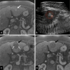
Images in a participant with a 2.3-cm hepatocellular carcinoma in
segment III of the liver. (A) Preprocedure contrast-enhanced MRI scan in the
late arterial phase shows hyperenhancing hepatocellular carcinoma (arrow).
(B) Intraprocedural US image shows defined treatment volume; the orange
circle outlines the hyperechoic targeted tumor and the red circle outlines
the planned margin. The treatment effect is visible as an echogenic bubble
cloud at the red crosshair, the focal point of the transducer. The robotic
arm moves the focal point through the planned treatment volume to create the
volumetric treatment. (C) Contrast-enhanced MRI scan obtained less than 36
hours postprocedure in the late arterial phase shows the nonenhancing
treatment zone (black arrow). Note patent blood vessels (dashed white
arrows). (D) Contrast-enhanced MRI scan obtained 30 days postprocedure in
the late arterial phase shows partial involution of the treatment zone
(arrow).
Figure 2:
Images in a participant with a 2.3-cm hepatocellular carcinoma in segment III of the liver. (A) Preprocedure contrast-enhanced MRI scan in the late arterial phase shows hyperenhancing hepatocellular carcinoma (arrow). (B) Intraprocedural US image shows defined treatment volume; the orange circle outlines the hyperechoic targeted tumor and the red circle outlines the planned margin. The treatment effect is visible as an echogenic bubble cloud at the red crosshair, the focal point of the transducer. The robotic arm moves the focal point through the planned treatment volume to create the volumetric treatment. (C) Contrast-enhanced MRI scan obtained less than 36 hours postprocedure in the late arterial phase shows the nonenhancing treatment zone (black arrow). Note patent blood vessels (dashed white arrows). (D) Contrast-enhanced MRI scan obtained 30 days postprocedure in the late arterial phase shows partial involution of the treatment zone (arrow).
Graphs show primary end points of (A) technical success and (B) safety
of procedure-related major complications. * = Estimated by bootstrap
sampling with replacement method to account for potential within-subject
lesion correlations. Subject was the bootstrap sampling unit,
1 000 000 iterations for bootstrap resampling were performed,
and the bootstrap method was the bias-corrected and accelerated method.
† = Two-sided 95% Wilson score CI.
Figure 3:
Graphs show primary end points of (A) technical success and (B) safety of procedure-related major complications. * = Estimated by bootstrap sampling with replacement method to account for potential within-subject lesion correlations. Subject was the bootstrap sampling unit, 1 000 000 iterations for bootstrap resampling were performed, and the bootstrap method was the bias-corrected and accelerated method. † = Two-sided 95% Wilson score CI.
Images show a 1.3-cm metastatic colorectal cancer in segment III of
the liver. (A) Preprocedure contrast-enhanced MRI scan shows tumor (arrow).
(B) Contrast-enhanced MRI scan obtained fewer than 36 hours postprocedure
with treatment zone (solid arrow) encompassing the site of the tumor and a
margin. Patent blood vessel (dashed arrow) traverses the treatment zone. (C)
Contrast-enhanced MRI scan obtained at 30 days postprocedure shows
involution of the treatment zone (solid arrow) with maintained patent vessel
(dashed arrow).
Figure 4:
Images show a 1.3-cm metastatic colorectal cancer in segment III of the liver. (A) Preprocedure contrast-enhanced MRI scan shows tumor (arrow). (B) Contrast-enhanced MRI scan obtained fewer than 36 hours postprocedure with treatment zone (solid arrow) encompassing the site of the tumor and a margin. Patent blood vessel (dashed arrow) traverses the treatment zone. (C) Contrast-enhanced MRI scan obtained at 30 days postprocedure shows involution of the treatment zone (solid arrow) with maintained patent vessel (dashed arrow).

References

    1. Xu Z , Hall TL , Vlaisavljevich E , Lee FT Jr . Histotripsy: the first noninvasive, non-ionizing, non-thermal ablation technique based on ultrasound . Int J Hyperthermia 2021. ; 38 ( 1 ): 561 – 575 . - PMC - PubMed
    1. Vidal-Jove J , Serres X , Vlaisavljevich E , et al . First-in-man histotripsy of hepatic tumors: the THERESA trial, a feasibility study . Int J Hyperthermia 2022. ; 39 ( 1 ): 1115 – 1123 . - PubMed
    1. Bray F , Ferlay J , Soerjomataram I , Siegel RL , Torre LA , Jemal A . Global cancer statistics 2018: GLOBOCAN estimates of incidence and mortality worldwide for 36 cancers in 185 countries . CA Cancer J Clin 2018. ; 68 ( 6 ): 394 – 424 . [Published correction appears in CA Cancer J Clin 2020;70(4):313.] - PubMed
    1. Heimbach JK , Kulik LM , Finn RS , et al . AASLD guidelines for the treatment of hepatocellular carcinoma . Hepatology 2018. ; 67 ( 1 ): 358 – 380 . - PubMed
    1. Reig M , Forner A , Rimola J , et al . BCLC strategy for prognosis prediction and treatment recommendation: The 2022 update . J Hepatol 2022. ; 76 ( 3 ): 681 – 693 . - PMC - PubMed

Publication types