Case Reports
. 2024 Sep 3;148(1):35.
doi: 10.1007/s00401-024-02792-0.

Glioblastoma, IDH-wildtype with primarily leptomeningeal localization diagnosed by nanopore sequencing of cell-free DNA from cerebrospinal fluid

Affiliations
Case Reports

Glioblastoma, IDH-wildtype with primarily leptomeningeal localization diagnosed by nanopore sequencing of cell-free DNA from cerebrospinal fluid

Nik Sol et al. Acta Neuropathol. .
No abstract available


Figures

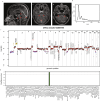
Fig. 1
Fig. 1
Clinical diagnostics. a Sagittal and axial post-contrast T1-weighted MRI images show contrast enhancement around the folia of the cerebellum and the pons (indicated arrows in I, II). Axial post-contrast T1-weighted image shows contrast enhancement of cranial nerve VII and VIII on both sides (arrows in III). b Size distribution of the sequence reads obtained from CSF DNA sequencing. c CNV profile obtained from nanopore sequencing. d Sturgeon classification with a confident (> 0.95) classification as Glioblastoma—MID
Fig. 2
Fig. 2
Autopsy findings. A coronal brain section showed leptomeningeal opacification and thickening (a; white arrowheads), the parasagittal part with a rough surface partly due to the presence of granulations of Pacchioni. Microscopically, atypical cells are diffusely spread throughout the leptomeningeal compartment (b). In several regions, such cells were also (but relatively subtly) present in the subependymal/periventricular region, e.g., near the hippocampus (c, d; square box), with more dense accumulation of these cells in an adjacent, deeply invaginating cerebral sulcus (*). Immunohistochemical staining for Olig2 (e) and Ki-67 (f) confirmed the glial nature of the tumor and its moderate proliferative activity, respectively. L: leptomeninges; CTX: molecular layer of the cerebral cortex; HE: hematoxylin and eosin staining

References

    1. Afflerbach A-K, Rohrandt C, Brändl B, Sönksen M, Hench J, Frank S et al (2024) Classification of brain tumors by nanopore sequencing of cell-free DNA from cerebrospinal fluid. Clin Chem 70:250–260 10.1093/clinchem/hvad115 - DOI - PubMed
    1. Capper D, Jones DTW, Sill M, Hovestadt V, Schrimpf D, Sturm D et al (2018) DNA methylation-based classification of central nervous system tumours. Nature 555:469–474 10.1038/nature26000 - DOI - PMC - PubMed
    1. El Zarif T, Machaalani M, Nawfal R, Nassar AH, Xie W, Choueiri TK et al (2024) TERT promoter mutations frequency across race, sex, and cancer type. Oncologist 29(1):8–14 10.1093/oncolo/oyad208 - DOI - PMC - PubMed
    1. Katsuhara T, Moro N, Ohta T, Homma T, Yoshino A (2018) Solitary primary intracranial leptomeningeal glioblastoma invading the normal cortex: case report. Mol Clin Oncol 8:466–470 - PMC - PubMed
    1. Louis DN, Perry A, Wesseling P, Brat DJ, Cree IA, Figarella-Branger D et al (2021) The 2021 WHO classification of tumors of the central nervous system: a summary. Neuro Oncol 23:1231–1251 10.1093/neuonc/noab106 - DOI - PMC - PubMed

MeSH terms

Substances

LinkOut - more resources