. 2024 Sep 3;18(9):e0012452.
doi: 10.1371/journal.pntd.0012452. eCollection 2024 Sep.

Paracoccidioides lutzii Infects Galleria mellonella Employing Formamidase as a Virulence Factor

Affiliations

Paracoccidioides lutzii Infects Galleria mellonella Employing Formamidase as a Virulence Factor

Elisa Dias Pereira et al. PLoS Negl Trop Dis. .

Abstract

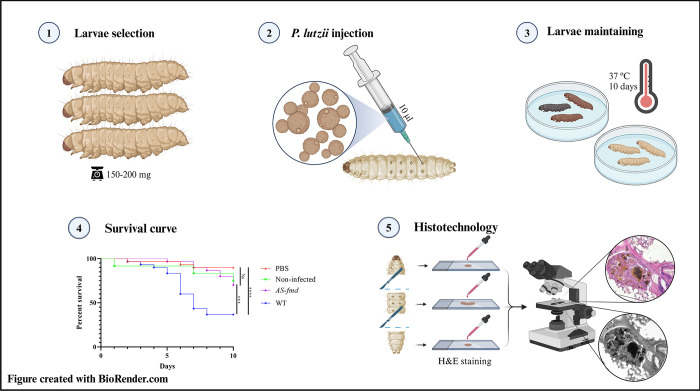
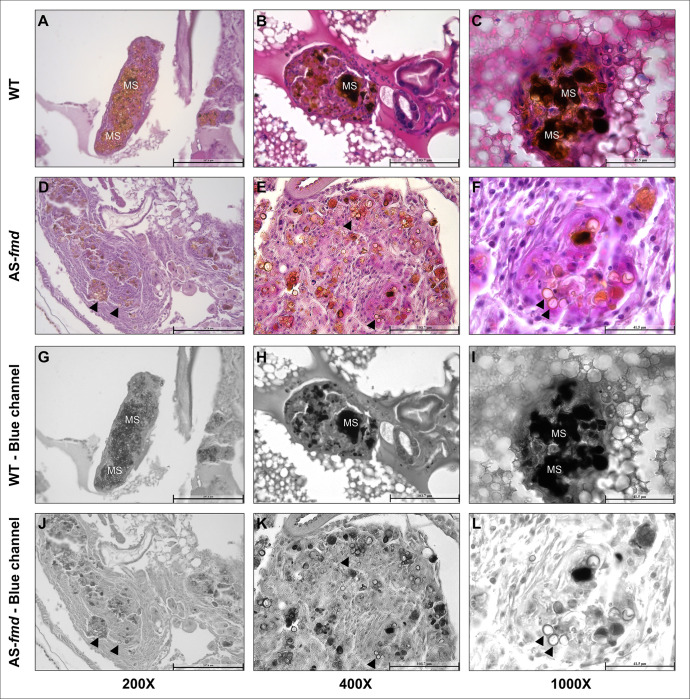
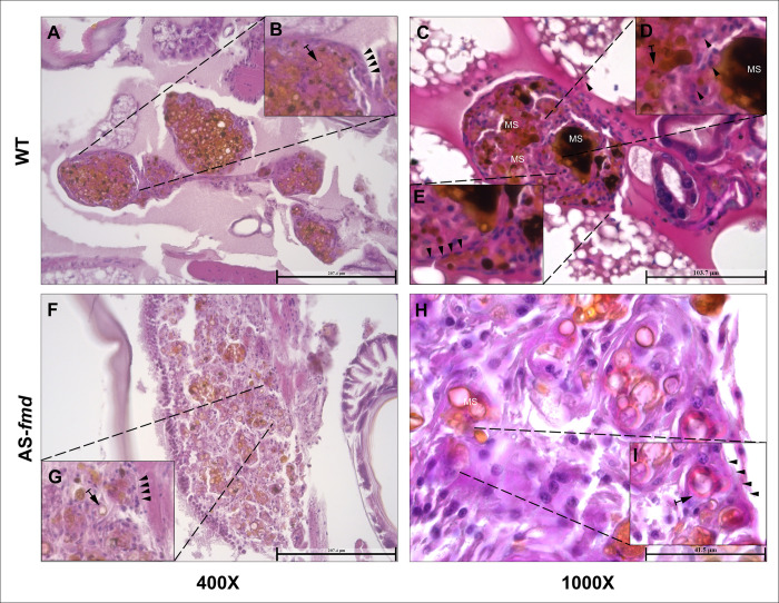
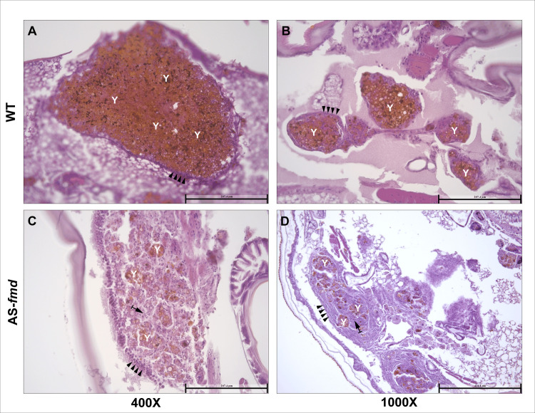
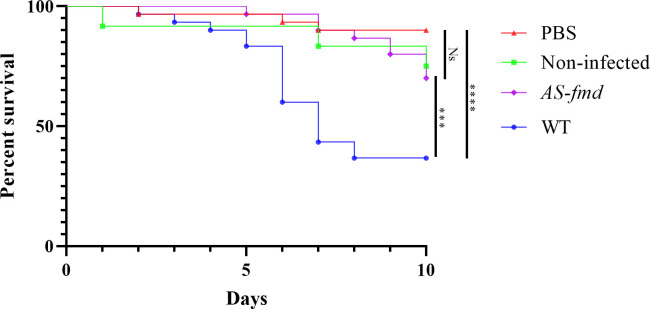
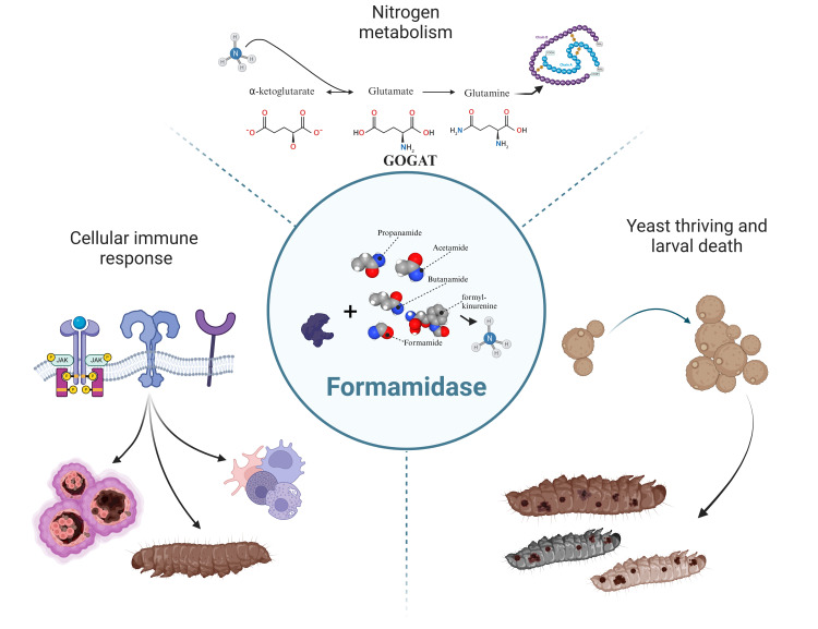
The formamidase (FMD) enzyme plays an important role in fungal thriving by releasing a secondary nitrogen source as a product of its activity. In Paracoccidioides species, previous studies have demonstrated the upregulation of this enzyme in a wide range of starvation and infective-like conditions. However, Paracoccidioides lutzii formamidase has not yet been defined as a virulence factor. Here, by employing in vivo infections using an fmd-silenced strain in Galleria mellonella larvae model, we demonstrate the influence of formamidase in P. lutzii's immune stimulation and pathogenicity. The formamidase silencing resulted in improper arrangement of the nodules, poor melanogenesis and decreased fungal burden. Thus, we suggest that formamidase may be a piece composing the process of molecular recognition by Galleria immune cells. Furthermore, formamidase silencing doubled the observed survival rate of the larvae, demonstrating its importance in fungal virulence in vivo. Therefore, our findings indicate that formamidase contributes to Galleria's immune incitement and establishes the role of this enzyme as a P. lutzii virulence factor.


Conflict of interest statement

The authors have declared that no competing interests exist.

Figures

Fig 1
Fig 1. Experimental workflow for larval infection and histotechnology.
– The larvae were infected with two strains of P. lutzii and the death events were assessed for ten days to build the Kaplan-Meyer survival curve. Histotechnological analyses were performed to investigate the effects causing larval death. Figure created with BioRender.com.
Fig 2
Fig 2. –Melanization patterns of larvae infected with wild-type and AS-fmd P. lutzii strains.
Tissue slides displaying nodules from three different specimens of G. mellonella infected with wild-type (A-C) and AS-fmd strains (D-F) then stained with HE. The images of the blue channel split highlight the melanin spots (G-L). A-C) The three different nodules enclosing P. lutzii wild-type cells show well-defined structures, tight cellular organization, and high intensity of dark regions derived from melanin stain (MS), demonstrating increased levels of melanin. D-F) The mutant cells of P. lutzii are encompassed by three distinct nodules that have softly defined structures, loose cellular disposition, and clear-brown regions, indicating low levels of melanin. G-L) The wild-type and AS-fmd images after digital splitting of the blue channel to evidence melanin. The darker regions of each image indicate high melanized sites, while the clear-brown regions bordering the cell wall indicate low melanized sites (MS). AS-fmd images show yeast cells with a slightly darker color delineating the cell wall (arrowheads).
Fig 3
Fig 3. –Structure of the nodular tissues from infected larvae.
Tissue slides from four different animals stained with HE. Visualization with 400x and 1000x magnification. A and C) The structures of nodules from two different specimens infected with the wildtype strain are finely arranged with a tight cell disposition. B, D and E) There is a precise BM-like layer of plasmatocytes strictly delimiting the nodular boundaries (arrowheads), and numerous yeasts surrounded by defense cells (arrows). F and H) Tissue from two different specimens infected with the AS-fmd strain. Both nodules exhibit a loose arrangement and cellular sparseness. G and I) There is a poorly defined and incontiguous BM-like layer (arrowheads) with impaired plasmatocyte discrimination. Yeast cells are present in low numbers surrounded by hemocytes (arrows).
Fig 4
Fig 4. –Fungal burden inside the nodules from both groups.
Fungal burden was assessed inside the nodules of both groups. Tissue slides from the wild-type and AS-fmd group were HE-stained and viewed at 400X magnification. A and B) Tissue slides of nodules from distinct animals displaying yeast predominance (Y) in their interior. The BM-like monolayer of plasmatocytes is continuously surrounding the nodule and accurately defined (arrowheads). C and D) Nodules from two distinct animals showing a predominance of hemocytes (arrow). The BM-like layer is composed of several hemocytes and is disconnected in some areas (arrowheads), indicating improper formation. The yeast cell content is found in small clusters, demonstrating a low fungal load (Y).
Fig 5
Fig 5. –Survival curve of G. mellonella infected with P. lutzii strains.
Four groups of larvae were injected with PBS (red triangle), non-infected (green square), AS-fmd (purple diamond), and wild-type (blue circle) suspensions. Death events were recorded and counted over a 10-days period. The collected data was plotted, and a Kaplan-Meyer curve was generated to analyze the results. The statistical difference between pairs of groups was assessed using the Gehan-Breslow-Wilcoxon test. The survival rates of the PBS and non-infected groups were not significantly different (p-value = 0.2303), serving as controls. The wild-type and PBS groups were highly different (p-value < 0.0001). The AS-fmd showed no significant difference when compared to either PBS (p-value = 0.095) or non-infected larvae (p-value = 0.8607). However, the AS-fmd and wild-type groups were significantly different (p-value 0.0006). The figure was created using GraphPad Prism 8 software. We use (****) to indicate p-value < 0.0001; (***) for p-value < 0.001; and (Ns) for non-significant difference.
Fig 6
Fig 6. –Probable impacts of formamidase during P. lutzii infection in G. mellonella model.
Top) The FMD enzyme is closely related to ammonium insertion into the glutamate and glutamine synthesis. It serves then as a source of these amino acids to the yeast protein metabolism, increasing fungal survival or even multiplication. Right) The generated ammonium may act as a cellular signal stimulating yeast-yeast communication to growth. This role played by the FMD-generated ammonium is a step towards larval death. Left) Immune recognition by PRRs induces nodulation, humoral response, fungal burden, and body melanization. Figure created with BioRender.com.

References

    1. Brummer E, Castaneda E, Restrepo A, Microbiologia GD, Bogota SD. Paracoccidioidomycosis: an Update. 1993;6(2):89–117. doi: 10.1128/CMR.6.2.89 - DOI - PMC - PubMed
    1. Muñoz JF, Farrer RA DC, Gallo JE, Sykes S, Sakthikumar S ME, Whiston EA, Bagagli E, Soares CMA T, MDM, Taylor JW, MJ Clay OK, Cuomo C CA. Genome Diversity, Recombination, and Virulence across the Major Lineages of Paracoccidioides. mSphere. 2016;1(5):1–18. - PMC - PubMed
    1. Turissini DA, Gomez OM, Teixeira MM, McEwen JG, Matute DR. Species boundaries in the human pathogen Paracoccidioides. Fungal Genet Biol. 2017;106(April):9–25. doi: 10.1016/j.fgb.2017.05.007 - DOI - PMC - PubMed
    1. Martinez R. New trends in paracoccidioidomycosis epidemiology. J Fungi. 2017;3(1):1–13. doi: 10.3390/jof3010001 - DOI - PMC - PubMed
    1. Shoham S, Levitz SM. The immune response to fungal infections. Br J Haematol. 2005;129(5):569–82. doi: 10.1111/j.1365-2141.2005.05397.x - DOI - PubMed

LinkOut - more resources