. 2024 Sep 10;121(37):e2403421121.
doi: 10.1073/pnas.2403421121. Epub 2024 Sep 3.

Cryo-EM structures of a mycobacterial ABC transporter that mediates rifampicin resistance

Affiliations

Cryo-EM structures of a mycobacterial ABC transporter that mediates rifampicin resistance

Yinan Wang et al. Proc Natl Acad Sci U S A. .

Abstract

Drug-resistant Tuberculosis (TB) is a global public health problem. Resistance to rifampicin, the most effective drug for TB treatment, is a major growing concern. The etiological agent, Mycobacterium tuberculosis (Mtb), has a cluster of ATP-binding cassette (ABC) transporters which are responsible for drug resistance through active export. Here, we describe studies characterizing Mtb Rv1217c-1218c as an ABC transporter that can mediate mycobacterial resistance to rifampicin and have determined the cryo-electron microscopy structures of Rv1217c-1218c. The structures show Rv1217c-1218c has a type V exporter fold. In the absence of ATP, Rv1217c-1218c forms a periplasmic gate by two juxtaposed-membrane helices from each transmembrane domain (TMD), while the nucleotide-binding domains (NBDs) form a partially closed dimer which is held together by four salt-bridges. Adenylyl-imidodiphosphate (AMPPNP) binding induces a structural change where the NBDs become further closed to each other, which downstream translates to a closed conformation for the TMDs. AMPPNP binding results in the collapse of the outer leaflet cavity and the opening of the periplasmic gate, which was proposed to play a role in substrate export. The rifampicin-bound structure shows a hydrophobic and periplasm-facing cavity is involved in rifampicin binding. Phospholipid molecules are observed in all determined structures and form an integral part of the Rv1217c-1218c transporter system. Our results provide a structural basis for a mycobacterial ABC exporter that mediates rifampicin resistance, which can lead to different insights into combating rifampicin resistance.

Keywords: ABC transporter; Mycobacterium tuberculosis; drug resistance; rifampicin.


Conflict of interest statement

Competing interests statement:The authors declare no competing interest.

Figures
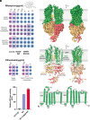
Fig. 1.
Fig. 1.
Functional characterization and overall structure of Rv1217c–1218c. (A) Top: Overexpression of recombinant WT Rv1217c–1218c in M. smeg mc2155 shows rifampicin resistance with MIC of 8 μg/mL, while the E161Q mutant restored the MIC value back to that for the vector control (1.25 μg/mL). Bottom: Ethambutol was used as an additional control drug. Pink color indicates active bacteria and blue killed bacteria. Middle: Expression of (Left) WT and (Right) EQ mutant Rv1217c–1218c were confirmed by western-blot using 6*His-Tag monoclonal antibody. (B) Normalized ATPase activity of WT Rv1217c–1218c and the E161Q mutant purified in GDN. ATPase activity was further stimulated in the presence of rifampicin (RIF). All data points represent the mean and SDs of two separate experiments with each performed in triplicate. (C) (Top) EM density for Rv1217–1218c and (Bottom) overall structure in ribbon presentation. One Rv1217c molecule is composed of the two TMDs in green, and the two Rv1218c molecules that contain the NBDs which are colored in gold and salmon. (D) Topology diagram for the secondary structure of Rv1217c.
Fig. 2.
Fig. 2.
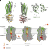
Structural features of the TMDs and dimerized NBDs in the nucleotide-free state. (A) (Middle) Overall structure of the TMDs reveal the bound lipids and the periplasmic gate, respectively. Details of (Left) the polar interactions formed by CL and R120 and R209 on TMD1, and (Right) EH1 on TMD1 and EH5 on TMD2 form antiparallel helices, that seal the TMD in the periplasm. (B) Linear arrangement of sequence motifs of Rv1218c. (C) Left: Top view of the dimerized NBDs in the nucleotide-free state in cartoon representation. The two NBDs are colored as in Fig. 1C. The different motifs are colored as in (B). Right: Zoom-in view of the four salt bridges formed by the two NBDs.
Fig. 3.
Fig. 3.
The structure of Rv1217–1218c in the presence of AMPPNP and Mg2+. (A) Ribbon diagram of AMPPNP-bound Rv1217c–1218c structure. The two AMPPNPs are shown as solid spheres, and the CLs as stick models. (B) Top: Structure of the two CLs in the TMDs, CL1 is fully inside the TMDs and CL2 is partially outside the TMDs. TMD1 is omitted for clarity. Bottom: EM density map for AMPPNP.Mg2+. (C) Ribbon diagram of the fully dimerized NBDs with the bound AMPPNP.Mg2+, typical motifs are colored as in 2B. AMPPNP is shown as a stick model and Mg2+ ions as spheres. (D) Close-up view of the AMPPNP.Mg2+ binding site in one NBD. Sidechains of key residues and mainchain amide of G139 making interactions with AMPPNP.Mg2+ are shown as stick models. (E) Top view of the NBDs in the nucleotide-free (gray) and AMPPNP-bound (gold and salmon) structures shown in surface presentation, the two structures are superimposed on the canonical NBD regions.
Fig. 4.
Fig. 4.
Nucleotide binding induced conformational changes and the proposed transport mechanism. (A) Comparison of TMD1. NBD1 in the AMPPNP-bound (green and gold) and unbound (gray) structures upon superimposing the NBD1. The rotation of TMD1 is revealed and the rotation axis of TMD1 is indicated as a black arrow. (B) Comparison of TMD2. NBD2 in the AMPPNP-bound (green and salmon) and the unbound (gray) structures upon superimposing the NBD2, revealing a minor change to TMD2. (C) Closing of the TM cavity upon TMD1 rotation. The structures of AMPPNP bound (green) and unbound (gray) are aligned based on TMD2. (D) Proposed working model of Rv1217c–1218c. See detailed description in the text. Left: In the ground state, lipids fill the cytoplasmic leaflet; middle: substrate enters the translocation cavity halfway up the TMDs that was sealed by the periplasmic gate; Right: upon ATP binding, the NBDs are fully dimerized and the TMD cavity is collapsed, the periplasmic gate open to facilitate substrate release. Pi release upon ATP hydrolysis may reset Rv1217c–1218c to the ground state.

References

    1. Chakraborty S., Rhee K. Y., Tuberculosis drug development: History and evolution of the mechanism-based paradigm. Cold Spring Harb. Perspect. Med. 5, a021147 (2015). - PMC - PubMed
    1. WHO, “Global tuberculosis report 2023” (World Health Organization, Geneva, 2023).
    1. Telenti A., et al. , Detection of rifampicin-resistance mutations in Mycobacterium tuberculosis. Lancet 341, 647–650 (1993). - PubMed
    1. Peek J., et al. , Rifamycin congeners kanglemycins are active against rifampicin-resistant bacteria via a distinct mechanism. Nat. Commun. 9, 4147 (2018). - PMC - PubMed
    1. Lilic M., et al. , The antibiotic sorangicin A inhibits promoter DNA unwinding in a Mycobacterium tuberculosis rifampicin-resistant RNA polymerase. Proc. Natl. Acad. Sci. U.S.A. 117, 30423–30432 (2020). - PMC - PubMed

MeSH terms

LinkOut - more resources