. 2024 Sep 10;121(37):e2401531121.
doi: 10.1073/pnas.2401531121. Epub 2024 Sep 3.

Qki5 safeguards spinal motor neuron function by defining the motor neuron-specific transcriptome via pre-mRNA processing

Affiliations

Qki5 safeguards spinal motor neuron function by defining the motor neuron-specific transcriptome via pre-mRNA processing

Yoshika Hayakawa-Yano et al. Proc Natl Acad Sci U S A. .

Abstract

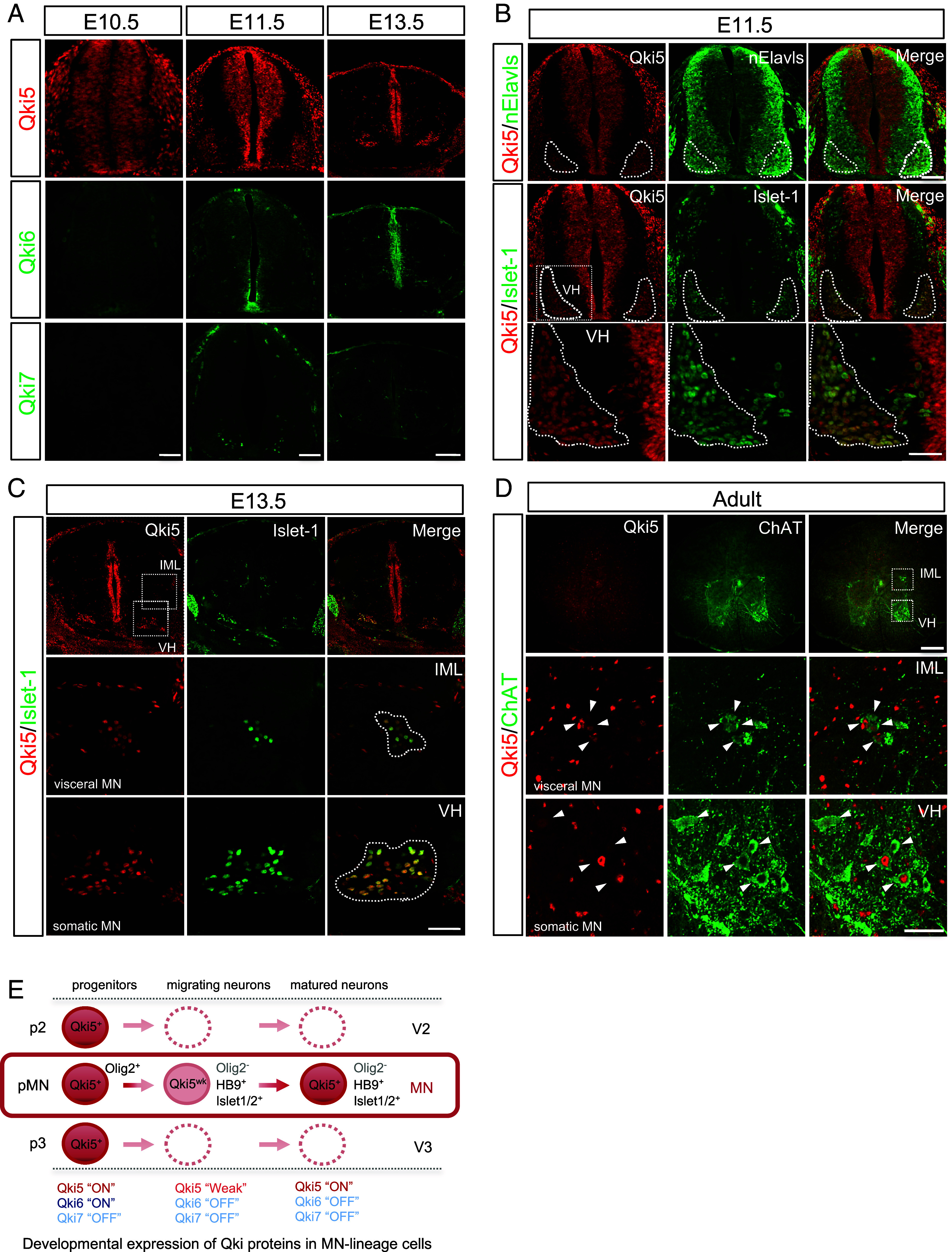
Many RNA-binding proteins (RBPs) are linked to the dysregulation of RNA metabolism in motor neuron diseases (MNDs). However, the molecular mechanisms underlying MN vulnerability have yet to be elucidated. Here, we found that such an RBP, Quaking5 (Qki5), contributes to formation of the MN-specific transcriptome profile, termed "MN-ness," through the posttranscriptional network and maintenance of the mature MNs. Immunohistochemical analysis and single-cell RNA sequencing (scRNA-seq) revealed that Qki5 is predominantly expressed in MNs, but not in other neuronal populations of the spinal cord. Furthermore, comprehensive RNA sequencing (RNA-seq) analyses revealed that Qki5-dependent RNA regulation plays a pivotal role in generating the MN-specific transcriptome through pre-messenger ribonucleic acid (mRNA) splicing for the synapse-related molecules and c-Jun N-terminal kinase/stress-activated protein kinase (JNK/SAPK) signaling pathways. Indeed, MN-specific ablation of the Qki5 caused neurodegeneration in postnatal mice and loss of Qki5 function resulted in the aberrant activation of stress-responsive JNK/SAPK pathway both in vitro and in vivo. These data suggested that Qki5 plays a crucial biological role in RNA regulation and safeguarding of MNs and might be associated with pathogenesis of MNDs.

Keywords: Quaking5; RNA-binding protein; alternative splicing; degeneration; motor neuron.


Conflict of interest statement

Competing interests statement:H.O. is a paid member of the Scientific Advisory Boards of San Bio Co., Ltd., and K Pharma Inc. M. Yano is a paid member of the Scientific Advisory Board of K Pharma Inc.

Figures

Fig. 1.
Fig. 1.
Qki5 protein is expressed in MNs in the mouse spinal cord. (A) Transverse sections of mouse spinal cords at each developmental age embryonic day (E) 10.5, E11.5, and E13.5 were immunostained using isoform-specific antibodies (Qki5, Qki6, and Qki7), showing a specific expression pattern of Qki5 among proteins encoded by the Qk gene. (Scale bar, 50 μm for E10.5, 100 μm for E11.5, and 150 μm for E13.5.) (B) Detailed expression of Qki5 and lineage marker proteins of MNs in the E11.5 mouse spinal cord. Qki5 (red) emerged in nElavls-positive (green, Top) and Islet-1-positive (green, Middle, and Bottom) MN pools. The Bottom panels represent the enlarged views of the ventral horn (VN) indicated by Insets in the Middle-Left panel. Note that Qki5 is expressed in MNs of the ventral horn, but not in other types of neurons. (Scale bar, 100 μm for Top and Middle and 50 μm for Bottom.) (C) Qki5 (red) is expressed in both somatic MNs of the ventral horn (VH) and visceral MNs of the intermediolateral nucleus (IML) labeled with Islet-1 (green) in the E13.5 mouse spinal cord. The Middle and Bottom panels represent the enlarged view of the IML and ventral horn (VN) indicated by Insets in the Top-Left panel. (Scale bar, 150 μm for the Top and 50 μm for the Middle and Bottom.) (D) Transverse sections of the adult mouse spinal cord were immunostained using anti-Qki5 (red) and ChAT (green) antibodies. The Middle and Bottom panels represent the enlarged views of the IML and ventral horn (VH), respectively, indicated by the Insets in the Top panel. (Scale bar, 200 μm for the Left and 50 μm for the Top and Middle/Bottom panels.) (E) Schematic illustrations of the expression patterns of Qki proteins in MN-lineage cells. Qki5 is expressed in embryonic NSCs of progenitor domains, including the pMN domain labeled with Olig2. After that, its expression is weakened in migrating Olig2-negative, Islet-1, and HB9-positive MNs and restored in both somatic and visceral MNs, which are settled in the final destination. In contrast, the Qki6 and Qki7 proteins were not detected in any neuronal population.
Fig. 2.
Fig. 2.
Qki5 expression in mouse and human spinal MNs. (A) RNA-seq analyses were performed using mouse-MN-like NSC-34 cells, mouse OL precursor cells (mOPCs), and hiPSC-MNs. Integrative Genomics Viewer (IGV) image of alternatively spliced Qk transcripts (5, 6, and 7) in NSC-34 cells, mOPCs, and hiPSC-MNs. mMNs and hiPSC-MNs predominantly express Qk5 and QKI5, respectively. In contrast, mOPCs equally express all three Qk isoforms. The alternative splicing pattern of each isoform is shown on the Right. (B) UMAP of scRNA-seq using hiPSC-MNs showing six different clusters. QKI and ISL1 are dominantly expressed in cluster 2, whereas MAP2 is expressed in all clusters. (C) Seurat’s dot plot showing expression of QKI and 16 marker genes in the scRNA-seq. Dot color intensity and dot size represent average expression of the gene and the percentage of cells expressing the gene, respectively in a given cluster.
Fig. 3.
Fig. 3.
Qki5 regulates pre-mRNA splicing of a subset of transcripts in mouse and human MNs. (A) Scatterplot of the Log10 RPKM of siNC vs. siQk RNA-seq (expression levels: logCPM > 0) in NSC-34 cells, hiPSC-MNs, and mOPCs. Each dot in the plot indicates an individual transcript, and significant hits for transcript level change (P < 0.01 and FDR < 0.1) are indicated by red color. Only four transcripts and 87 transcripts were changed in Qki5-expressing NSC-34 cells and hiPSC-MNs, respectively, relative to the dramatic change in mOPCs. Each point represents the mean RPKM value obtained from three biological replicates for an individual gene. Each R-squared value is denoted in the graphs. (B) Scatterplot of the exon inclusion of siNC vs. siQk RNA-seq significant hits (P < 0.01 and FDR < 0.01) with absolute fold changes of 1.5 or more. Each point represents the ratio of read counts obtained from three replicates for an individual alternative splicing event. The number of changed alternative splicing events is denoted in each graph. (C) Representative IGV view of alternative splicing changes in the Daam2 gene in siNC and siQk NSC-34 cells with Qki5 HITS-CLIP cluster. siQk cells show that a cryptic exon induces nonsense-mediated decay (NMD). Gel images for RT–PCR validation of alternative splicing between exon 15 and 16. Bar graphs indicate the qRT-PCR assay of the relative expression of Daam2 transcript adjusted to the internal control Gapdh using siNC and siQk NSC-34 cells with or without cycloheximide (CHX) treatment. Data represent the mean ± SD from three independent biological replicates, two-tailed Student’s t test. (D) A dendrogram and heat-map overview of the hierarchical cluster analysis of alternative exon usage in genes from mOPC-, siNC-, or siQk-transfected NSC-34 cells and mouse cortical neurons (CNs) (n = 3 for each) using 28 Qki5-regulated OPC-specific exons (Top). Columns represent individual samples, and rows represent each exon. Each cell in the matrix represents the expression level of an alternative exon in an individual sample. The color key and histogram indicate that red and yellow in cells reflect high and low expression levels, respectively.
Fig. 4.
Fig. 4.
Aberrant activation of JNK/SAPK signaling in MNs with Qk knockdown. (A) Double immunostaining of Qki5 (red) and SMI-32 (green) in differentiated NSC-34 cells transfected with siQk or siNC. Forty-eight hours after siRNA transfection, Qki5 protein expression was markedly decreased in siQk cells, and SMI-32-positive inclusions were evident in the neurites of the cells (indicated by arrowheads) (Left). (Scale bar, 50 μm.) Bar graph showing that significant increase of SMI-32-positive inclusions in Qk KD differentiated NSC-34. Data represent the mean of five independent experiments (total 189 cells and 168 cells counted for siNC and siQk, respectively) Each dots represent average number of SMI-32 positive inclusion adjusted for the cell number in each replicate. Student’s t test. (B) Qki5-dependent AS regulation in mouse Map4k4 and human MAP4K4. Schematic representation of Qki5 binding position-dependent alternative exon usage (tandem cassette exons, exon 16 and exon 17) in the mouse transcripts in the Top-Right panel. IGV image of Qki5 HITS CLIP clusters and expression levels of the transcript in siNC and siQk NSC-34 cells in the Top Left panel. Qki5 HITS-CLIP clusters were found upstream and on alternative exon 16 of the transcripts and inhibited exon 16 usage. RT–PCR validation assays were performed to monitor the effect of Qki5 on Map4k4 (Bottom Left). Similar regulation was observed for human MAP4K4. Qki5-dependent exon exclusion was confirmed by three independent biological replicates. (C) Time course assay for activation of JNK/SAPK signaling with TNF-α stimulation in control and Qk-KD cells. Western blot indicated elevated JNK/SAPK signal transduction in Qk-KD cells under basal and TNF-α-treated conditions. Qk-KD causes increased phosphorylation of TAK1 (S412), JNKs (T183/Y185), and c-Jun (S63). Line graphs showing significant upregulation of phosphorylated (P)-TAK1, JNK, c-Jun normalized with β-actin. Data represent mean ± SD. Two-way ANOVA. (D) Western blot analysis of γH2AX expression in siNC and siQk cells. Note that Qk deletion induced γH2AX expression (Top). The bar graph shows the quantification of the γH2AX protein relative to α-tubulin (Bottom). Values were normalized to α-tubulin to obtain relative densitometric intensity. Data represent the mean ± SD of three independent experiments, two-tailed Student’s t test.
Fig. 5.
Fig. 5.
Qki5 deletion causes MN degeneration in the mouse spinal cord. (A) Double immunostaining analysis using antibodies against ChAT (green) and Qki5 (red) in control (Qkfl/fl or Qkfl/+) and HB9-cre/+; Qkfl/fl (cKO) spinal cords at 1 mo of age. Note that Qki5 expression was specifically down-regulated in the MN in cKO. White and yellow arrowheads indicate Qki5/ChAT double-positive and Qki5-negative and ChAT-weakly positive cells, respectively. (Scale bar, 20 μm.) (B) Bar graph showing the number of ChAT-positive MNs of the thoracic spinal cord was significantly decreased in 1-mo-old HB9-cre/+; Qkfl/fl mice compared to littermate controls (Qkfl/fl or Qkfl/+). Bars represent the mean ± SD for each group (mouse N = 4 each genotype). Dots represent for mean of MN number/section (N = 3 to 6 sections for each mouse), two-tailed Student’s t test. (C) Box blot showing the size of MNs in ctrl and cKO. The box represents the mean ± SD for each group (mouse N = 4 each genotype). Dots represent for the size for control; N = 160 MNs and cKO; N = 130 MNs- Welch two-sample t test. (D) The nuclear membrane was visualized by immunostaining with anti-LaminB1 antibodies (green). The nuclear membrane seemed to be disorganized within the nucleus. Arrowheads indicate abnormal nuclear membrane structures. (Scale bar, 10 μm.) (E) Bar graph showing the rate of aberrant structured laminB1 of MNs in ctrl and cKO. Bars represent the mean ± SD for each group (mouse N = 3 each genotype). Dots represent for mean of MNs with abnormal nuclear membrane structure of total MNs /section (N = 2 to 4 sections for each mouse, N = 88 MNs for ctrl and N = 78 for cKO). two-tailed Student’s t test. (F) Enhancement of the DNA damage response, as indicated by γH2AX immunostaining (green), was observed in Qki5-deleted MNs. (Scale bar, 10 μm.) (G) Bar graph showing the number of γH2AX-positive signals in MNs of the thoracic spinal cord in 1-mo-old HB9-cre/+; Qkfl/fI mice and littermate controls (Qkfl/fl or Qkfl/+). Data represent the mean ± SD, two-tailed Student’s t test. (H) Double immunostaining analysis using antibodies against SMI-32 (green) and Phospho-cJun (red) in control and cKO. (Scale bar, 50 μm.) (I) Bar graph showing the number of P-cJun-positive nuclei in SMI-32 positive MNs in 1-mo-old HB9-cre/+; Qkfl/fI mice and littermate controls (Qkfl/fl or Qkfl/+) section. Data represent the mean ± SD, two-tailed Student’s t test. (J) Bar graph indicates average body weight in male mice (Left, n = 13 for cKO mice and n = 9 for littermate controls) at 2 to 3 mo, 6 mo old, and 1 y old of age. Error bars show the SD, assessed with a two-tailed Student’s t test. (K) Bar graphs showing the grip strength in male mice shown in F. Error bars show the SD, assessed with a two-tailed Student’s t test. (L) Representative images (Top, 1 y old of mouse age) and CT scanning (Bottom, 1.5 y old of mouse age) for kyphosis assessment of cKO and littermate control mice.

References

    1. Gerstberger S., Hafner M., Tuschl T., A census of human RNA-binding proteins. Nat. Rev. Genet. 15, 829–845 (2014). - PMC - PubMed
    1. Okano H. J., Darnell R. B., A hierarchy of Hu RNA binding proteins in developing and adult neurons. J. Neurosci. 17, 3024–3037 (1997). - PMC - PubMed
    1. Yano M., Hayakawa-Yano Y., Mele A., Darnell R. B., Nova2 regulates neuronal migration through an RNA switch in disabled-1 signaling. Neuron 66, 848–858 (2010). - PMC - PubMed
    1. Conn S. J., et al. , The RNA binding protein quaking regulates formation of circRNAs. Cell 160, 1125–1134 (2015). - PubMed
    1. Wang Y., Vogel G., Yu Z., Richard S., The QKI-5 and QKI-6 RNA binding proteins regulate the expression of microRNA 7 in glial cells. Mol. Cell Biol. 33, 1233–1243 (2013). - PMC - PubMed