. 2024 Sep 3;15(1):7682.
doi: 10.1038/s41467-024-52101-2.

Substrate specificity of a branch of aromatic dioxygenases determined by three distinct motifs

Affiliations

Substrate specificity of a branch of aromatic dioxygenases determined by three distinct motifs

Chengsen Cui et al. Nat Commun. .

Abstract

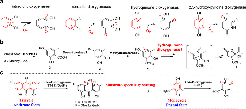
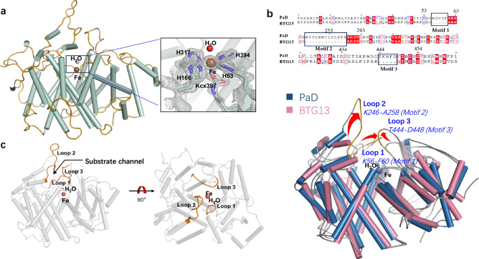
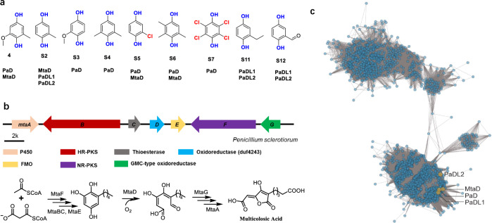
The inversion of substrate size specificity is an evolutionary roadblock for proteins. The Duf4243 dioxygenases GedK and BTG13 are known to catalyze the aromatic cleavage of bulky tricyclic hydroquinone. In this study, we discover a Duf4243 dioxygenase PaD that favors small monocyclic hydroquinones from the penicillic-acid biosynthetic pathway. Sequence alignments between PaD and GedK and BTG13 suggest PaD has three additional motifs, namely motifs 1-3, distributed at different positions in the protein sequence. X-ray crystal structures of PaD with the substrate at high resolution show motifs 1-3 determine three loops (loops 1-3). Most intriguing, loops 1-3 stack together at the top of the pocket, creating a lid-like tertiary structure with a narrow channel and a clearly constricted opening. This drastically changes the substrate specificity by determining the entry and binding of much smaller substrates. Further genome mining suggests Duf4243 dioxygenases with motifs 1-3 belong to an evolutionary branch that is extensively involved in the biosynthesis of natural products and has the ability to degrade diverse monocyclic hydroquinone pollutants. This study showcases how natural enzymes alter the substrate specificity fundamentally by incorporating new small motifs, with a fixed overall scaffold-architecture. It will also offer a theoretical foundation for the engineering of substrate specificity in enzymes and act as a guide for the identification of aromatic dioxygenases with distinct substrate specificities.


Conflict of interest statement

The authors declare no competing interests.

Figures

Fig. 1
Fig. 1. Microorganism-derived aromatic dioxygenases.
a Aromatic cleavage by intradiol dioxygenase, extradiol dioxygenase, hydroquinone dioxygenase, and 2,5-hydroxy-pyridine dioxygenase. b Biosynthetic pathway of penicillic acid proposed by previous studies. A nonreducing polyketide synthase (NR-PKS) generates orsellinic acid (OA, 2), followed by a decarboxylase and methyltransferase catalyze the formation of hydroquinone 4 (6-methylbenzene-1,2,4-triol). c Substrate-specificity shifting of different Duf4243 dioxygenases with their specific substrates. BTG and GedK catalyze the oxidative ring opening of the tricycle hydroquinone, while PaD accepts the monocyclic hydroquinone.
Fig. 2
Fig. 2. Proposed biosynthetic pathway of penicillic acid (1).
a Penicillic acid (1) biosynthetic gene cluster (BGC) in A. westerdijkiae. NR-PKS, nonreducing polyketide synthase; FMO, flavin-containing monooxygenase; OMeT, O-methyltransferase; SDR, short chain reductase; MFS, major facilitator superfamily; TF, transcription factor. b Product profiles of wild-type and mutants (ΔpaA, ΔpaD, and ΔpaE) of A. westerdijkiae. c In vitro assays of PaB and PaC with 2. d In vitro assays of PaD with 4 or 8. e LC-MS analysis of product profiles of compound 4 fed to S. cerevisiae expressing paD and paE. S. cerevisiae containing empty pXW55 and pXW06 vectors was used as a control. f Proposed biosynthetic pathway of 1.
Fig. 3
Fig. 3. Crystal structure of PaD with a lid-like tertiary structure.
a Crystal structure of PaD (PDB: 8Z4Q). H63, H166, H317, H394, K397, and one water molecule coordinate the iron. The electron density 2Fo-Fc maps of the ligands coordinated with Fe are shown with a gray-colored mesh, and contoured at 2.5 σ. b Sequence and structure comparison of PaD with BTG13. Structural comparison was depicted by the overlay of the overall structure of BTG13 and PaD. The three distinct motifs 1-3 are marked with black boxes. The three loops 1-3 determined by motifs 1-3 are marked in gold. c The locations of loops 1-3 in PaD structure viewed from different positions.
Fig. 4
Fig. 4. Crystal structure analysis of PaD-4 complex.
a Overall structure of PaD-4 complex (PDB: 8Z4R). Loops 1-3 are marked in gold. b The active site view of PaD-4 complex. The electron density 2Fo-Fc maps of substrate 4 in PaD are shown with a gray-colored mesh, and contoured at 1 σ. c Relative activities of PaD mutants located on the loops 1-3. The yield of 9 is quantified, defining the activity of wild-type PaD at 100%. Three biological parallel (n = 3) replicates are performed and presented as triangular points. The error bars are presented as standard deviation (SD). ND: not detected. Source data are provided as a Source Data file. d Comparison of the active-site shape and the substrate-binding mode in PaD and BTG13. The BTG13 complex structure was stimulated by Discovery Studio Client v19.1.0. e Proposed catalytic mechanism for PaD.
Fig. 5
Fig. 5. Characterizing PaD-like dioxygenases (PaDLs) as a family of hydroquinone dioxygenases.
a Promiscuity profile substrate scope of PaD, MtaD, PaDL1, and PaDL2. PaDL1 and PaDL2 were selected from the same cluster of PaD. The active proteins are marked below the tested hydroquinones. b The BGC and proposed biosynthetic pathway of multicolosic acid. c Sequence similarity network (SSN) analysis of PaDLs. The SSN uses 2730 PaDLs obtained from uniport database by searching for Duf4243-domain containing proteins.
Fig. 6
Fig. 6. Crystal structure analysis of PaD-S6.
a The overall structure and substrate-binding site view of PaD-S6 complex (PDB: 8Z4S). Loops 1-3 are marked in gold. The cleavage-site of PaD toward S6 and the generated product is shown at the top of the PaD-S6 structure complex, which was proposed based on the structure and mass spectroscopy analysis. The electron density 2Fo-Fc maps of the bound hydroquinone S6 in PaD are shown with a gray-colored mesh, and contoured at 1 σ. b The comparison of the spatial distances from the bound hydroquinone to key amino acids in the PaD-4 and PaD-S6 structure complexes.

References

    1. Jakoobi, M. & Sergeev, A. G. Transition-metal-mediated cleavage of C-C bonds in aromatic rings. Chem.-Asian J.14, 2181–2192 (2019). 10.1002/asia.201900443 - DOI - PubMed
    1. Qiu, X. et al. Cleaving arene rings for acyclic alkenylnitrile synthesis. Nature597, 64–69 (2021). 10.1038/s41586-021-03801-y - DOI - PubMed
    1. Harayama, S., Kok, M. & Neidle, E. L. Functional and evolutionary relationships among diverse oxygenases. Annu. Rev. Microbiol.46, 565–601 (1992). 10.1146/annurev.mi.46.100192.003025 - DOI - PubMed
    1. Tang, H. et al. Systematic unraveling of the unsolved pathway of nicotine degradation in Pseudomonas. PLoS Genet9, e1003923 (2013). 10.1371/journal.pgen.1003923 - DOI - PMC - PubMed
    1. Fuchs, G., Boll, M. & Heider, J. Microbial degradation of aromatic compounds—from one strategy to four. Nat. Rev. Microbiol.9, 803–816, (2011). 10.1038/nrmicro2652 - DOI - PubMed

Publication types

LinkOut - more resources