. 2024 Sep 3;15(1):7671.
doi: 10.1038/s41467-024-51194-z.

Optoelectronic synapses with chemical-electric behaviors in gallium nitride semiconductors for biorealistic neuromorphic functionality

Affiliations

Optoelectronic synapses with chemical-electric behaviors in gallium nitride semiconductors for biorealistic neuromorphic functionality

Xin Liu et al. Nat Commun. .

Abstract

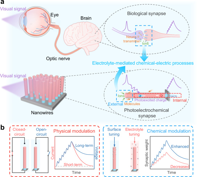
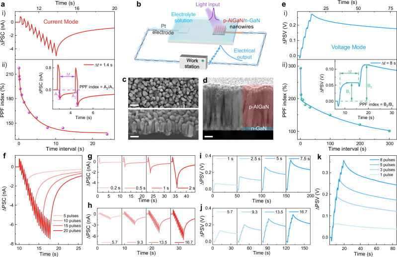
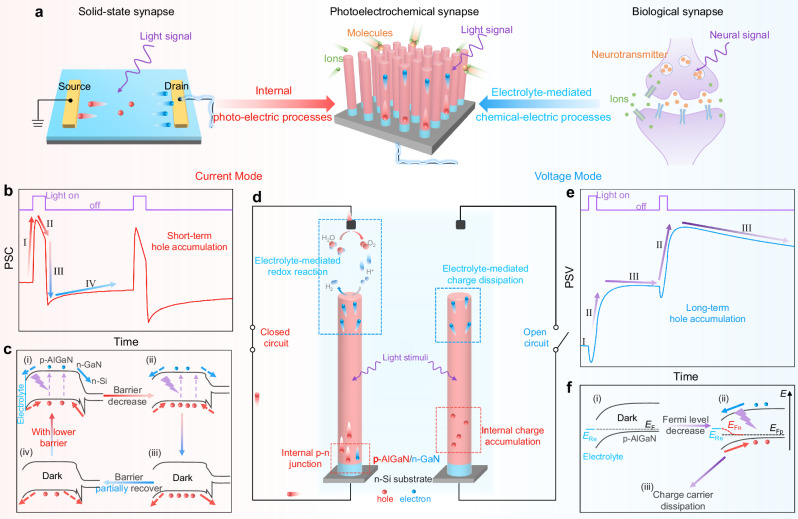
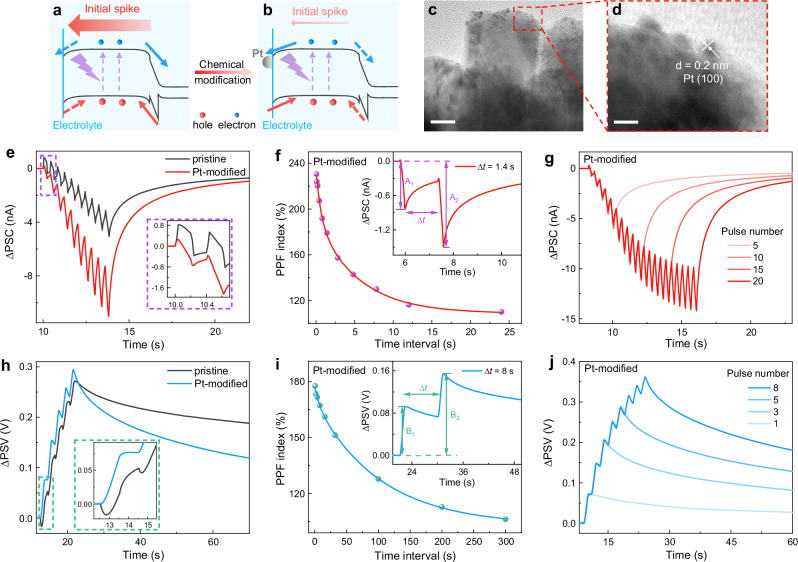
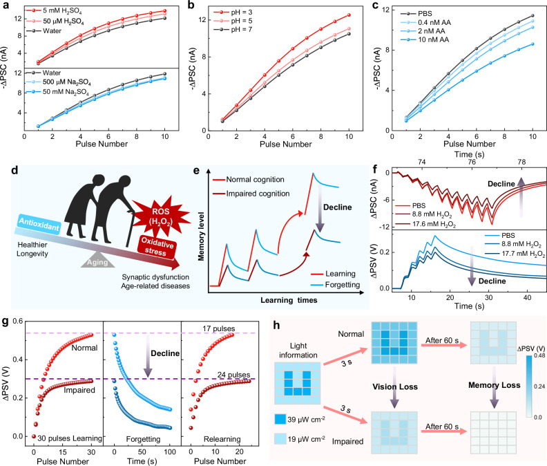
Optoelectronic synapses, leveraging the integration of classic photo-electric effect with synaptic plasticity, are emerging as building blocks for artificial vision and photonic neuromorphic computing. However, the fundamental working principles of most optoelectronic synapses mainly rely on physical behaviors while missing chemical-electric synaptic processes critical for mimicking biorealistic neuromorphic functionality. Herein, we report a photoelectrochemical synaptic device based on p-AlGaN/n-GaN semiconductor nanowires to incorporate chemical-electric synaptic behaviors into optoelectronic synapses, demonstrating unparalleled dual-modal plasticity and chemically-regulated neuromorphic functions through the interplay of internal photo-electric and external electrolyte-mediated chemical-electric processes. Electrical modulation by implementing closed or open-circuit enables switching of optoelectronic synaptic operation between short-term and long-term plasticity. Furthermore, inspired by transmembrane receptors that connect extracellular and intracellular events, synaptic responses can also be effectively amplified by applying chemical modifications to nanowire surfaces, which tune external and internal charge behaviors. Notably, under varied external electrolyte environments (ion/molecule species and concentrations), our device successfully mimics chemically-regulated synaptic activities and emulates intricate oxidative stress-induced biological phenomena. Essentially, we demonstrate that through the nanowire photoelectrochemical synapse configuration, optoelectronic synapses can be incorporated with chemical-electric behaviors to bridge the gap between classic optoelectronic synapses and biological synapses, providing a promising platform for multifunctional neuromorphic applications.


Conflict of interest statement

The authors declare no competing interests.

Figures

Fig. 1
Fig. 1. Schematics of the biological visual system and the photoelectrochemical synapse.
a Schematic of biological visual system (top) and photoelectrochemical synapse (bottom). Both biological synapses and photoelectrochemical synapses operate through electrolyte-mediated chemical-electric processes in aqueous environments. Additionally, the photoelectrochemical synapses also rely on internal physical (photo-electric) processes involving hole/electron transfer behaviors. b Schematic of the synaptic activity modulation by physical (left) and chemical approaches (right) in the photoelectrochemical synapses. By manipulating the physical dynamics, the device can switch between current mode and voltage mode, showing short-term and long-term plasticity, respectively. Through the application of chemical methods, including surface chemical modification and varying the electrolyte environment, synaptic responses can be regulated.
Fig. 2
Fig. 2. Structure and dual-modal synaptic behaviors of the photoelectrochemical synapse.
a i): Postsynaptic current (ΔPSC) in response to ten light pulses (tp = Δt = 0.2 s); ii): PPF index versus pulse interval, with the inset showing the PPF effect and its definition. b Schematic of the photoelectrochemical synaptic device. c Top-view SEM image (top; scale bar, 150 nm) and 45°-tilted SEM image of the as-grown nanowires (bottom; scale bar, 150 nm). d STEM image of the as-grown nanowires (scale bar, 50 nm). e i): Postsynaptic voltage (ΔPSV) induced by five light pulses (tp = Δt = 1 s); ii): PPF index versus pulse interval, with the inset showing the PPF effect and its definition. f-h Correlations between ΔPSC and pulse number (f), pulse duration (g), and pulse intensity (h). ik Correlations between ΔPSV and pulse number (k), pulse duration (i), and pulse intensity (j). Light pulses of 12 µW cm−2 were used, except for the pulse intensity-dependent tests. The inset numbers in (h) and (j) represent different light intensities from 5.7 to 16.7 µW cm−2.
Fig. 3
Fig. 3. Working mechanisms of the photoelectrochemical synapse.
a Comparison of our photoelectrochemical synapse with solid-state and biological synapses. b Representative postsynaptic current (PSC) triggered by two sequential pulses. c Schematic of band bending and charge carrier behaviors in current mode during the light stimulation (i & ii) and the dark recovery phase (iii & iv), illustrating short-term hole accumulation and barrier height modulation. Arrows are used to indicate the direction of charge carrier transport, with solid arrows representing the dominant charge carrier transfer direction. d Schematic of the charge carrier dynamics upon light stimulation in closed (left) and open (right) circuit, involving the internal nanowire and electrolyte-mediated charge behaviors. e Typical postsynaptic voltage (PSV) induced by two successive pulses. f Schematic of band bending and charge carrier behaviors in voltage mode during the initial dark state (i), the light stimulation (ii), and the post-stimulation recovery in the dark (iii).
Fig. 4
Fig. 4. Synaptic plasticity modulation via surface Pt modification.
a, b Schematic illustrations of the charge carrier behaviors before (a) and after (b) anchoring Pt nanoparticles on the nanowire surface. Arrows are used to indicate the direction of charge carrier transport, with solid arrows representing the dominant charge carrier transfer direction. c TEM image of Pt-modified nanowires (scale bar, 25 nm). d HRTEM image of the red-outlined area (scale bar, 5 nm). e, h PSC curves (e) and PSV curves (h) of pristine device and Pt-modified device with the inset exhibiting an enlarged view of the synaptic responses. f, i PPF curves in current mode (f) and voltage mode (i) with Pt modification, with the inset showing the PPF effect. g, j Synaptic responses with different pulse numbers in current mode (g) and voltage mode (j). Light pulses of 255 nm and 12 µW cm−2 were used. In current mode, tp = Δt = 0.2 s. In voltage mode, tp = Δt = 1 s.
Fig. 5
Fig. 5. Electrolyte-regulated synaptic plasticity and simulation of oxidative stress-associated biological activities.
a ΔPSC responses to various concentrations of H2SO4 solution (top) and Na2SO4 solution (bottom). b ΔPSC responses in 0.1 M Na2SO4 solution at different pH values. c ΔPSC responses in PBS solution with various concentrations of ascorbic acid (AA). d Schematic illustration of the balance between reactive oxygen species (ROS) and antioxidants in humans. Excessive ROS leads to synaptic dysfunction and age-related diseases. e Schematic illustration of cognitive decline caused by excessive ROS. f Synaptic responses in PBS solution with different concentrations of H2O2. g Learning-forgetting-relearning behavior under normal and impaired conditions simulated by the photoelectrochemical synapse. h The simulation of visual perception and memory under normal and impaired conditions using the photoelectrochemical synapse. Light pulses of 255 nm and 12 µW cm−2 were used. In current mode, tp = Δt = 0.2 s. In voltage mode, tp = Δt = 1 s. Note that we take the absolute value of the postsynaptic current (-ΔPSC) for performance comparison in Figs. 5a, b, and c.

Similar articles

Cited by

References

    1. Yang, X. et al. Bioinspired neuron-like electronics. Nat. Mater.18, 510–517 (2019). 10.1038/s41563-019-0292-9 - DOI - PMC - PubMed
    1. Jiang, L. et al. Flexible ultrasound-induced retinal stimulating piezo-arrays for biomimetic visual prostheses. Nat. Commun.13, 3853 (2022). 10.1038/s41467-022-31599-4 - DOI - PMC - PubMed
    1. Ling, Y. et al. Bioinspired elastomer composites with programmed mechanical and electrical anisotropies. Nat. Commun.13, 524 (2022). 10.1038/s41467-022-28185-z - DOI - PMC - PubMed
    1. Kim, Y. et al. A bioinspired flexible organic artificial afferent nerve. Science360, 998–1003 (2018). 10.1126/science.aao0098 - DOI - PubMed
    1. Liao, F. et al. Bioinspired in-sensor visual adaptation for accurate perception. Nat. Electron.5, 84–91 (2022).10.1038/s41928-022-00713-1 - DOI

LinkOut - more resources