. 2024 Sep 3;15(1):275.
doi: 10.1186/s13287-024-03805-1.

Nucleoporin 153 deficiency in adult neural stem cells defines a pathological protein-network signature and defective neurogenesis in a mouse model of AD

Affiliations

Nucleoporin 153 deficiency in adult neural stem cells defines a pathological protein-network signature and defective neurogenesis in a mouse model of AD

Claudia Colussi et al. Stem Cell Res Ther. .

Abstract

Background: Reduction of adult hippocampal neurogenesis is an early critical event in Alzheimer's disease (AD), contributing to progressive memory loss and cognitive decline. Reduced levels of the nucleoporin 153 (Nup153), a key epigenetic regulator of NSC stemness, characterize the neural stem cells isolated from a mouse model of AD (3×Tg) (AD-NSCs) and determine their altered plasticity and gene expression.

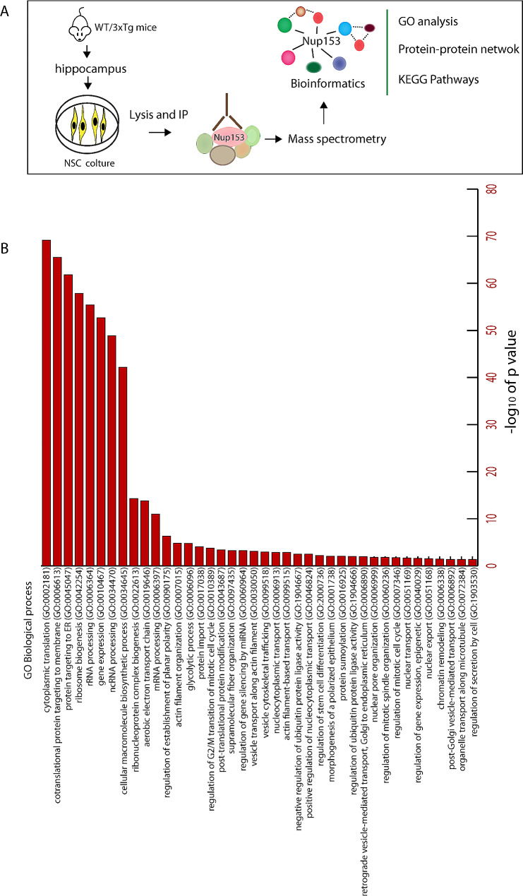
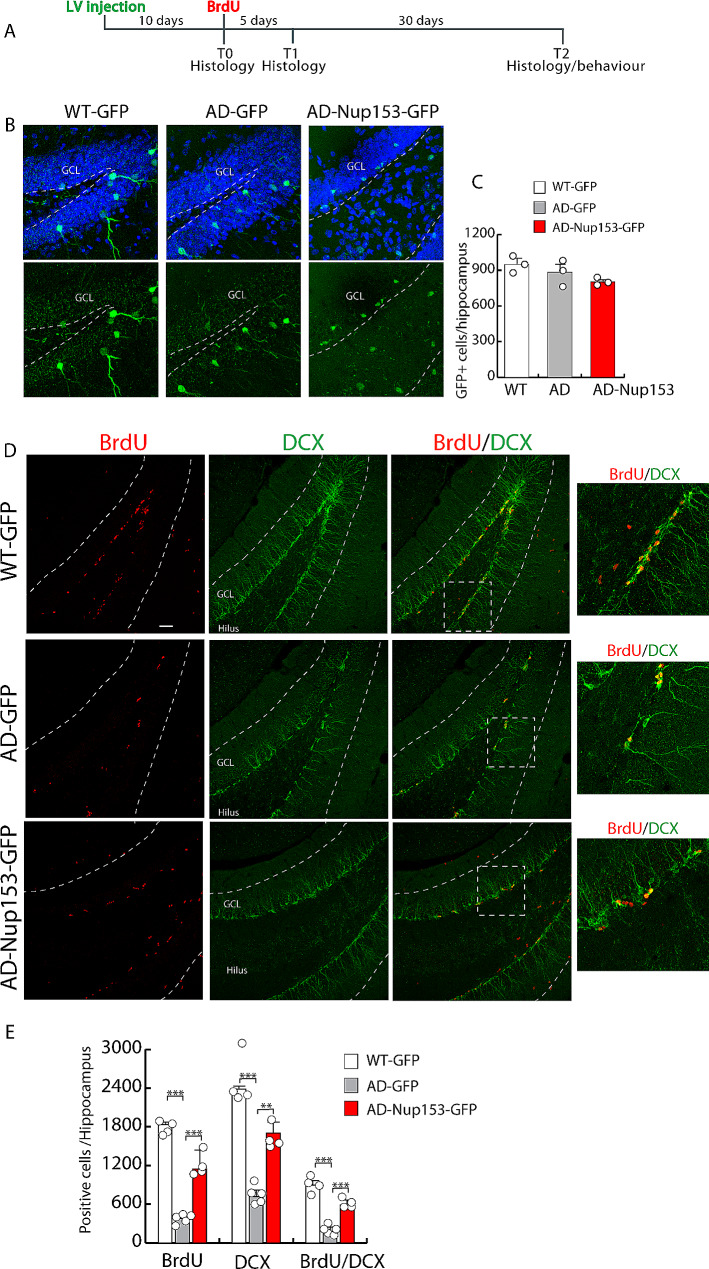
Methods: Nup153-regulated mechanisms contributing to NSC function were investigated: (1) in cultured NSCs isolated from AD and wild type (WT) mice by proteomics; (2) in vivo by lentiviral-mediated delivery of Nup153 or GFP in the hippocampus of AD and control mice analyzing neurogenesis and cognitive function; (3) in human iPSC-derived brain organoids obtained from AD patients and control subjects as a model of neurodevelopment.

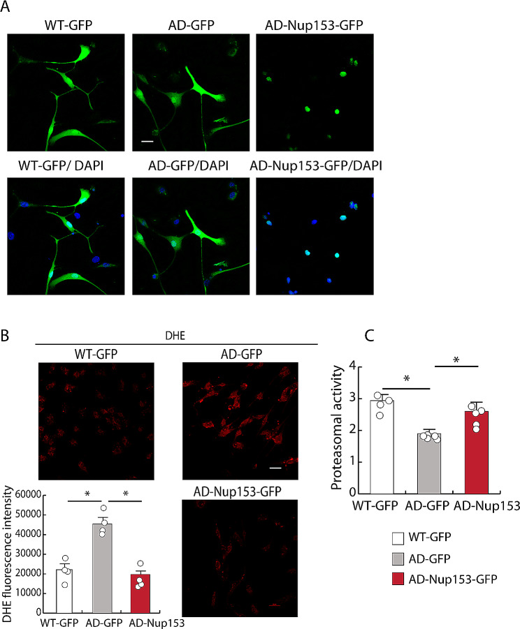
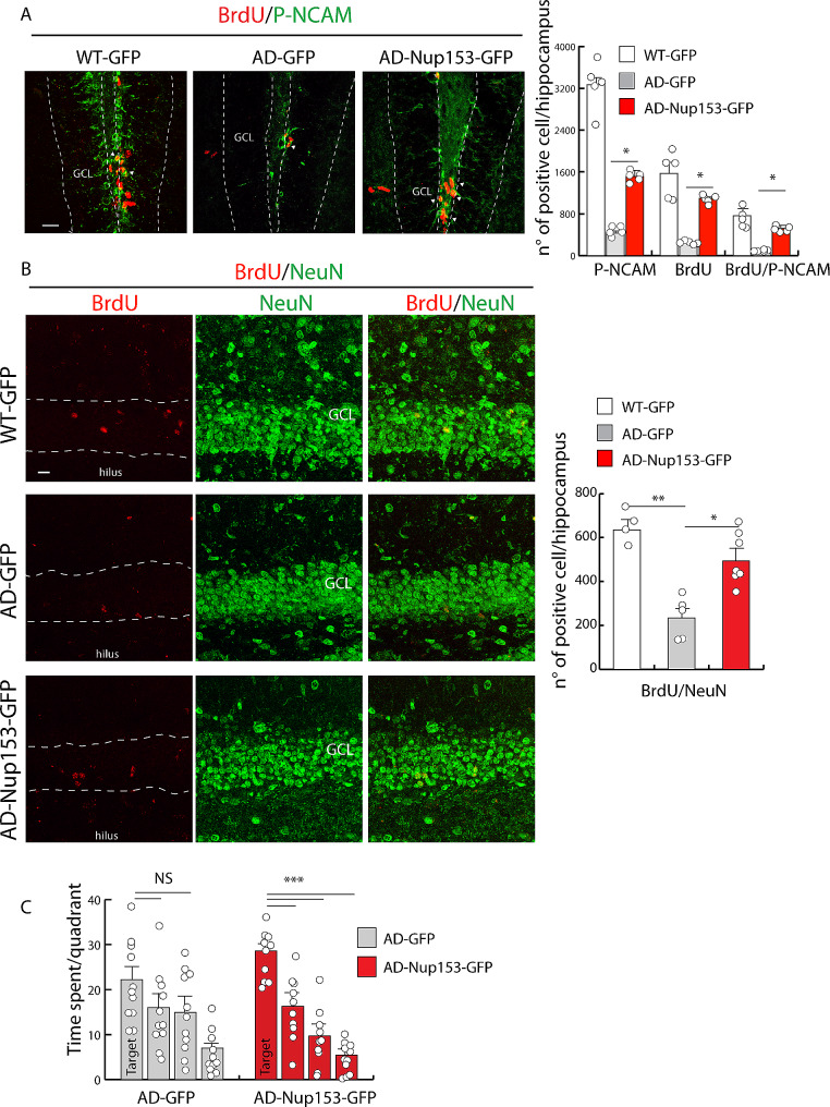
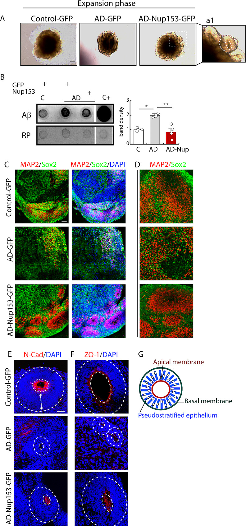
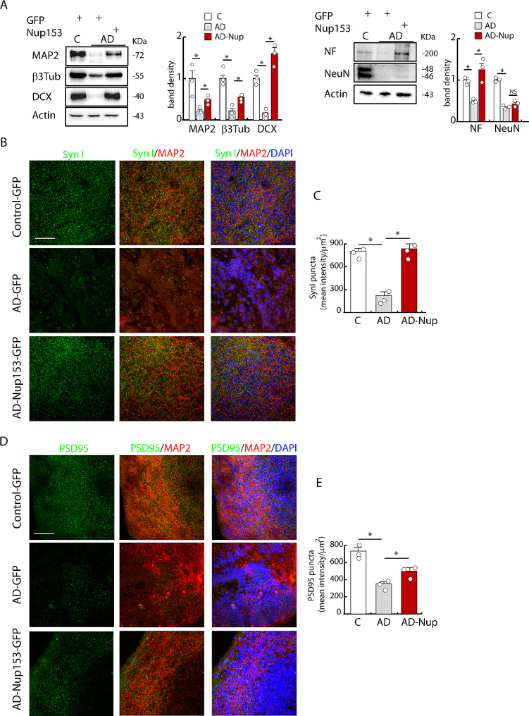
Results: Proteomic approach identified Nup153 interactors in WT- and AD-NSCs potentially implicated in neurogenesis regulation. Gene ontology (GO) analysis showed that Nup153-bound proteins in WT-NSCs were involved in RNA metabolism, nuclear import and epigenetic mechanisms. Nup153-bound proteins in AD-NSCs were involved in pathways of neurodegeneration, mitochondrial dysfunction, proteasomal processing and RNA degradation. Furthermore, recovery of Nup153 levels in AD-NSCs reduced the levels of oxidative stress markers and recovered proteasomal activity. Lentiviral-mediated delivery of Nup153 in the hippocampal niche of AD mice increased the proliferation of early progenitors, marked by BrdU/DCX and BrdU/PSANCAM positivity and, later, the integration of differentiating neurons in the cell granule layer (BrdU/NeuN+ cells) compared with GFP-injected AD mice. Consistently, Nup153-injected AD mice showed an improvement of cognitive performance in comparison to AD-GFP mice at 1 month after virus delivery assessed by Morris Water Maze. To validate the role of Nup153 in neurogenesis we took advantage of brain organoids derived from AD-iPSCs characterized by fewer neuroepithelial progenitor loops and reduced differentiation areas. The upregulation of Nup153 in AD organoids recovered the formation of neural-like tubes and differentiation.

Conclusions: Our data suggest that the positive effect of Nup153 on neurogenesis is based on a complex regulatory network orchestrated by Nup153 and that this protein is a valuable disease target.

Keywords: Alzheimer’s disease; Neurogenesis; Nucleoporin; Organoids.


Conflict of interest statement

The authors declare no conflict of interest.

Figures

Fig. 1
Fig. 1
(A) Cartoon depicting the experimental plan for the in vitro experiments. (B) Schematic illustrating the main biological processes from Gene ontology analysis of Nup153-protein network in WT-NSCs
Fig. 2
Fig. 2
Nup153 overexpression in AD-NSCs counteracts oxidative stress and recovers proteasomal activity. (A) Immunofluorescence showing GFP expression in AD-NSCs transfected with GFP or GFP-Nup153 vectors at 72 h. (B) DHE labelling of NSCs in the above conditions to reveal the level of oxidative stress. The graph indicates the mean fluorescence intensity for DHE (n = 4). (C) Proteasomal activity measured in WT-, AD-GFP- and AD-GFP-Nup153-NSCs (n = 5). *P < 0.05. Scale bar 20 μm
Fig. 3
Fig. 3
Nup153 overexpression in the hippocampus of AD mice stimulates proliferation and early differentiation. (A) Experimental plan for the in vivo experiments. (B) Representative confocal images showing GFP positive cells in the hippocampal niche of WT and AD mice injected with a lentivirus coding for GFP (WT and AD) or GFP-Nup153 at 10 days (T0). (C) Quantification of the GFP + cells in WT-GFP, AD-GFP and AD-GFP-Nup153 injected mice. Nuclei were counterstained with DAPI (n = 3, scale bar 20 μm). (D) Representative confocal images showing hippocampal cells positive for BrdU (red) and DCX (green) from WT-GFP, AD-GFP and AD-GFP-Nup153 injected mice at T1. Arrows indicate BrdU/DCX double positive cells. (E) The graph shows the number of BrdU+, DCX+ and BrdU/DCX double positive cells in WT-GFP, AD-GFP and AD-GFP-Nup153 injected mice (n = 4, scale bar 50 μm). GCL = granule cell layer, ***P < 0.001, **P < 0.01
Fig. 4
Fig. 4
Nup153 overexpression in the hippocampus of AD mice promotes neurogenesis and cognitive performance. (A) Representative confocal images showing hippocampal cells positive for BrdU (red) and P-NCAM (green) from WT-GFP, AD-GFP and AD-GFP-Nup153 injected mice at T1. Arrows indicate BrdU/P-NCAM positive cells. The graph shows the number of BrdU+, P-NCAM+ and BrdU/P-NCAM double positive cells in the above conditions, n = 5 scale bar 20 μm. (B) Representative confocal images showing BrdU+ cells (red) in the granule layer of the hippocampus identified by NeuN+ cells in WT-GFP, AD-GFP and AD-GFP-Nup153 injected mice at T2 (scale bar 10 μm). The graph shows the number of BrdU/NeuN double positive cells in the hippocampus in the above conditions (n = 4–7). (C) Assessment of memory performance by the Morris Water Maze test (MWM). The graph shows the time spent in the four quadrants during the probe test performed on day 5 of MWM. *P < 0.05, **P < 0.01, ***P < 0.001
Fig. 5
Fig. 5
Nup153 transduction in AD-iPSCs improves the maturation and organization of brain cortical organoids. (A) Phase contrast representative images of control, AD and AD-Nup153 organoids in the expansion phase. a1) Enlargement showing a detail of the circular neuroepithelium-like structure surrounded by the apical and basal membranes indicated by the dotted lines. (B) Aβ levels by dot blot analysis (n = 3–4). Each lysate was obtained from the pool of 3–4 individual organoids. Hippocampal lysate from 9-month-old 3×Tg mice was used as positive control. Red ponceau (RP) staining was used as loading index and used to normalize samples. C-F) Confocal analysis of MAP2/Sox2 (C and D at different magnification) and N-Cadherin (E) and ZO-1 (F) in control, AD and AD-Nup153 brain organoids. Nuclei were counterstained with DAPI, scale bar 50 μm. The inner dotted white circles indicate the ring-like N-cadherin and ZO-1 distribution at the apical membrane. The double arrow indicates the pseudostratified neuroepithelium starting from the cavity to the basal membrane. (G) Scheme representing the organization of the pseudostratified epithelium between the inner apical membrane and the outer basal membrane in the organoid. *P < 0.05, **P < 0.01
Fig. 6
Fig. 6
Nup153 transduction in AD-iPSCs improves the differentiation of brain cortical organoids. (A) Western blot analysis of MAP2, β3 tubulin, DCX, NeuN and NF proteins in brain organoids (n = 3) and relative quantification based on the expression of actin. Each lysate was obtained from the pool of 3–4 individual organoids. (B, D) Representative images of syn I and PSD95 expression from control, AD and AD-Nup153 brain organoids analyzed by confocal analysis and counterstained with MAP2 and DAPI (scale bar 50 μm) at 2 months of differentiation. (C, E) Quantification of mean fluorescence intensity of SynI and PSD95 puncta/area relative to control, AD and AD-Nup153 brain organoids (n = 3). *P < 0.05

References

    1. Snyder JS, Kee N, Wojtowicz JM. Effects of adult neurogenesis on synaptic plasticity in the rat dentate gyrus. J Neurophysiol. 2001;85(6):2423–31. - PubMed
    1. Toni N, Schinder AF. Maturation and Functional Integration of New Granule Cells into the adult Hippocampus. Cold Spring Harb Perspect Biol. 2015;8(1):a018903. - PMC - PubMed
    1. Deng W, Aimone JB, Gage FH. New neurons and new memories: how does adult hippocampal neurogenesis affect learning and memory? Nat Rev Neurosci. 2010;11(5):339–50. - PMC - PubMed
    1. Kempermann G, Gage FH. Genetic determinants of adult hippocampal neurogenesis correlate with acquisition, but not probe trial performance, in the water maze task. Eur J Neurosci. 2002;16(1):129–36. - PubMed
    1. Jurkowski MP, Bettio L, Patten EKW, Yau A, Gil-Mohapel SY. Beyond the Hippocampus and the SVZ: adult neurogenesis throughout the brain. Front Cell Neurosci. 2020;14:576444. - PMC - PubMed

Substances