. 2024 Sep 3;25(1):330.
doi: 10.1186/s12931-024-02958-0.

Recombinant thrombomodulin and recombinant antithrombin attenuate pulmonary endothelial glycocalyx degradation and neutrophil extracellular trap formation in ventilator-induced lung injury in the context of endotoxemia

Affiliations

Recombinant thrombomodulin and recombinant antithrombin attenuate pulmonary endothelial glycocalyx degradation and neutrophil extracellular trap formation in ventilator-induced lung injury in the context of endotoxemia

Kenichiro Kikuchi et al. Respir Res. .

Abstract

Background: Vascular endothelial damage is involved in the development and exacerbation of ventilator-induced lung injury (VILI). Pulmonary endothelial glycocalyx and neutrophil extracellular traps (NETs) are endothelial protective and damaging factors, respectively; however, their dynamics in VILI and the effects of recombinant thrombomodulin and antithrombin on these dynamics remain unclear. We hypothesized that glycocalyx degradation and NETs are induced by VILI and suppressed by recombinant thrombomodulin, recombinant antithrombin, or their combination.

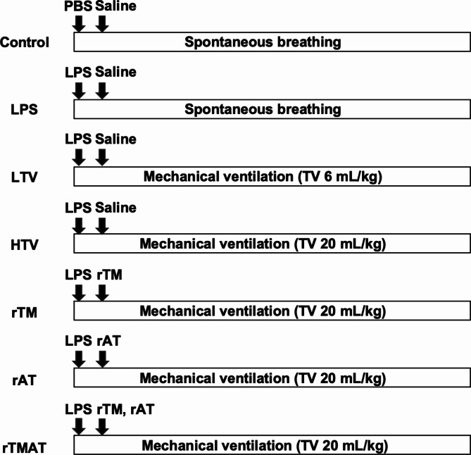
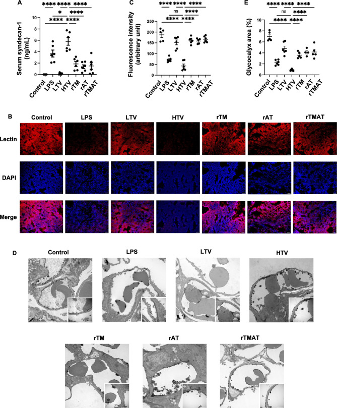
Methods: VILI was induced in male C57BL/6J mice by intraperitoneal lipopolysaccharide injection (20 mg/kg) and high tidal volume ventilation (20 mL/kg). In the intervention groups, recombinant thrombomodulin, recombinant antithrombin, or their combination was administered at the start of mechanical ventilation. Glycocalyx degradation was quantified by measuring serum syndecan-1, fluorescence-labeled lectin intensity, and glycocalyx-occupied area in the pulmonary vascular lumen. Double-stranded DNA in the bronchoalveolar fluid and fluorescent areas of citrullinated histone H3 and myeloperoxidase were quantified as NET formation.

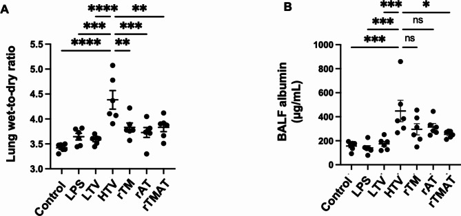
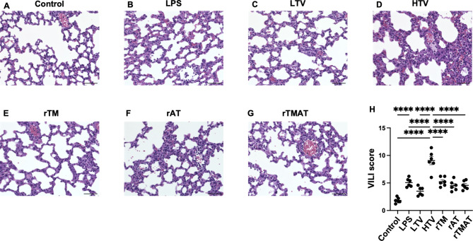
Results: Serum syndecan-1 increased, and lectin fluorescence intensity decreased in VILI. Electron microscopy revealed decreases in glycocalyx-occupied areas within pulmonary microvessels in VILI. Double-stranded DNA levels in the bronchoalveolar lavage fluid and the fluorescent area of citrullinated histone H3 and myeloperoxidase in lung tissues increased in VILI. Recombinant thrombomodulin, recombinant antithrombin, and their combination reduced glycocalyx injury and NET marker levels. There was little difference in glycocalyx injury and NET makers between the intervention groups.

Conclusion: VILI induced glycocalyx degradation and NET formation. Recombinant thrombomodulin and recombinant antithrombin attenuated glycocalyx degradation and NETs in our VILI model. The effect of their combination did not differ from that of either drug alone. Recombinant thrombomodulin and antithrombin have the potential to be therapeutic agents for biotrauma in VILI.

Keywords: Biotrauma; Glycocalyx; Neutrophil extracellular traps; Recombinant antithrombin; Recombinant thrombomodulin; Ventilator-induced lung injury.


Conflict of interest statement

The authors declare no competing interests.

Figures

Fig. 1
Fig. 1
Experimental timeline for each group. The control and LPS groups breathed spontaneously, whereas the other groups were mechanically ventilated for 4 h. The LTV group was mechanically ventilated with a tidal volume of 6 mL/kg and a respiratory rate of 140/min. The HTV, rTM, rAT, and rTMAT groups were mechanically ventilated with tidal volume of 20 mL/kg and respiratory rate of 70/min. LPS (20 mg/kg) was administered immediately after the initiation of mechanical ventilation. rTM and/or rAT were administered immediately after LPS injection. PBS = phosphate-buffered saline; TV = tidal volume; LPS = lipopolysaccharide; rTM = recombinant thrombomodulin; rAT = recombinant antithrombin; LTV = low tidal volume ventilation; HTV = high tidal volume ventilation
Fig. 2
Fig. 2
Systemic and pulmonary inflammation. A and B) Serum TNF-α (N = 5–6) and HMGB-1 levels (N = 6) after 4 h of mechanical ventilation or spontaneous breathing. C) BALF TNF-α levels (N = 5–6). D) BALF HMGB-1 levels (N = 6). E) Total inflammatory cell counts in BALF (N = 6). F) The percentage of neutrophils in BALF (N = 6). ns = not significant. *P ≤ 0.05, **P ≤ 0.01, ***P ≤ 0.001, ****P ≤ 0.0001. BALF = bronchoalveolar lavage fluid
Fig. 3
Fig. 3
Pulmonary vascular permeability. A) Lung wet-to-dry ratio (N = 6). Lung wet-to-dry ratio was calculated as (wet weight – dry weight) / (dry weight) of each sample. B) BALF albumin levels (N = 6). ns = not significant. *P ≤ 0.05, **P ≤ 0.01, ***P ≤ 0.001, ****P ≤ 0.0001. BALF = bronchoalveolar lavage fluid
Fig. 4
Fig. 4
Lung histology in VILI. A–G) Representative images of hematoxylin-eosin staining of the lungs in each group. Alveolar wall thickening, neutrophilic infiltration, and hemorrhage were observed in the LPS group (B) and were more severe in the HTV group (D). These features were reduced in the rTM, rAT, and rTMAT groups (E–G). Scale bar = 50 μm. H) Quantitative analysis of lung injury was performed using VILI score (N = 6). The degree of lung injury was quantitatively assessed by adding up the scores, ranging from 0 (minimal damage) to 12 (maximal damage). *P ≤ 0.05, **P ≤ 0.01, ***P ≤ 0.001, ****P ≤ 0.0001. LPS = lipopolysaccharide; rTM = recombinant thrombomodulin; rAT = recombinant antithrombin; HTV = high tidal volume ventilation; VILI = ventilator-induced lung injury
Fig. 5
Fig. 5
Changes in peak inspiratory pressure and dynamic lung compliance. (A) Hourly changes in PIP from the start of mechanical ventilation to 4 h (N = 6). PIP at the end of mechanical ventilation was higher in the HTV group than in the rTMAT group (*P < 0.05). PIP at the beginning of mechanical ventilation was lower than at any other time point in all groups. PIP at 4 h was higher than that at 1 h in the HTV group (+P < 0.01). (B) Hourly changes in DLP from the start of mechanical ventilation to 4 h (N = 6). There was no difference in DLP between groups at any time point. DLP was higher at the beginning of mechanical ventilation than at any other time point in all groups. DLP at 4 h was lower than that at 1 h in the HTV group (#P < 0.05). PIP = peak inspiratory pressure; DLP = dynamic lung compliance; HTV = high tidal volume ventilation
Fig. 6
Fig. 6
Degradation of the endothelial glycocalyx. (A) Serum syndecan-1 levels (N = 6). (B) Representative images of fluorescent lectin staining of the lung for each group. The lectin-bound glycocalyx is shown in red, and the DNA is shown in blue. Scale bar = 50 μm. (C) Lectin fluorescence intensity for each group (N = 5–6). (D) Representative images of the glycocalyx by transmission electron microscopy. A continuous glycocalyx layer was observed in the control group. The glycocalyx was removed from the endothelium, and the glycocalyx layer was thin and disrupted in the LPS and HTV groups. Spherical aggregation of the glycocalyx layer was observed in the HTV group. The deviation and thinning of the glycocalyx decreased in the rTM, rAT, and rTMAT groups. Low and high magnification images for each group. Scale bar = 1 μm. (E) The percentage of glycocalyx area in the vascular lumen (N = 4–6). ns = not significant. *P ≤ 0.05, **P ≤ 0.01, ***P ≤ 0.001, ****P ≤ 0.0001. HTV = high tidal volume ventilation; LPS = lipopolysaccharide; rTM = recombinant thrombomodulin; rAT = recombinant antithrombin
Fig. 7
Fig. 7
NET formation in VILI. (A) BALF ds-DNA levels (N = 5–6). (B) Representative immunofluorescence images of NET formation in the lung. Citrullinated histone H3 is shown in green (Cit-H3), MPO in red, and DNA in blue (DAPI). There was extensive colocalization of citrullinated histone H3 and MPO in the HTV group and weak colocalization in the LTV and LPS groups. Colocalization was significantly reduced in the rTM, rAT, and rTMAT groups compared with that in the HTV group. Scale bar = 50 μm. C, D) The percentage of Cit-H3-positive (N = 6) and MPO-positive (N = 6) area. ns = not significant; Cit-H3 = citrullinated histone H3. *P ≤ 0.05, **P ≤ 0.01, ***P ≤ 0.001, ****P ≤ 0.0001. HTV = high tidal volume ventilation; LTV = low tidal volume ventilation; LPS = lipopolysaccharide; rTM = recombinant thrombomodulin; rAT = recombinant antithrombin

Similar articles

References

    1. Slutsky AS, Ranieri VM. Ventilator-induced lung injury. N Engl J Med. 2013;369:2126–36. 10.1056/NEJMra1208707 - DOI - PubMed
    1. Ricard JD, Dreyfuss D, Saumon G. Ventilator-induced lung injury. Eur Respir J Suppl. 2003;42:s2–9.10.1183/09031936.03.00420103 - DOI - PubMed
    1. Curley GF, Laffey JG, Zhang H, Slutsky AS. Biotrauma and ventilator-induced lung injury: clinical implications. Chest. 2016;150:1109–17. 10.1016/j.chest.2016.07.019 - DOI - PubMed
    1. Moore KH, Murphy HA, George EM. The glycocalyx: a central regulator of vascular function. Am J Physiol Regul Integr Comp Physiol. 2021;320:R508–18. 10.1152/ajpregu.00340.2020 - DOI - PMC - PubMed
    1. Yildiz C, Palaniyar N, Otulakowski G, Khan MA, Post M, Kuebler WM, et al. Mechanical ventilation induces neutrophil extracellular trap formation. Anesthesiology. 2015;122:864–75. 10.1097/ALN.0000000000000605 - DOI - PubMed

MeSH terms

LinkOut - more resources