. 2024 Sep 4;15(1):281.
doi: 10.1186/s13287-024-03911-0.

A two-step strategy to expand primary human hepatocytes in vitro with efficient metabolic and regenerative capacities

Affiliations

A two-step strategy to expand primary human hepatocytes in vitro with efficient metabolic and regenerative capacities

Huangfan Xie et al. Stem Cell Res Ther. .

Abstract

Background: Primary human hepatocytes (PHHs) are highly valuable for drug-metabolism evaluation, liver disease modeling and hepatocyte transplantation. However, their availability is significantly restricted due to limited donor sources, alongside their constrained proliferation capabilities and reduced functionality when cultured in vitro. To address this challenge, we aimed to develop a novel method to efficiently expand PHHs in vitro without a loss of function.

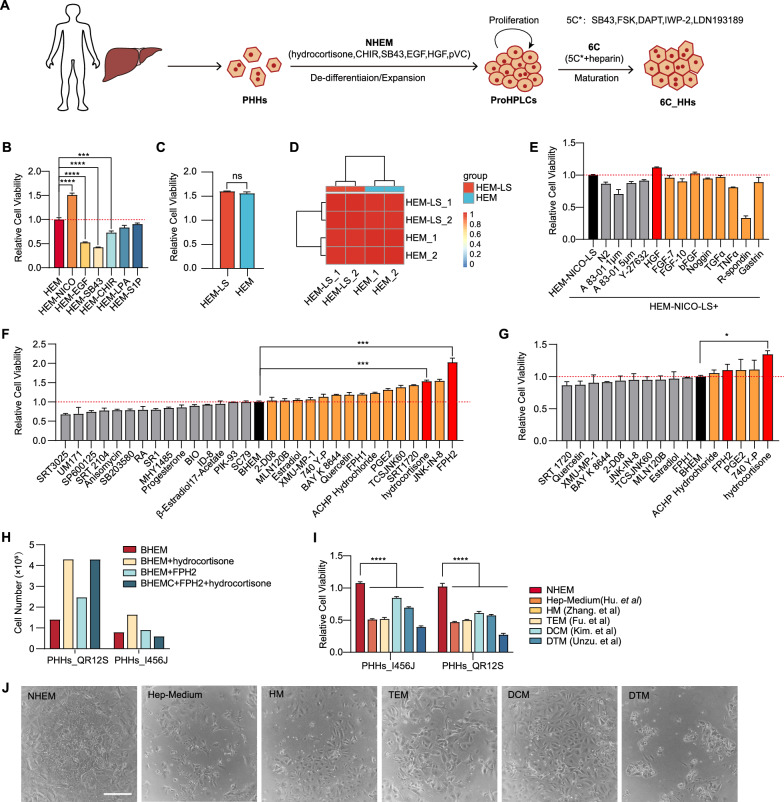
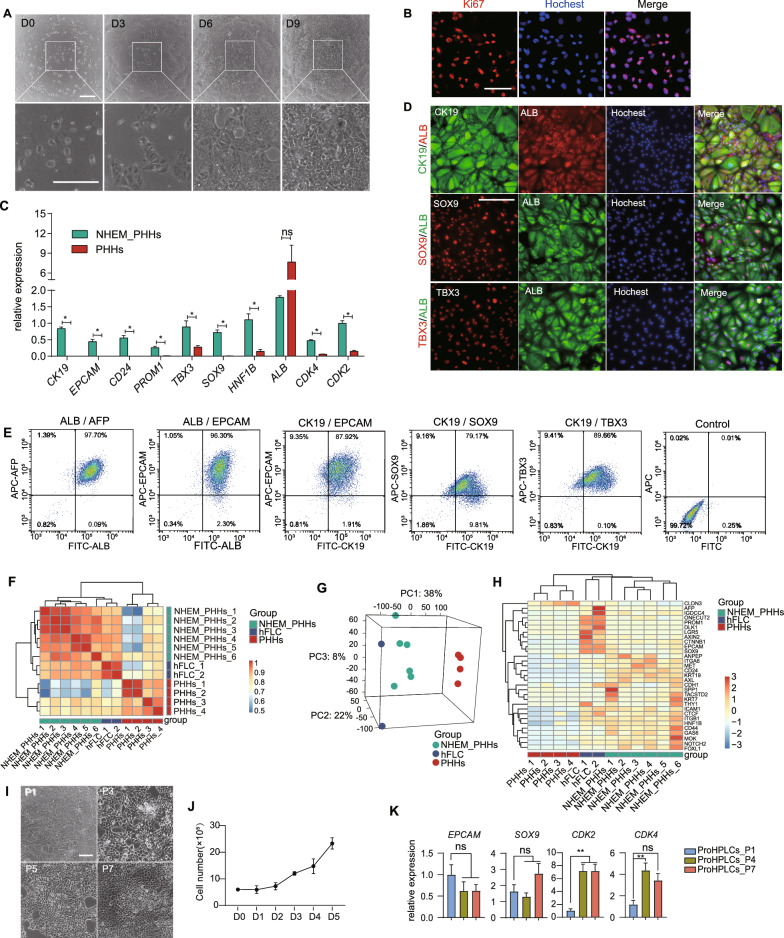
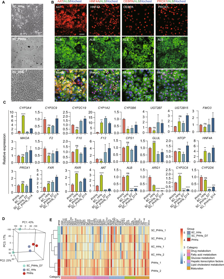
Methods: By mimicking the in vivo liver regeneration route, we developed a two-step strategy involving the de-differentiation/expansion and subsequent maturation of PHHs to generate abundant functional hepatocytes in vitro. Initially, we applied SiPer, a prediction algorithm, to identify candidate small molecules capable of activating liver regenerative transcription factors, thereby formulating a novel hepatic expansion medium to de-differentiate PHHs into proliferative human hepatic progenitor-like cells (ProHPLCs). These ProHPLCs were then re-differentiated into functionally mature hepatocytes using a new hepatocyte maturation condition. Additionally, we investigated the underlying mechanism of PHHs expansion under our new conditions.

Results: The novel hepatic expansion medium containing hydrocortisone facilitated the de-differentiation of PHHs into ProHPLCs, which exhibited key hepatic progenitor characteristics and demonstrated a marked increase in proliferation capacity compared to cells cultivated in previously established expansion conditions. Remarkably, these subsequent matured hepatocytes rivaled PHHs in terms of transcriptome profiles, drug metabolizing activities and in vivo engraftment capabilities. Importantly, our findings suggest that the enhanced expansion of PHHs by hydrocortisone may be mediated through the PPARα signaling pathway and regenerative transcription factors.

Conclusions: This study presents a two-step strategy that initially induces PHHs into a proliferative state (ProHPLCs) to ensure sufficient cell quantity, followed by the maturation of ProHPLCs into fully functional hepatocytes to guarantee optimal cell quality. This approach offers a promising means of producing large numbers of seeding cells for hepatocyte-based applications.

Keywords: Hepatocyte proliferation; Hydrocortisone; PPARα; Primary human hepatocytes; Regenerative transcription factors.


Conflict of interest statement

The authors declare that there are no competing interests.

Figures

Fig. 1
Fig. 1
Optimization of Medium for Expanding Primary Human Hepatocytes. A. Schematic diagram of the two-step strategy of de-differentiation/expansion and maturation of PHHs in vitro. B. Relative cell viability of PHHs cultured in hepatic expansion medium (HEM) compared to HEM without NICO, EGF, SB43, CHIR, LPA, or S1P over a 9-day period measured by CCK8 analysis. n = 3. P values were determined by one-way ANOVA. C. Relative cell viability of PHHs cultured in HEM with or without LPA and S1P (HME-LS) for 9 days measured by CCK8 analysis. n = 3. P values were determined by one-way ANOVA. D. Transcriptome correlation analysis of PHHs cultured in HEM with or without LPA and S1P (HME-LS) for 9 days. E. Relative cell viability of hepatocytes cultured in HEM-NICO-LS (HEM without NICO, LPA and S1P) with the addition of various reported small molecules or growth factors for 7 days measured by CCK8 analysis. n = 2. F Round 1 screening of candidate small molecule compounds for PHHs expansion based on BHEM through analyzing the cell viability of PHHs_ C567D cultured for 9 days measured by CCK8 analysis. Compounds with better effects were designated as round 1 hits (orange and red columns). n = 3. P values were determined by one-way ANOVA. G. Round 2 screening of round 1 hits for PHHs expansion based on BHEM through analyzing the cell viability of PHHs_NO45P cultured for 9 days measured by CCK8 analysis. Compounds with better effects were designated as round 2 hits (orange and red columns). n = 2. P values were determined by one-way ANOVA. H. Cell number of PHHs cultured in BHEM with FPH2 and/or hydrocortisone for 9 days. I. Comparison of the cell viability of PHHs cultured in NHEM and previously reported expansion media (Hep-Medium, HM, TEM, DCM and DTM) for 9 days measured by CCK8 analysis (n = 4 for PHHs_I456J, n = 2 for PHHs_QR12S). P values were determined by two-way ANOVA. J. Representative images of PHHs cultured in NHEM and reported expansion media for 9 days. Scale bar, 100 μm. All data were presented as mean ± SEM
Fig. 2
Fig. 2
Characterization of ProHPLCs. A. Bright field images of PHHs_QR12S cultured in NHEM at day 0, 3, 6, and 9. Scale bar, 100 μm B. IF staining of Ki67 in PHHs cultured in NHEM for 7 days. Scale bar, 100 μm. C. RT-qPCR analysis of the expression of hepatic progenitor-associated signature genes and cyclin-dependent kinase genes in NHEM-cultured PHHs for 9 days compared with fresh PHHs. n = 4. P values were determined by multiple t-tests. D. IF staining of hepatic progenitor-associated markers SOX9, CK19, TBX3 and ALB in PHHs cultured in NHEM for 9 days. Scale bar, 50 μm. E. Flow cytometric analysis of hepatic progenitor-associated protein expression of PHHs cultured in NHEM for 9 days. F. Transcriptome correlation analysis of PHHs cultured in NHEM (n = 6), hFLC (n = 2), and fresh PHHs (n = 4). G. Principal components analysis of PHHs cultured in NHEM (n = 6), hFLC (n = 2), and fresh PHHs (n = 4). H. Heatmap of hepatic progenitor gene expression in PHHs cultured in NHEM (n = 6), hFLC (n = 2), and fresh PHHs (n = 4). I. Representative images of ProHPLCs at passage 1 (P1), passage 3 (P3), passage 5 (P5), and passage 7 (P7) in NHEM. Scale bar, 50 μm. J. The growth curve of ProHPLCs at passage 7. n = 3. K. qRT-PCR analysis of the expression of EPCAM, SOX9, CDK2, and CDK4 in ProHPLCs at P1, P4, and P7 in NHEM. n = 4. P values were determined by one-way ANOVA. All data were presented as mean ± SEM
Fig. 3
Fig. 3
Maturation of ProHPLCs in 6C medium. A. Representative bright field images of 5C_HHs (ProHPLCs cultured in 5C medium for 7 days), 6C_HHs (ProHPLCs cultured in 6C medium for 7 days), and 5C_PHHs (PHHs cultured in 5C medium for 7 days). Scale bar, 100 μm. B. IF staining images of mature hepatocyte markers in 6C_HHs. Scale bar, 200 μm. C. qRT-PCR analysis of the expression of hepatocyte functional genes and TFs in PHHs, 6C_HHs, and 5C_PHHs cultured in 5C for 7 days (5C_PHHs_7D) and 14 days (5C_PHHs_14D). n = 3. P values were determined by one-way ANOVA (compared with PHHs). D. Principal components analysis of global genes in 6C_HHs (n = 2), 5C_PHHs (n = 2) and PHHs (n = 2). E. Heatmap of hepatocyte functional gene sets expression in 6C_HHs (n = 2), 5C_PHHs (n = 2) and PHHs (n = 2). All data were presented as mean ± SEM
Fig. 4
Fig. 4
Functional characterization of 6C_HHs. A. Lipid accumulation analysis in 6C_ HHs by oil red O staining. Scale bar, 50 μm B. Glycogen storage analysis in 6C_HHs by periodic acid- Schiff staining (PAS). Scale bar, 50 μm. C. Dil-LDL uptake analysis in 6C_HHs. Scale bar,100 μm. D. CDFDA staining images of 6C_HHs. Scale bar, 100 μm. E. The albumin secretion and urea biosynthesis in 6C_HHs (n = 7) and 5C_PHHs cultured for 7 days (n = 3) and 14 days (n = 3), respectively. P values were determined by one-way ANOVA. F. IF staining of major CYP450 proteins in 6C_HHs cultured in 6C for 7 days. Scale bar, 50 μm. G. LC–MS/MS analysis of drug-metabolizing activities of core CYP450 enzymes in 6C_HHs_KL178M, 6C_HHs_AZ17S and PHHs_mix (1:1 mixture of PHHs_KL178M and PHHs_AZ17S) (n = 3 for 6C_HHs, n = 6 for PHHs). P values were determined by one-way ANOVA. All data were presented as mean ± SEM
Fig. 5
Fig. 5
Characterization of cells transplanted into URG mouse livers. A. Secretion of human ALB in mouse serum from 5 to 8 weeks post-transplantation in mice transplanted with 6C_HHs (n = 7), ProHPLCs (n = 5), and PHHs (n = 5 for 5w and 6w, n = 4 for 7w and 8w). P values were determined by multiple t-tests. B. Engraftment rate of transplanted 6C_HHs, ProHPLCs, and PHHs measured by percentage of hALB-positive cells in mouse livers at 8 weeks post-transplantation. Around 3–6 sections of each mouse liver were chosen for analysis, and a total of 26 sections were chosen for each cell type. n = 26. P values were determined by one-way ANOVA. C. Representative IF images of whole liver of mice transplanted with 6C_HHs, ProHPLCs, and PHHs at 8 weeks post-transplantation. Scale bar, 800 μm. DE. IF staining of functional hepatocyte markers AAT, CYP3A4, CYP1A2, CYP2D6 and CYP2C9, a key hepatocyte TF HNF4A, and hepatic progenitor marker EPCAM in mouse livers transplanted with ProHPLCs (D) and 6C_HHs (E) at 8 weeks post-transplantation, Scale bar, 100 μm
Fig. 6
Fig. 6
Hydrocortisone promotes human hepatocytes proliferation via PPARα signaling pathway. A. Cell viability of PHHs cultured with different glucocorticoids based on BHEM for 6 days measured by CCK8 analysis. n = 3. P values were determined by one-way ANOVA. B. Heatmap depicting the top 50 differentially expressed genes in PHHs treated with or without hydrocortisone based on BHEM for 9 days. C. Volcano plot displaying differentially expressed genes in PHHs treated with or without hydrocortisone based on BHEM for 9 days. The horizontal dashed line represents an adjusted P value of 0.05, while the vertical dashed lines are fold changes of -1 and 1. D. KEGG analysis of activated signaling pathways in PHHs treated with or without hydrocortisone based on BHEM for 9 days. E. GSEA of PPARα signaling pathway in PHHs treated with or without hydrocortisone based on BHEM for 9 days. F. Cell viability of PHHs cultured in NHEM, and NHEM with PPARγ inhibitor GW9662, PPARγ agonist troglitazone, PPARα inhibitor GW6471, and PPARα agonists fenofibrate and GW7647 from day 2 to day 12 measured by CCK8 analysis. n = 2. P values were determined by two-way ANOVA. G. Cell viability of PHHs cultured in BHEM, BHEM with PPARγ inhibitor GW9662, PPARγ agonist troglitazone, PPARα inhibitor GW6471, and PPARα agonists fenofibrate and GW7647 from day 2 to day 12 by CCK8 analysis. n = 2. P values were determined by two-way ANOVA. H. Cell viability analysis of PPARα knockdown in PHHs cultured in NHEM for 10 days measured by CCK8 analysis. n = 3. P values determined by two-way ANOVA. I. Gene expression of regenerative TFs in PHHs cultured in BHEM with and without hydrocortisone. n = 3. P values were determined by one-way ANOVA. All data were presented as mean ± SEM

References

    1. Devarbhavi H, Asrani SK, Arab JP, Nartey YA, Pose E, Kamath PS. Global burden of liver disease: 2023 update. J Hepatol. 2023;79:516–37. 10.1016/j.jhep.2023.03.017 - DOI - PubMed
    1. Yamaguchi T, Matsuzaki J, Katsuda T, Saito Y, Saito H, Ochiya T. Generation of functional human hepatocytes in vitro: current status and future prospects. Inflamm Regen. 2019;39:13. 10.1186/s41232-019-0102-4 - DOI - PMC - PubMed
    1. Afshari A, Shamdani S, Uzan G, Naserian S, Azarpira N. Different approaches for transformation of mesenchymal stem cells into hepatocyte-like cells. Stem Cell Res Ther. 2020;11:54. 10.1186/s13287-020-1555-8 - DOI - PMC - PubMed
    1. Sun Z, Yuan X, Wu J, Wang C, Zhang K, Zhang L, Hui L. Hepatocyte transplantation: the progress and the challenges. Hepatol Commun. 2023;7(10):e0266. 10.1097/HC9.0000000000000266 - DOI - PMC - PubMed
    1. Mirahmad M, Sabourian R, Mahdavi M, Larijani B, Safavi M. In vitro cell-based models of drug-induced hepatotoxicity screening: progress and limitation. Drug Metab Rev. 2022;54:161–93. 10.1080/03602532.2022.2064487 - DOI - PubMed

LinkOut - more resources