Review
. 2024 Aug;40(4):384-411.
doi: 10.3393/ac.2024.00388.0055. Epub 2024 Aug 30.

Essential knowledge and technical tips for total mesorectal excision and related procedures for rectal cancer

Affiliations
Review

Essential knowledge and technical tips for total mesorectal excision and related procedures for rectal cancer

Min Soo Cho et al. Ann Coloproctol. 2024 Aug.

Abstract

Total mesorectal excision (TME) has greatly improved rectal cancer surgery outcomes by reducing local recurrence and enhancing patient survival. This review outlines essential knowledge and techniques for performing TME. TME emphasizes the complete resection of the mesorectum along embryologic planes to minimize recurrence. Key anatomical insights include understanding the rectal proper fascia, Denonvilliers fascia, rectosacral fascia, and the pelvic autonomic nerves. Technical tips cover a step-by-step approach to pelvic dissection, the Gate approach, and tailored excision of Denonvilliers fascia, focusing on preserving pelvic autonomic nerves and ensuring negative circumferential resection margins. In Korea, TME has led to significant improvements in local recurrence rates and survival with well-adopted multidisciplinary approaches. Surgical techniques of TME have been optimized and standardized over several decades in Korea, and minimally invasive surgery for TME has been rapidly and successfully adopted. The review emphasizes the need for continuous research on tumor biology and precise surgical techniques to further improve rectal cancer management. The ultimate goal of TME is to achieve curative resection and function preservation, thereby enhancing the patient's quality of life. Accurate TME, multidisciplinary-based neoadjuvant therapy, refined sphincter-preserving techniques, and ongoing tumor research are essential for optimal treatment outcomes.

Keywords: Pelvic anatomy; Rectal cancer; Total mesorectal excision.


Conflict of interest statement

Conflict of interest

Nam Kyu Kim is an Editor-in-Chief Emeritus of Annals of Coloproctology, but was not involved in the reviewing or decision process of this manuscript. No other potential conflict of interest relevant to this article was reported.

Figures

Fig. 1.
Fig. 1.
Sagittal and coronal views of rectal magnetic resonance imaging. Sagittal view showed (A) a huge mass (cT3N2), (B) a large, bulky tumor in close proximity to the seminal vesicle (arrow), and (C) the left levator ani muscle (arrow). Additionally, numerous enlarged lymph nodes are identified within the mesorectum.
Fig. 2.
Fig. 2.
A whole-mount section of a rectal specimen fixed in formalin reveals the detailed shape of the mesorectum, facilitating a clear understanding of the circumferential resection margin.
Fig. 3.
Fig. 3.
The mesorectum is well-developed on the posterolateral side of the rectum. It tapers down, ending 2 to 3 cm above the level of the levator ani muscle. The arrow indicates a junction of the rectum and levator ani muscle. Adapted from Kim [35], available under the Creative Commons License.
Fig. 4.
Fig. 4.
Gimbap, a popular takeaway food in Korea, can be likened to the anatomical structure of the rectum during total mesorectal excision. In this analogy, the outer layer of seaweed represents the rectal proper fascia. If the inner rice and vegetable contents push against or penetrate the outer seaweed layer (blue dotted circles), it threatens the circumferential resection margin (CRM). Similarly, during total mesorectal excision, if a surgeon accidentally tears the outer layer of fascia in the deep pelvic cavity, it could lead to tumor spillage, compromising the surgical outcome.
Fig. 5.
Fig. 5.
A schematic sagittal view of the pelvis illustrating the various pelvic fasciae. The rectal proper fascia enveloped the mesorectum. Below the peritoneal reflection, the anterior Denonvilliers fascia, a dense membrane located between the rectum and seminal vesicle, is depicted. Posteriorly, the rectosacral fascia (Waldeyer fascia), a dense connective tissue between the posterior part of the rectal proper fascia and the presacral fascia at the S3 and S4 level, is shown. The presacral fascia covers the periosteum of the sacral bone. Adapted from Lee and Kim [34], available under the Creative Commons License.
Fig. 6.
Fig. 6.
The surgical anatomy of rectosacral fascia. (A) Cadaveric dissection of a hemisectioned pelvis reveals the retrorectal space, displaying the rectosacral fascia at the level of the 4th sacral vertebra when the dissection follows the rectal proper fascia. Reprinted from Kim [35], available under the Creative Commons License. (B) The rectosacral fascia is encountered during deep posterior dissection in robotic surgery.
Fig. 7.
Fig. 7.
Topographic anatomy of Denonvilliers fascia (DVF) and neurovascular bundle. (A) The apron-shaped DVF is located between the seminal vesicle (SV), prostate (P), and the rectum (R). It is attached to the prostate gland more densely, but loosely attached to the SV. (B) Each neurovascular bundle is found at the 10 and 2 o’clock direction. The neurovascular bundle is highlighted with a red box (Masson trichome stain, ×50). (C) A histological study of the cadaveric specimen revealed that the neurovascular bundle to the genitalia was located at the posterolateral side of the prostate gland, posterior to the DVF (Masson trichome stain, ×100). Panels B and C are reproduced from Yang et al. [40], available under the Creative Commons License.
Fig. 8.
Fig. 8.
A cadaveric dissection of a hemisectioned pelvis shows the inferior hypogastric nerve descending along the pelvic sidewall. It converges with the sacral parasympathetic nerves, which arise from the 2nd, 3rd, and 4th sacral segments near the piriformis muscle. The inferior hypogastric nerve forms the pelvic plexus at the lateral pelvic wall after merging with these sacral parasympathetic nerves. Nerve bundles from the pelvic plexus then run to the genitourinary organs along the seminal vesicle in men. Reprinted from Kim [35], available under the Creative Commons License.
Fig. 9.
Fig. 9.
From the above, anterior to the ischial spine, the pelvic diaphragm includes the areas through which the vagina, rectum, and urethra pass. The pelvic floor consists of the pubococcygeus, puborectalis, and iliococcygeus muscles.
Fig. 10.
Fig. 10.
After removal of the sacrum, the posterior midline view in a cadaveric study reveals the levator ani muscle. (A, B) The U-shaped puborectalis muscle encircles the rectum, and the pubococcygeus muscle is also clearly visible. (C) The levator ani muscle, attached to the pelvic sidewall, resembles a membranous sheet adhering to the rectal proper fascia, which envelops the mesorectum (reprinted from Kim [42], available under the Creative Commons License).
Fig. 11.
Fig. 11.
Technical tips for abdominoperineal resection with negative circumferential resection margin. (A) A schematic coronal view of low rectal cancer invading the external anal sphincter (EAS) and levator ani muscle (LA). The red dotted line indicates the extralevator abdominoperineal excision (ELAPE) line. (B) Specimen after ELAPE for low rectal cancer invading the LA and EAS. The red arrow indicates excised portion of LA. IAS, internal anal sphincter.
Fig. 12.
Fig. 12.
Histological study (hematoxylin–eosin, ×10) showed the internal anal sphincter (IAS; smooth muscle) and external anal sphincter (EAS; skeletal muscle) muscles, along with the longitudinal muscle between them.
Fig. 13.
Fig. 13.
Histological study (hematoxylin–eosin, ×10) shows boundary between the rectum and levator ani muscle, where smooth and skeletal muscle intermingled. IAS, internal anal sphincter; EAS, external anal sphincter.
Fig. 14.
Fig. 14.
Basic concept of intersphincteric resection and pathologic specimen. (A) Coronal view depicting the suggested dissection line (red dotted line) of the transanal approach for intersphincteric resection for T1/T2 low rectal cancer. (B) Completely resected low rectal cancer with intersphincteric resection. (C) A histological study (hematoxylin–eosin, ×50) shows the tumor confined to the rectal muscle propria, with pT2 staging and a sufficient circumferential resection margin obtained. (D) Cadaveric dissection shows a clear cleavage plane between the internal anal sphincter (IAS) and external anal sphincter (EAS); reprinted from Varela and Kim [44], available under the Creative Commons License. LA, levator ani muscle; R, rectum.
Fig. 15.
Fig. 15.
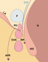
A schematic view of the relationship between the rectum (R) and rectourethralis muscle (RU) and suggested anterior dissection line. Sagittal view depicting the suggested dissection line (red dotted line) of intersphincteric resection (ISR) and abdominoperineal resection (APR) for low rectal cancer. Denonvilliers fascia (DVF) end to the RU. Identifying the RU is key for avoiding damage to the urethra. EAS, external anal sphincter; IAS, internal anal sphincter; LA, levator ani muscle; P, prostate.
Fig. 16.
Fig. 16.
Schematic images. (A) Red dotted lines depict anterolateral dissection behind the Denonvilliers fascia (DVF), and this dissection plane meets with posterolateral pelvic dissection along the parietal pelvic fascia (PPF), while preserving the hypogastric nerve (HGN) and division of rectosacral fascia. (B) The orange dotted line indicates the proposed dissection line. DVF and its lateral border meet with the PPF. Underneath this fascia, the neurovascular bundle (NVB) and HGN are present. IHN; inferior hypogastric nerve; MRF, mesorectal fascia; PP, pelvic plexus; R, rectum; MR, mesorectum; SV, seminal vesicle.
Fig. 17.
Fig. 17.
A schematic image of the Gate approach illustrates one way to obtain a complete total mesorectal excision specimen in a deep narrow pelvic cavity. The red box area depicts a dissection area with the Gate approach. There will be a sharp pelvic dissection between the mesorectal fascia and pelvic floor. PP, pelvic plexus. Adapted and reproduced from Kim et al. [51], available under the Creative Commons License.
Fig. 18.
Fig. 18.
The concept of customized Denonvilliers fascia (DVF) excision. (A) A schematic sagittal view of the pelvis illustrating the anterior dissection plane. The red dotted line depicts the suggested dissection plane for early stage tumor (e.g., T1, T2), preserving the DVF and reducing the risk of nerve damage while ensuring circumferential margin negativity. (B) For more advanced tumor, especailly those anteriorly located, excising the DVF might be necessary to achieve circumferential resection margin negativity. (C) Under third robotic arm upward traction of the seminal vesicle, the whitish membrane-like DVF was exposed, robotic scissor incised it, and sharp dissection proceeded behind the DVF. MR, mesorectum; P, prostate; R, rectum; SV, seminal vesicle.
Fig. 19.
Fig. 19.
The rectal proper fascia adheres to the mesh-like pelvic plexus at the lateral pelvic wall. The fine branches from the pelvic plexus enter the rectal wall.
Fig. 20.
Fig. 20.
The blue dotted line depicts a division line of partial levator ani muscle. On the left side, the part of the levator ani muscle invaded by the tumor is excised; however, on the right side, the levator ani muscle is preserved. DL, dentate line; IAS, internal anal sphincter; EAS, external anal sphincter; ISS, intersphincteric space. Adapted from Varela and Kim [44], available under the Creative Commons License.
Fig. 21.
Fig. 21.
Comparison of rectal magnetic resonance imaging before and after partial excision of the involved levator ani muscle (PELM) shows an absence of right-side levator ani muscle plate. (A) Before PELM, tumor involved the right side of levator ani muscle partially. (B) After PELM, left side levator ani muscle can be identified, while right side of levator ani already excised.
Fig. 22.
Fig. 22.
(A, B) Local recurrence near the anastomosis site after total mesorectal excision. Red arrows indicate recurrence near the anastomosis.

References

    1. Ferlay J, Ervik M, Lam F, Colombet M, Mery L, Piñeros M, et al. International Agency for Research on Cancer (IARC); Global cancer observatory: cancer today [Internet] [cited 2024 Jun 3]. Available from: https://gco.iarc.who.int/today/en.
    1. Siegel RL, Miller KD, Jemal A. Cancer statistics, 2019. CA Cancer J Clin. 2019;69:7–34. - PubMed
    1. Hur H, Oh CM, Won YJ, Oh JH, Kim NK. Characteristics and survival of Korean patients with colorectal cancer based on data from the Korea central cancer registry data. Ann Coloproctol. 2018;34:212–21. - PMC - PubMed
    1. Kang MJ, Jung KW, Bang SH, Choi SH, Park EH, Yun EH, et al. Cancer statistics in Korea: incidence, mortality, survival, and prevalence in 2020. Cancer Res Treat. 2023;55:385–99. - PMC - PubMed
    1. Heald RJ, Husband EM, Ryall RD. The mesorectum in rectal cancer surgery: the clue to pelvic recurrence? Br J Surg. 1982;69:613–6. - PubMed

LinkOut - more resources