. 2024 Jul 25;27(9):110585.
doi: 10.1016/j.isci.2024.110585. eCollection 2024 Sep 20.

Reiterated male-to-female violence disrupts hippocampal estrogen receptor β expression, prompting anxiety-like behavior

Affiliations

Reiterated male-to-female violence disrupts hippocampal estrogen receptor β expression, prompting anxiety-like behavior

Jacopo Agrimi et al. iScience. .

Abstract

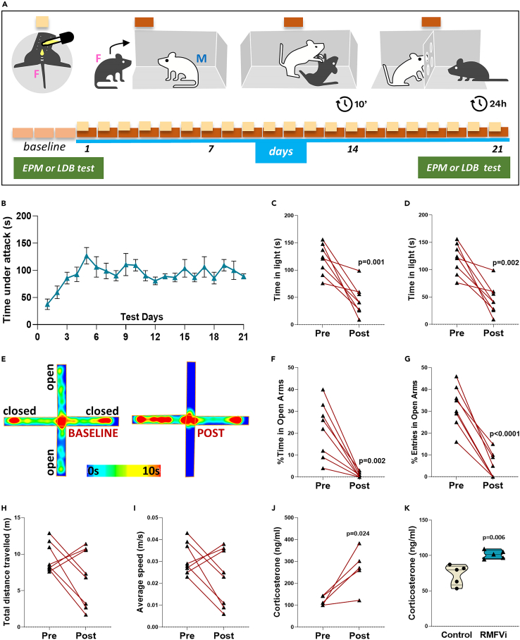
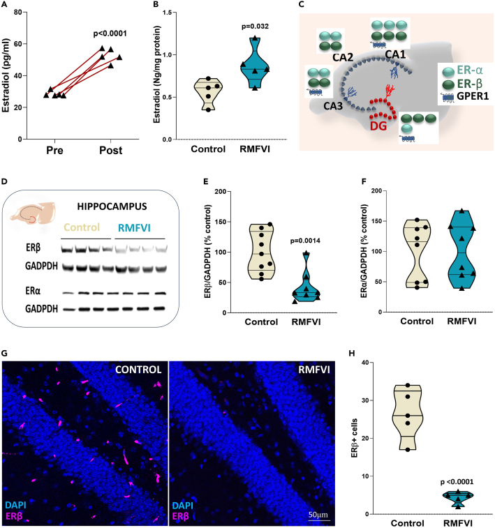
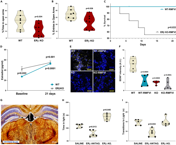
Intimate partner violence (IPV) is a significant public health concern whose neurological/behavioral sequelae remain to be mechanistically explained. Using a mouse model recapitulating an IPV scenario, we evaluated the female brain neuroendocrine alterations produced by a reiterated male-to-female violent interaction (RMFVI). RMFVI prompted anxiety-like behavior in female mice whose hippocampus displayed a marked neuronal loss and hampered neurogenesis, namely reduced BrdU-DCX-positive nuclei and diminished dendritic arborization in the dentate gyrus (DG): effects paralleled by a substantial downregulation of the estrogen receptor β (ERβ). After RMFVI, the DG harbored reduced brain-derived neurotrophic factor (BDNF) pools and tyrosine kinase receptor B (TrkB) phosphorylation. Accordingly, ERβ knockout (KO) mice had heightened anxiety and curtailed BDNF levels at baseline while dying prematurely during the RMFVI procedure. Strikingly, injecting an ERβ antagonist or agonist into the wild-type (WT) female hippocampus enhanced or reduced anxiety, respectively. Thus, reiterated male-to-female violence jeopardizes hippocampal homeostasis, perturbing the ERβ/BDNF axis and ultimately instigating anxiety and chronic stress.

Keywords: Behavioral neuroscience; Molecular neuroscience; Neuroscience.


Conflict of interest statement

The authors declare no conflicts of interests.

Figures

None
Graphical abstract
Figure 1
Figure 1
RMFVI induces anxiety-like behavior (A) Reiterated male-to-female violent interaction (RMFVI), explicative scheme. (B) Daily time under males attack spent by the females during RMFVI procedure. (C and D) Light-dark box test (LDB), (C) number of transitions from the dark to the light compartment, RMFVI vs. control, n = 8, p < 0.0001 (paired t-test), (D) time spent by mice in the light compartment, RMFVI vs. control, n = 8, p < 0.0001 (paired t-test). (E–I) Elevated plus maze test (EPM), (E) heat plot of time spent in different parts of the maze at baseline (left) and after RMFVI (right), (F) percentage of entries in the open arms, pre-RMFVI vs. 21 days RMFVI, n = 8, p < 0.0001 (paired t-test), (G) percentage of time spent in the open arms, pre- RMFVI vs. 21 days RMFVI, n = 8, p = 0.002 (paired t-test), (H) total distance traveled by the animals, pre- RMFVI vs. 21 days RMFVI, n = 8 and (I) average speed of the animal movements, pre- RMFVI vs. 21 days RMFVI, n = 8. (J) Circulating corticosterone, pre-RMFVI vs. 21 days RMFVI, n = 5, p = 0.024 (Wilcoxon signed rank test). (K) Central corticosterone, pre-RMFVI vs. 21 days RMFVI, n = 5, p = 0.006 (Mann-Whitney U test).
Figure 2
Figure 2
RMFVI triggers apoptosis while impairing neurogenesis in the DG (A) Scheme of the hippocampal formation morphology redrawn from Toda T. et al. (B) Representative images of IF with TUNEL assay in the DG of controls and RMFVI-inflicted mice. (C) TUNEL positive nuclei quantification, control vs. RMFVI, n = 5, p = 0.08 (Mann-Whitney U test). (D) Representative images of IF staining for DAPI, BrdU, and DCX in the DG of controls and RMFVI-inflicted mice. (E) BrdU/DCX positive cells quantification, control vs. RMFVI, n = 5, p = 0.08 (Mann-Whitney U test). (F) Representative scheme of DCX positive cell differentiation. (G) Representative images of IF staining for DAPI and DCX in the DG of controls and RMFVI-inflicted mice. (H) DCX positive cells quantification, control vs. RMFVI, n = 5. (I) DCX positive cells (classified as postmitotic) quantification, control vs. RMFVI, n = 5, p = 0.039 (Mann-Whitney U test). (J) Percentage of DCX postmitotic positive cells (on total DCX positive cells), control vs. RMFVI, n = 5, p = 0.008 (Mann-Whitney U test). (K) Average dendrite length of DCX postmitotic positive cells, control vs. RMFVI, n = 5, p = 0.008 (Mann-Whitney U test).
Figure 3
Figure 3
RMFVI jeopardizes ERβ expression in the female hippocampus (A) Circulating 17-beta-estradiol measured by ELISA at baseline and after 21 days of RMFVI, n = 5, p < 0.0001 (Wilcoxon signed rank test). (B) 17-beta-estradiol measured by ELISA in lysates of whole brain from controls and RMFVI-exacted mice, n = 5, p = 0.022 (Mann-Whitney U test). (C) Scheme of estrogen receptor expression in different hippocampal areas. (D–F) Western blot for ERα and ERβ in the hippocampus of controls and RMFVI-imposed mice, (D) representative immunoblotting in the hippocampus for ERα, Erβ, and GAPDH; (E) quantification of ERβ (normalized to GADPDH content), control vs. RMFVI, n = 8, p = 0.0014 (unpaired t-tet); (F) quantification of ERα (normalized to GADPDH content), control vs. RMFVI, n = 8. (G) Representative images of IF staining for ERβ in the DG, control, and RMFVI-inflicted mouse. (H) Quantification of IF staining for ERβ in the DG, control vs. RMFVI, n = 5, p < 0.0001 (Mann-Whitney U test).
Figure 4
Figure 4
RMFVI alters BDNF/TrkB signaling in the DG (A) Representative images of IF staining for P-ERK in the DG, control, and RMFVI mice. (B) Quantification of IF staining for P-ERK+ cells in the DG, control vs. RMFVI, n = 5, p = 0.0079 (Mann-Whitney U test). (C) Representative images of IF staining for BDNF in the DG, control, and RMFVI-inflicted mice. (D) Quantification of IF staining for BDNF in the DG, control vs. RMFVI, n = 5, p = 0.008 (Mann-Whitney U test). (E) Representative images of IF staining for P-TrkB in the DG, control, and RMFVI mice. (F) Quantification of IF staining for P-TrkB in the DG, control vs. RMFVI, n = 5, p = 0.0079 (Mann-Whitney U test).
Figure 5
Figure 5
Manipulation of ERβ signaling reproduces RMFVI-ignited behavioral perturbations and estradiol and BDNF decline (A and B) Elevated plus maze test for WT and ERβ KO, (A) percentage of time spent in the open arms from WT and ERβ KO mice, n = 8, p = 0.034 (unpaired t-test) (B) percentage of entries in the open arms from WT and ERβ KO mice, n = 8, p = 0.026 (unpaired t-test). (C) Survival Kaplan-Meir curves of WT and ERβ KO mice during RMFVI procedure, n = 9, p = 0.023. (D) Circulating 17-beta-estradiol measured by ELISA at baseline and after 21 days RMFVI in WT and ERβ KO mice, n = 5, baseline WT vs. baseline ERβ KO mice p = 0.033, WT baseline vs. 21 days RMFVI, p < 0.0001, ERβ KO mice baseline vs. 21 days RMFVI, p = 0.001 (Friedman's test). (E) Representative images of IF staining for BDNF in the DG (hilar cells), WT, and ERβ KO mice before and after RMFVI. (F) Quantification of IF staining for BDNF in the DG, control WTs vs. ERβ KO mice, before and after RMFVI, n = 5; WT vs. WT-RMFVI, p = 0.0004, WT vs. KO, p = 0.0002, WT vs. KO-RMFVI p < 0.0001 (Kruskal-Wallis one-way test). (G) Stereotaxic injection in the hippocampal formation: site of injection stained with trypan blue. (H) Light-dark box test, time spent by mice in the light compartment, n = 5; saline vs. PHTPP (antagonist), p = 0.013; Saline vs. DPN (agonist), p = 0.045 (Kruskal-Wallis one-way test). (I) Light-dark box test, transition to the light compartment, n = 5; saline vs. PHTPP (antagonist), p = 0.033 (Kruskal-Wallis one-way test).

Similar articles

Cited by

References

    1. Miller E., McCaw B. Intimate Partner Violence. N. Engl. J. Med. 2019;380:850–857. doi: 10.1056/NEJMra1807166. - DOI - PubMed
    1. Barbier A., Chariot P., Lefèvre T. Intimate partner violence against ever-partnered women in Europe: Prevalence and associated factors—Results from the violence against women EU-wide survey. Front. Public Health. 2022;10 doi: 10.3389/fpubh.2022.1033465. - DOI - PMC - PubMed
    1. Oram S., Fisher H.L., Minnis H., Seedat S., Walby S., Hegarty K., Rouf K., Angénieux C., Callard F., Chandra P.S., et al. The Lancet Psychiatry Commission on intimate partner violence and mental health: advancing mental health services, research, and policy. Lancet Psychiatr. 2022;9:487–524. doi: 10.1016/S2215-0366(22)00008-6. - DOI - PubMed
    1. Harris A.Z., Atsak P., Bretton Z.H., Holt E.S., Alam R., Morton M.P., Abbas A.I., Leonardo E.D., Bolkan S.S., Hen R., Gordon J.A. A Novel Method for Chronic Social Defeat Stress in Female Mice. Neuropsychopharmacology. 2018;43:1276–1283. doi: 10.1038/npp.2017.259. - DOI - PMC - PubMed
    1. Anacker C., Hen R. Adult hippocampal neurogenesis and cognitive flexibility — linking memory and mood. Nat. Rev. Neurosci. 2017;18:335–346. doi: 10.1038/nrn.2017.45. - DOI - PMC - PubMed