

Case Reports
doi: 10.1016/j.radcr.2024.07.108.
eCollection 2024 Nov.
Chronic invasive fungal sinusitis mimicking pseudotumor: A case report
Affiliations
- PMID: 39228933
- PMCID: PMC11366909
- DOI: 10.1016/j.radcr.2024.07.108
Item in Clipboard
Case Reports
Chronic invasive fungal sinusitis mimicking pseudotumor: A case report
Radiol Case Rep.
.
Abstract
Fungal sinusitis encompasses a wide range of diseases, including both invasive and noninvasive, acute, and chronic forms. The incidence of invasive sinusitis is on the rise, primarily affecting immunocompromised individuals and diabetics. This case report highlights a patient who developed invasive fungal sinusitis despite no other apparent cause of immunosuppression. Imaging studies suggested the diagnosis, confirmed by presence of Aspergillus flavus on mycological culture.
Keywords: Aspergillus flavus; CT; Invasive fungal sinusitis; MRI.
© 2024 The Authors. Published by Elsevier Inc. on behalf of University of Washington.
Figures
Axial slice CT scan in parenchymal window demonstrating heterogeneous spontaneously hyperdense filling, obstructing the left nasosinus cavities. The mass exerts pressure on the nasal septum, displacing it to the right, and contributes to grade I left exophthalmos.

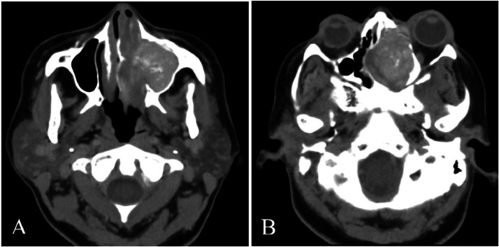
CT axial section in parenchymal window (A) and a coronal reconstruction in bone window (B). It demonstrates the filling of the left nasosinus cavities with calcifications, associated with bone lysis affecting the sinus walls and the roof of the left orbit.

MRI in coronal T2 FATSAT sequence with signal voids within the frontal and left maxillary sinus opacities (red arrows) (A), and an axial T1 FATSAT sequence post-Gadolinium injection demonstrating contrast enhancement (blue arrow) (B).
References
-
- Lassausaie A, Barthélémy I. Rhinosinusites fongiques des sinus maxillaires. Chirurgie Orale Et Maxillo-Faciale. 2021;34(1):1–11.
-
- Raz E, Win W, Hagiwara M, Lui YW, Cohen B, Fatterpekar GM. Fungal sinusitis. Neuroimaging Clin N Am. 2015;25(4):569–576. - PubMed
Publication types
LinkOut - more resources
Full Text Sources
