[Preprint]. 2024 Aug 23:2024.08.22.609143.
doi: 10.1101/2024.08.22.609143.

Multiplexed Glycan Immunofluorescence Identification of Pancreatic Cancer Cell Subpopulations in Both Tumor and Blood Samples

Affiliations

Multiplexed Glycan Immunofluorescence Identification of Pancreatic Cancer Cell Subpopulations in Both Tumor and Blood Samples

Braelyn Binkowski et al. bioRxiv. .

Update in

Abstract

Pancreatic ductal adenocarcinoma (PDAC) tumor heterogeneity impedes the development of biomarker assays suitable for early disease detection that would improve patient outcomes. The CA19-9 glycan is currently used as a standalone biomarker for PDAC. Furthermore, previous studies have shown that cancer cells may display aberrant membrane-associated glycans. We therefore hypothesized that PDAC cancer cell subpopulations could be distinguished by aberrant glycan signatures. We used multiplexed glycan immunofluorescence combined with pathologist annotation and automated image processing to distinguish between PDAC cancer cell subpopulations within tumor tissue. Using a training-set/test-set approach, we found that PDAC cancer cells may be identified by signatures comprising 4 aberrant glycans (VVL, CA19-9, sTRA, and GM2) and that there are three glycan-defined PDAC tumor types: sTRA type, CA19-9 type, and intermixed. To determine whether the aberrant glycan signatures could be detected in blood samples, we developed hybrid glycan sandwich assays for membrane-associated glycans. In both patient-matched tumor and blood samples, the proportion of aberrant glycans detected was consistent. Furthermore, our multiplexed glycan immunofluorescent approach proved to be more sensitive and more specific than CA19-9 alone. Our results provide proof of concept for a novel methodology to improve early PDAC detection and patient outcomes.


Conflict of interest statement

Conflict-of-interest statement: The authors have declared that no conflict of interest exists.

Figures
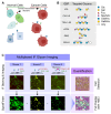
Figure 1.
Figure 1.
Identification and quantification of cellular glycan signatures. A) Model of cell surface glycan signatures. B) Glycan-binding proteins (GBP) and their targets. C) Multiplexed immunofluorescence with automated signal thresholding by SignalFinder, followed by assignments of glycan signatures to individual cells.
Figure 2.
Figure 2.
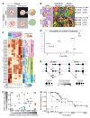
Training set identification of cancer-association glycan signatures. A) Training design. B) Quantification of cells with each glycan signature in ROIs. C) Distributions of cell surface glycan signatures among acinar and PDAC cells. D) Volcano plots of pairwise comparisons between cell types. E) Cancer-associated and non-cancer-associated glycan signatures. F) Representative images and quantifications of non-cancer and cancer ROIs. The asterisk marks normal islet cells and arrowhead marks a normal duct.
Figure 3.
Figure 3.
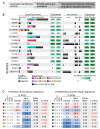
Test set validation of PDAC-associated glycan signatures. A) Test set design. B) Patterns of positivity of the putative cancer-associated glycan signatures compared to CA19-9 in individual ROIs in each specimen. C) Prevalence and association with cancer of the predicted cancer-associated (left) and non-cancer-associated glycan signatures.
Figure 4.
Figure 4.
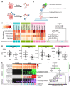
Unequal distribution of glycan-defined subpopulations among spatial clusters and tumors. A) Cluster and tumor heterogeneity. Clusters may be classified as uniform or mixed, and tumors may be classified as heterogeneous or heterogeneous. B) Two examples of within-cluster subpopulation purity, or uniformity, in cancer-cell clusters. C) Tumor type classifications based on glycan-signature type—CA19-9 exclusive, sTRA exclusive, or dual expressing—present in the tumor. D) Probability of association with pure, or uniform, clusters. E) Apparent lineages of glycan-defined PDAC cell subpopulations in the cell clusters and tumors. F) Relationship between average subpopulations purity of the clusters and the average centroid differences between clusters within each tumor. Each data point is a tumor, color coded by tumor type from panel C. The dashed lines are the median values in each axis. G) Kaplan-Meijer plot of PDAC patients in the different groups defined in panel F.
Figure 5.
Figure 5.
Complementary detection of tumors with divergent PDAC subpopulations using plasma assays. A) Model of co-secretion of glycans. B) Hybrid glycan sandwich assays. C) Correspondence of combined plasma assays to tumor types. D) Cancer-associated relative fluorescent unit (RFU) elevations in individual assays. E) Complementary detection of PDACs. F) Classification and stratification decision tree.

References

    1. Siegel RL, et al. Cancer statistics, 2023. CA: A Cancer J Clin. 2023;73(1):17–48. - PubMed
    1. Collisson EA, et al. Subtypes of Pancreatic Ductal Adenocarcinoma and Their Differing Responses to Therapy. Nat Med. 2011;17(4):500–503. - PMC - PubMed
    1. Moffitt RA, et al. Virtual microdissection identifies distinct tumor- and stroma-specific subtypes of pancreatic ductal adenocarcinoma. Nat Genet. 2015;47(10):1168–1178. - PMC - PubMed
    1. Bailey P, et al. Genomic analyses identify molecular subtypes of pancreatic cancer. Nature. 2016;531(7592):47–52. - PubMed
    1. Chan-Seng-Yue M, et al. Transcription phenotypes of pancreatic cancer are driven by genomic events during tumor evolution. Nat Genet. 2020;52(2):231–240. - PubMed

Publication types

LinkOut - more resources