[Preprint]. 2024 Aug 24:2024.08.23.609423.
doi: 10.1101/2024.08.23.609423.

Non-canonical Wnt signaling triggered by WNT2B drives adrenal aldosterone production

Affiliations

Non-canonical Wnt signaling triggered by WNT2B drives adrenal aldosterone production

Kleiton S Borges et al. bioRxiv. .

Abstract

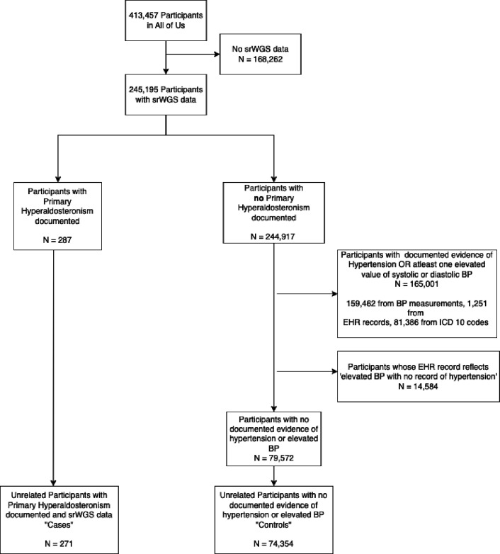
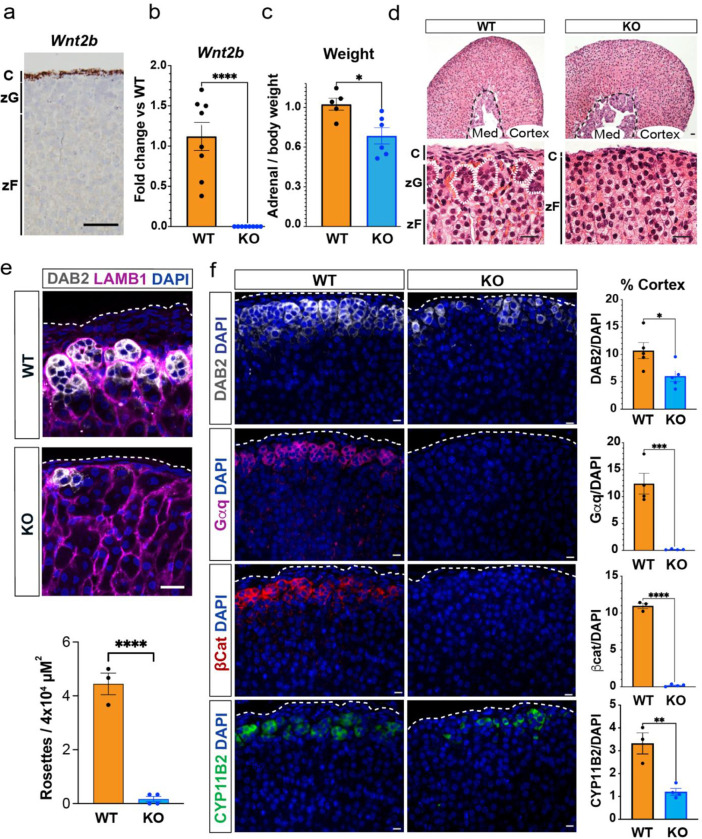
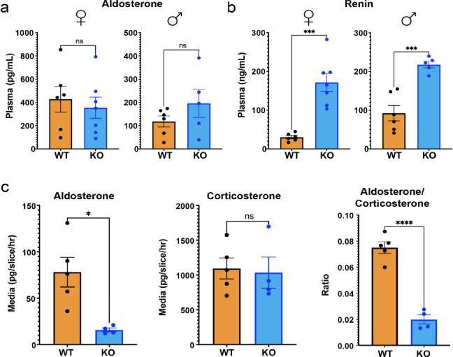
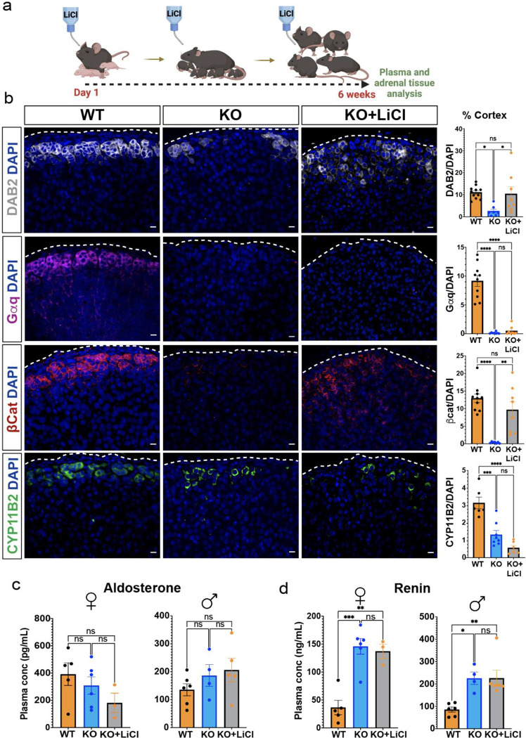
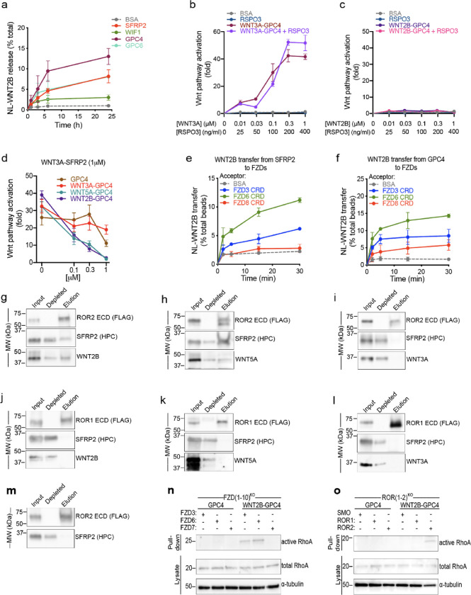
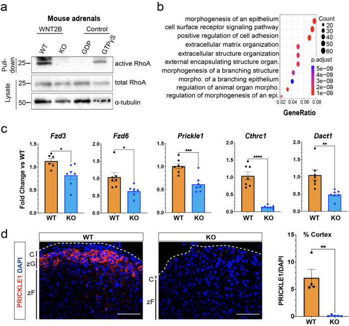
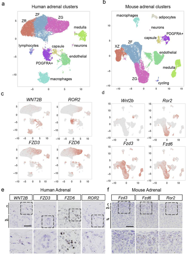
The steroid hormone aldosterone, produced by the zona glomerulosa (zG) of the adrenal gland, is a master regulator of plasma electrolytes and blood pressure. While aldosterone control by the renin-angiotensin system is well understood, other key regulatory factors have remained elusive. Here, we replicated a prior association between a non-coding variant in WNT2B and an increased risk of primary aldosteronism, a prevalent and debilitating disease caused by excessive aldosterone production. We further show that in both mice and humans, WNT2B is expressed in the mesenchymal capsule surrounding the adrenal cortex, in close proximity to the zG. Global loss of Wnt2b in the mouse results in a dysmorphic and hypocellular zG, with impaired aldosterone production. Similarly, humans harboring WNT2B loss-of-function mutations develop a novel form of Familial Hyperreninemic Hypoaldosteronism, designated here as Type 4. Additionally, we demonstrate that WNT2B signals by activating the non-canonical Wnt/planar cell polarity pathway. Our findings identify WNT2B as a key regulator of zG function and aldosterone production with important clinical implications.

Keywords: Familial Hyperreninemic Hypoaldosteronism; WNT2B; Wnt/PCP pathway; adrenal cortex; beta-catenin-independent signaling; hypoaldosteronism; non-canonical Wnt signaling; primary aldosteronism; rosette; zona glomerulosa.


Figures

Figure A1:
Figure A1:
Inclusion / Exclusion Criteria for the Study
Figure 1.
Figure 1.. WNT2B deficiency results in a dysmorphic zG in mice.
a. Representative image of Wnt2b expression using single molecule in situ hybridization (smISH) from an adult female adrenal. Scale bar: 100μm. C, capsule; zG, zona glomerulosa; zF, zona fasciculata. b. QRT-PCR was performed on WT and KO female adrenals (n=8 WT, n=8 KO). Two-tailed Student’s t-test. ****p < 0.0001. Data are represented as mean ± SEM. c. Adrenal weight normalized to body weight from female mice (n=5 WT, n=6 KO). Two-tailed Student’s t-test. *p < 0.05. Data are represented as mean fold change ± SEM. d. Representative H&E images of WT and KO female adrenals. Scale bar: 10μm. Dotted white line delineates rosette structures. C, capsule; zG, zona glomerulosa; zF, zona fasciculata. Med, medulla. e. Representative image and quantification of immunohistochemistry from WT and KO female adrenals stained for Laminin β1 (LAMB1, magenta), indicating the basement membrane surrounding distinct clusters of zG cells (DAB2, gray; DAPI, blue). Scale bar: 20μm. Staining delineates individual glomeruli, highlighting the loss of rosettes in KO adrenals. The number of DAB2+ clusters containing ≥5 cells (n=3 WT, n=4 KO). Two-tailed Student’s t-test. ****p < 0.0001. Data are represented as mean ± SEM. f. Representative images and quantification from female adrenals immunostained for DAB2 (gray, n=5 WT, n=5 KO), Gαq (magenta, n=4 WT, n=4 KO), β-catenin (β-cat, red, n=3 WT, n=4 KO) and CYP11B2 (green, n=3 WT, n=4 KO). Positive cells were quantified and normalized to nuclei (DAPI, blue) in the cortex. Scale bars: 10μm. Two-tailed Student’s t-test. *p < 0.05; **p < 0.01; ***p < 0.001; ****p < 0.0001. Data are represented as mean ± SEM.
Figure 2.
Figure 2.. WNT2B deficiency results in hypoaldosteronism in mice.
a. Quantification of plasma aldosterone levels (female, n=6 WT, n=7 KO; male, n=6 WT, n=5 KO). Two-tailed Student’s t-test. ns, not significant. Data are represented as mean ± SEM. b. Quantification of plasma renin levels (female, n=6 WT, n=7 KO; male n=6 WT, n=5 KO). Two-tailed Student’s t-test. ***p < 0.001. Data are represented as mean ± SEM. c. Aldosterone, corticosterone and aldosterone/corticosterone ratios produced from male adrenal slice preparations, ex vivo, plotted as mean of each mouse (n=5 WT, n=4 KO). Two-tailed Student’s t-test. *p < 0.05, ****p < 0.0001, ns, not significant. Data are represented as mean ± SEM.
Figure 3.
Figure 3.. Activation of β-catenin partially rescues zG morphology, but not function, in Wnt2b KO mice.
a. Treatment protocol with 0.06% lithium chloride (LiCl) or water from birth to 6 weeks of age. b. Representative images and quantification from 6-week-old adrenals stained for DAB2 (gray, n=11 WT, n=7 KO, n=8 KO+LiCl), Gαq (magenta, n=10 WT, n=7 KO, n=8 KO+LiCl), β-catenin (β-cat, red, n=10 WT, n=6 KO, n=8 KO+LiCl) and CYP11B2 (green, n=6 WT, n=8 KO, n=6 KO+LiCl) mice. Positive cells were quantified and normalized to nuclei (DAPI, blue) in the cortex. Scale bars: 10μm. One-way ANOVA with Tukey’s post-test. ns, not significant; *p < 0.05; **p < 0.01; ***p < 0.001; ****p < 0.0001. Data are represented as mean ± SEM. c, d. Quantification of (c) aldosterone levels from (female, n=5 WT, n=6 KO, n=3 KO+LiCl; male, n=6 WT, n=4 KO, n=5 KO+LiCl) and (d) plasma renin (female, n=5 WT, n=6 KO, n=3 KO+LiCl; male, n=6 WT, n=4 KO, n=6 KO+LiCl) mice. Data are represented as mean ± SEM. One-way ANOVA with Tukey’s post-test. ns, not significant; *p < 0.05; **p < 0.01; ***p < 0.001. Data are represented as mean ± SEM.
Figure 4.
Figure 4.. WNT2B released by SFRP2 and GPC4 activates the non-canonical Wnt/PCP pathway by binding to FZD3-CRD or FZD6-CRD, and ROR2-ECD.
a. HEK293 cells stably expressing NL-WNT2B were incubated with 1μM of purified SFRP2, WIF1, GPC4 or GPC6 ectodomains in serum-free media. NL-WNT2B release was measured at various time points by NanoLuc luciferase (NL) luminescence. Bovine serum albumin (BSA) served as negative control. WNT2B is released mainly by SFRP2, GPC4 and GPC6. Data represent the mean of two biological replicates, normalized to total NL-WNT in lysates, and error bars show SD. b. R-Spondin 3 (RSPO3; 0, 25, 100, 200 and 400ng/ml) or purified WNT3A-GPC4 complex (0, 0.01, 0.03, 0.1, 0.3 and 1μM with respect to WNT3A) with or without RSPO3 (400ng/ml) was added to Wnt reporter cells. After 24h, Wnt pathway activity was measured by luciferase assay. Incubation with BSA served as negative control. RSPO3 does not potentiate WNT3A-GPC4 activity. Points represent average activation for two biological replicates, normalized to untreated cells, and error bars represent SD. See also Supplemental Figure 4a–e for protein purification and activity of WNT5A-GPC4 complex and WNT3A-carrier or WNT2B-carrier conditioned media. c. As in (b), but with purified WNT2B-GPC4 complex. WNT2B-GPC4 complex is unable to activate canonical Wnt signaling, even with RSPO3. d. As in (b), but purified WNT3A-SFRP2 complex (1μM) was mixed with varying amounts of GPC4 alone or in complex with WNT3A, WNT5A or WNT2B (0.1, 0.3 and 1μM). Both WNT5A-GPC4 and WNT2B-GPC4 complexes abolish WNT3A-SFRP2 activity, in contrast to GPC4 alone or in complex with WNT3A. See Supplemental Figure 4f for a similar experiment using WNT3A-GPC4 complex. e. NL-WNT2B-SFRP2 complex was covalently captured on HaloLink beads from conditioned media, via HT7 fused to the C-terminus of SFRP2. The beads were then incubated with purified FZD-CRDs (5μM) and NL-WNT2B release was measured at different time points by NL luminescence. Incubation with BSA (5μM) served as negative control. WNT2B is preferentially transferred to FZD3-CRD and FZD6-CRD more than FZD8-CRD. Points represent average for two biological replicates, normalized by total NL-WNT on beads, and error bars represent SD. f. As in (e), but with NL-WNT2B-GPC4 on beads. g. Purified WNT2B-SFRP2 (5μM) was incubated with the extracellular domain (ECD) of ROR2 (2.5μM), followed by immunoprecipitation with antibodies against the FLAG tag attached to ROR. Samples were analyzed by SDS-PAGE and immunoblotting. WNT2B-SFRP2 complex interacts with ROR2-ECD. See also Supplemental Figure 4g–j for protein purification and a similar experiment using purified SFRP2. h. As in (g), but with purified WNT5A-SFRP2 complex. WNT5A-SFRP2 complex binds to ROR2-ECD. i. As in (g), but with purified WNT3A-SFRP2 complex. WNT3A-SFRP2 complex does not bind to ROR2-ECD. j. As in (g), but WNT2B-SFRP2 complex (5μM) was incubated with ROR1-ECD (2.5μM). WNT2B-SFRP2 does not bind to ROR1-ECD. k. As in (j), but with WNT5A-SFRP2 complex. WNT5A-SFRP2 does not bind to ROR1-ECD. l. As in (j), but with WNT3A-SFRP2 complex. WNT3A-SFRP2 does not bind to ROR1-ECD. m. As in (g) but using SFRP2 alone. SFRP2 is unable to interact with ROR2-ECD. n. Activity of RhoA in FZD(1–10)KO cells expressing FZD3, FZD6 or FZD7 was assessed by Rhotekin-RBD pull-down assay after 6h of treatment with GPC4 alone or in complex with WNT2B (2μM). RhoA endogenous levels are shown in the lysates. RhoA activity by WNT2B-GPC4, in contrast to GPC4 alone, is rescued in cells expressing FZD3 or FZD6, but not the canonical FZD7. Blotting for α-tubulin served as loading control. o. As in (n), but measuring activity of RhoA in ROR(1–2)KO cells expressing ROR1 or ROR2. WNT2B-GPC4, in contrast to GPC4 alone, activates RhoA only when ROR2 expression is rescued, not ROR1. Smoothened (SMO) transfection served as negative control.
Figure 5.
Figure 5.. WNT2B deficiency disrupts Wnt/PCP signaling in the adrenal.
a. Activity of RhoA in WT and KO adrenals was assessed by Rhotekin-RBD pull-down assay using adrenal lysates. GTPγS and GDP treated adrenal lysates served as positive and negative controls, respectively. Total RhoA and α-tubulin served as loading controls. b. Dot plot depicting Gene Ontology (GO) Gene Set enrichment analysis of genes downregulated in KO vs WT. c. QRT-PCR was performed in WT and KO adrenals for Fzd3 (n=6 WT, n=7 KO), Fzd6 (n=7 WT, n=7 KO), Prickle1 (n=7 WT, n=7 KO), Cthrc1 (n=7 WT, n=7 KO) and Dact1 (n=7 WT, n=7 KO) from female mice. Two-tailed Student’s t-test. *p<0.05; **p < 0.01 ***p < 0.001; ***p < 0.0001. Data are represented as mean ± SEM. d. Representative images and quantification from adrenals stained for PRICKLE1 (red, n=4 WT, n=5 KO). Positive cells were quantified and normalized to nuclei (DAPI, blue) in the cortex. Scale bars: 50μm. Two-tailed Student’s t-test. **p < 0.01. Data are represented as mean ± SEM. C, capsule; zG, zona glomerulosa; zF, zona fasciculata.
Figure 6.
Figure 6.. Components of Wnt/PCP signaling are conserved across mouse and human adrenals.
UMAP plots of snRNAseq from human (a) and mouse (b) adrenals depicting similarly diverse cell types including cortical and non-cortical cells. Expression patterns of WNT2B, ROR2, FZD3 and FZD6 projected over the UMAP projections from human (c), and mouse (d) adrenals. e. Representative smISH images of WNT2B, FZD3, FZD6 and ROR2 expression in the adrenal cortex of human adrenals. Scale bar: 25μm f. Representative smISH images of Fzd3, Fzd6 and Ror2 expression in the adrenal cortex of mouse adrenals. Scale bar: 25μm

References

    1. Hammer GD, Basham KJ. Stem cell function and plasticity in the normal physiology of the adrenal cortex. Mol Cell Endocrinol. 2021;519:111043. - PMC - PubMed
    1. Zhu Y, Zhang X, Hu C. Structure of rosettes in the zona glomerulosa of human adrenal cortex. J Anat. 2023;243(4):684–689. - PMC - PubMed
    1. Leng S, et al. β-Catenin and FGFR2 regulate postnatal rosette-based adrenocortical morphogenesis. Nat Commun. 2020;11(1):1680. - PMC - PubMed
    1. Zelander T. The ultrastructure of the adrenal cortex of the mouse. Zeitschrift für Zellforschung und Mikroskopische Anatomie. 1957;46(6):710–716. - PubMed
    1. Freedman BD, et al. Adrenocortical Zonation Results from Lineage Conversion of Differentiated Zona Glomerulosa Cells. Dev Cell. 2013;26(6):666–673. - PMC - PubMed

Publication types

LinkOut - more resources