. 2024 Aug 20;6(5):fcae282.
doi: 10.1093/braincomms/fcae282. eCollection 2024.

C9orf72 dipeptides activate the NLRP3 inflammasome

Affiliations

C9orf72 dipeptides activate the NLRP3 inflammasome

Jack Rivers-Auty et al. Brain Commun. .

Abstract

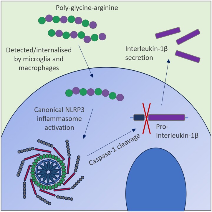
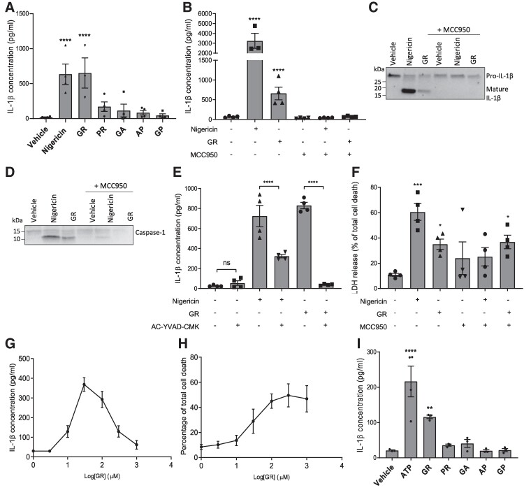
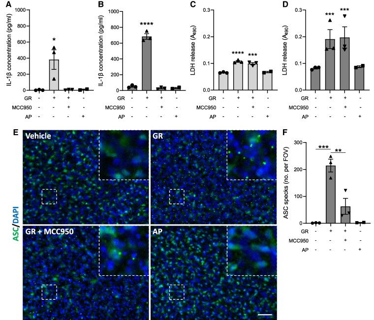
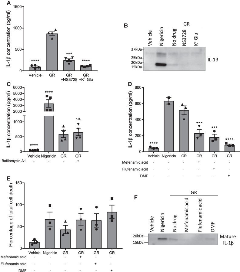
Frontotemporal dementia and amyotrophic lateral sclerosis are neurodegenerative diseases with considerable clinical, genetic and pathological overlap. The most common cause of both diseases is a hexanucleotide repeat expansion in C9orf72. The expansion is translated to produce five toxic dipeptides, which aggregate in patient brain. Neuroinflammation is a feature of frontotemporal dementia and amyotrophic lateral sclerosis; however, its causes are unknown. The nod-like receptor family, pyrin domain-containing 3 inflammasome is implicated in several other neurodegenerative diseases as a driver of damaging inflammation. The inflammasome is a multi-protein complex which forms in immune cells in response to tissue damage, pathogens or aggregating proteins. Inflammasome activation is observed in models of other neurodegenerative diseases such as Alzheimer's disease, and inflammasome inhibition rescues cognitive decline in rodent models of Alzheimer's disease. Here, we show that a dipeptide arising from the C9orf72 expansion, poly-glycine-arginine, activated the inflammasome in microglia and macrophages, leading to secretion of the pro-inflammatory cytokine, interleukin-1β. Poly-glycine-arginine also activated the inflammasome in organotypic hippocampal slice cultures, and immunofluorescence imaging demonstrated formation of inflammasome specks in response to poly-glycine-arginine. Several clinically available anti-inflammatory drugs rescued poly-glycine-arginine-induced inflammasome activation. These data suggest that C9orf72 dipeptides contribute to the neuroinflammation observed in patients, and highlight the inflammasome as a potential therapeutic target for frontotemporal dementia and amyotrophic lateral sclerosis.

Keywords: ALS; NLRP3; c9orf72; frontotemporal dementia; inflammasome.


Conflict of interest statement

The authors have no competing interests to declare.

Figures

Graphical Abstract
Graphical Abstract
Figure 1
Figure 1
GR activates the NLRP3 inflammasome in macrophages and microglia. (A) LPS-primed primary peritoneal macrophages from male and female WT mice were treated with 30 μM DPRs or controls for 24 h (n = 4). IL-1β was measured in the media by ELISA. (B) GR increased IL-1β release in LPS-primed BMDMs (n = 4) and was prevented by MCC950. (C) Western blot of culture media from GR-treated bone marrow-derived macrophages (BMDMs) showing pro-IL-1β cleavage to produce an active fragment (∼17 kDa). Nigericin was used as a positive control (n = 3). (D) Western blot of culture media from GR-treated bone marrow-derived macrophages (BMDMs) showing pro-caspase-1 cleavage to produce an active fragment (∼10 kDa). Nigericin was used as a positive control (n = 3). (E) GR-induced IL-1β secretion from LPS-primed BMDMs was prevented by AC-YVAD-CMK pre-treatment (n = 4). (F) Quantification of cell death in GR-treated BMDMs by LDH assay, 24 h post-treatment. (G) Dose–response curve of IL-1β detected in BMDM culture media 24 h after treatment with increasing concentrations of GR. (H) Dose–response curve of BMDM cell death measured by LDH assay, 24 h after treatment with increasing concentrations of GR. (I) IL-1β levels in culture media of LPS-primed primary WT mouse microglia 24 h post-treatment with 30 μM DPRs or 5 mM ATP (n = 3). All data analysed by one-way ANOVA with post hoc Dunnett’s or Tukey’s tests for multiple comparisons. **** indicates P < 0.0001, *** indicates P < 0.001, and ** indicates P 0.01. All values are mean ± SEM. Individual mice were considered the experimental unit. See Supplementary Fig. 1 for uncropped blots.
Figure 2
Figure 2
GR activates the NLRP3 inflammasome in hippocampal slice cultures. Mouse hippocampal slice cultures (from male and female mice) were LPS-primed and treated with GR ± MCC950 for 24 h (n = 3). AP treatment was also performed (n = 2). IL-1β concentrations in media collected from hippocampal slices 4 h (A) and 24 h (B) post-treatment. Cell death measured by LDH assay of media collected from hippocampal slices 4 h (C) and 24 h (D) post-treatment. (E) Immunofluorescence imaging of ASC in hippocampal slices. ASC specks are visible as intense puncta in the cytosol of cells in GR-treated hippocampal slices (top right). (F) Quantification of total ASC specks per field of vision. All values are mean ± SEM. Data were analysed by one-way ANOVA with post hoc Dunnett’s or Tukey’s tests for multiple comparisons. The scale bars represent 50 µm. Individual mice were considered the experimental unit.
Figure 3
Figure 3
Characterization of GR-induced NLRP3 inflammasome activation. (A), (B) LPS-primed primary mouse BMDMs (from male and female mice) were pre-treated for 15 min with a chloride channel inhibitor, NS3728, or potassium gluconate (K+ Glu) prior to a 24-h treatment with GR. IL-1β concentrations in the culture media were measured by ELISA (n = 4) (A) and cleavage of pro-IL-1β assessed by WB of the media (n = 3) (B). C LPS-primed BMDMs were pre-treated for 15 min with bafilomycin A1 or vehicle prior to a 24-h treatment with GR, and IL-1β secretion quantified by ELISA (n = 4). (D)(F) LPS-primed mouse primary BMDMs were pre-treated for 15 min with one of three clinically approved drugs which are known NLRP3 inhibitors: mefenamic acid, flufenamic acid, and dimethyl fumarate (DMF), before a 24-h treatment with GR. (D) Mean IL-1β concentrations in the culture media as detected by ELISA (n = 3). (E) Quantification of cell death in BMDMs assessed by LDH assay (n = 3). (F) Western blotting of the culture media showing cleavage of pro-IL-1β to produce mature IL-1β (17 kDa) (n = 3). All data were analysed by one-way ANOVA with post hoc Dunnett’s or Tukey’s tests for multiple comparisons. The error bars indicate SEM. Individual mice were considered the experimental unit. See Supplementary Fig. 2 for uncropped blots.

References

    1. Renton AE, Majounie E, Waite A, et al. A hexanucleotide repeat expansion in C9ORF72 is the cause of chromosome 9p21-linked ALS-FTD. Neuron. 2011;72(2):257–268. - PMC - PubMed
    1. DeJesus-Hernandez M, Mackenzie IR, Boeve BF, et al. Expanded GGGGCC hexanucleotide repeat in noncoding region of C9ORF72 causes chromosome 9p-linked FTD and ALS. Neuron. 2011;72(2):245–256. - PMC - PubMed
    1. Mori K, Weng SM, Arzberger T, et al. The C9orf72 GGGGCC repeat is translated into aggregating dipeptide-repeat proteins in FTLD/ALS. Science. 2013;339(6125):1335–1338. - PubMed
    1. Ash PEA, Bieniek KF, Gendron TF, et al. Unconventional translation of C9ORF72 GGGGCC expansion generates insoluble polypeptides specific to c9FTD/ALS. Neuron. 2013;77(4):639–646. - PMC - PubMed
    1. Callister B, Ryan J, Sim S, Rollinson J, Pickering-Brown S, M S. Modelling C9orf72 dipeptide repeat proteins of a physiologically relevant size. Hum Mol Genet. 2016;25(23):5069. - PMC - PubMed

LinkOut - more resources