

Atlas-based assessment of hypomyelination: Quantitative MRI in Pelizaeus-Merzbacher disease
- PMID: 39230009
- PMCID: PMC11372820
- DOI: 10.1002/hbm.70014
Atlas-based assessment of hypomyelination: Quantitative MRI in Pelizaeus-Merzbacher disease
Abstract
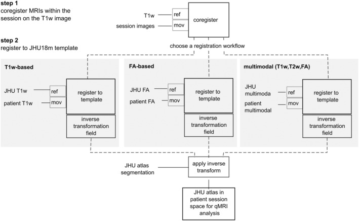
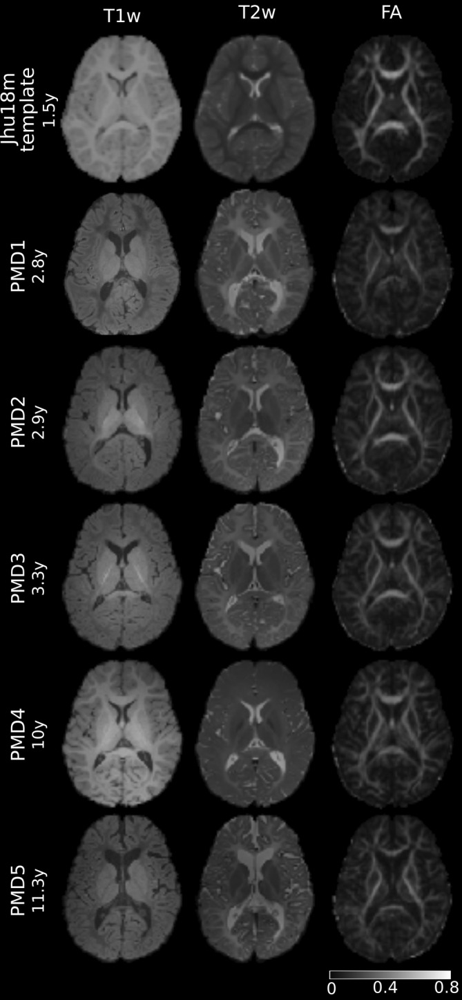
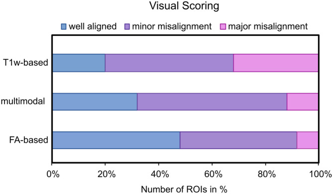
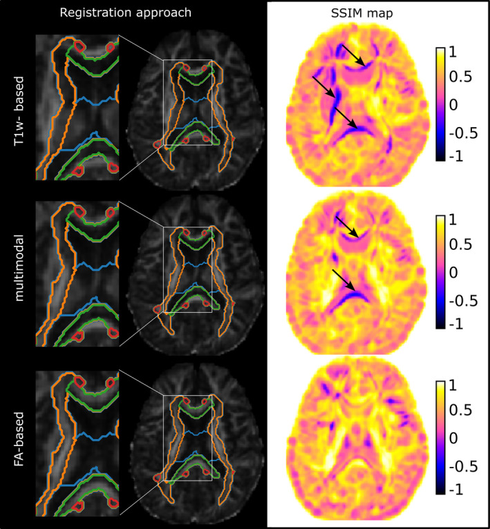
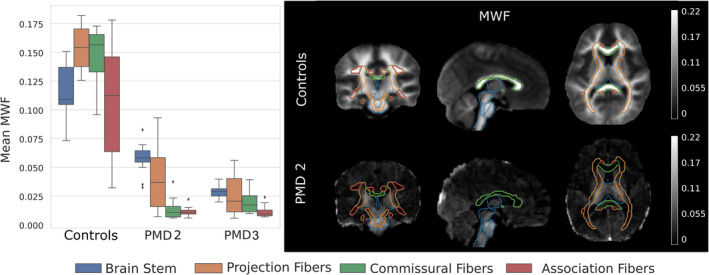
Pelizaeus-Merzbacher disease (PMD) is a rare childhood hypomyelinating leukodystrophy. Quantification of the pronounced myelin deficit and delineation of subtle myelination processes are of high clinical interest. Quantitative magnetic resonance imaging (qMRI) techniques can provide in vivo insights into myelination status, its spatial distribution, and dynamics during brain maturation. They may serve as potential biomarkers to assess the efficacy of myelin-modulating therapies. However, registration techniques for image quantification and statistical comparison of affected pediatric brains, especially those of low or deviant image tissue contrast, with healthy controls are not yet established. This study aimed first to develop and compare postprocessing pipelines for atlas-based quantification of qMRI data in pediatric patients with PMD and evaluate their registration accuracy. Second, to apply an optimized pipeline to investigate spatial myelin deficiency using myelin water imaging (MWI) data from patients with PMD and healthy controls. This retrospective single-center study included five patients with PMD (mean age, 6 years ± 3.8) who underwent conventional brain MRI and diffusion tensor imaging (DTI), with MWI data available for a subset of patients. Three methods of registering PMD images to a pediatric template were investigated. These were based on (a) T1-weighted (T1w) images, (b) fractional anisotropy (FA) maps, and (c) a combination of T1w, T2-weighted, and FA images in a multimodal approach. Registration accuracy was determined by visual inspection and calculated using the structural similarity index method (SSIM). SSIM values for the registration approaches were compared using a t test. Myelin water fraction (MWF) was quantified from MWI data as an assessment of relative myelination. Mean MWF was obtained from two PMDs (mean age, 3.1 years ± 0.3) within four major white matter (WM) pathways of a pediatric atlas and compared to seven healthy controls (mean age, 3 years ± 0.2) using a Mann-Whitney U test. Our results show that visual registration accuracy estimation and computed SSIM were highest for FA-based registration, followed by multimodal, and T1w-based registration (SSIMFA = 0.67 ± 0.04 vs. SSIMmultimodal = 0.60 ± 0.03 vs. SSIMT1 = 0.40 ± 0.14). Mean MWF of patients with PMD within the WM pathways was significantly lower than in healthy controls MWFPMD = 0.0267 ± 0.021 vs. MWFcontrols = 0.1299 ± 0.039. Specifically, MWF was measurable in brain structures known to be myelinated at birth (brainstem) or postnatally (projection fibers) but was scarcely detectable in other brain regions (commissural and association fibers). Taken together, our results indicate that registration accuracy was highest with an FA-based registration pipeline, providing an alternative to conventional T1w-based registration approaches in the case of hypomyelinating leukodystrophies missing normative intrinsic tissue contrasts. The applied atlas-based analysis of MWF data revealed that the extent of spatial myelin deficiency in patients with PMD was most pronounced in commissural and association and to a lesser degree in brainstem and projection pathways.
Keywords: Pelizaeus‐Merzbacher disease; hypomyelinating leukodystrophy; magnetic resonance imaging; medical image registration; neuroimaging; white matter disorder.
© 2024 The Author(s). Human Brain Mapping published by Wiley Periodicals LLC.
Conflict of interest statement
The authors declare no conflicts of interest.
Figures
Similar articles
-
Brain Myelin Water Fraction and Diffusion Tensor Imaging Atlases for 9-10 Year-Old Children.J Neuroimaging. 2020 Mar;30(2):150-160. doi: 10.1111/jon.12689. Epub 2020 Feb 16. J Neuroimaging. 2020. PMID: 32064721
-
The magnetic resonance imaging spectrum of Pelizaeus-Merzbacher disease: A multicenter study of 19 patients.Brain Dev. 2016 Jun;38(6):571-80. doi: 10.1016/j.braindev.2015.12.007. Epub 2016 Jan 13. Brain Dev. 2016. PMID: 26774704
-
Magnetic resonance fingerprinting-based myelin water fraction mapping for the assessment of white matter maturation and integrity in typical development and leukodystrophies.NMR Biomed. 2024 Jun;37(6):e5114. doi: 10.1002/nbm.5114. Epub 2024 Feb 23. NMR Biomed. 2024. PMID: 38390667
-
Inherited white matter disorders: Hypomyelination (myelin disorders).Handb Clin Neurol. 2024;204:197-223. doi: 10.1016/B978-0-323-99209-1.00014-4. Handb Clin Neurol. 2024. PMID: 39322379 Review.
-
Pelizaeus-Merzbacher Disease: Molecular and Cellular Pathologies and Associated Phenotypes.Adv Exp Med Biol. 2019;1190:201-216. doi: 10.1007/978-981-32-9636-7_13. Adv Exp Med Biol. 2019. PMID: 31760646 Review.
Cited by
-
A high-precision hierarchical registration approach for stain- and scanner-independent colocalization on whole slide images in histopathology.Health Inf Sci Syst. 2025 May 23;13(1):38. doi: 10.1007/s13755-025-00353-7. eCollection 2025 Dec. Health Inf Sci Syst. 2025. PMID: 40416515 Free PMC article.
References
-
- Al‐Saady, M. L. , Wolf, N. I. , & Pouwels, P. J. W. (2022). Segmentation of intrinsically very low contrast magnetic resonance brain images using tensor‐based DTI registration. Neuroimage: Reports, 2(4), 100120. 10.1016/j.ynirp.2022.100120 - DOI
-
- Deoni, S. C. L. , Dean, D. C. , O'Muircheartaigh, J. , Dirks, H. , & Jerskey, B. A. (2012). Investigating white matter development in infancy and early childhood using myelin water faction and relaxation time mapping. NeuroImage, 63(3), 1038–1053. 10.1016/j.neuroimage.2012.07.037 - DOI - PMC - PubMed
MeSH terms
Grants and funding
LinkOut - more resources
Full Text Sources
