. 2024 Oct 4;5(4):e230088.
doi: 10.1530/RAF-23-0088. Print 2024 Oct 1.

The efficacy and functional consequences of interactions between human spermatozoa and seminal fluid extracellular vesicles

Affiliations

The efficacy and functional consequences of interactions between human spermatozoa and seminal fluid extracellular vesicles

Cottrell T Tamessar et al. Reprod Fertil. .

Abstract

Abstract: Seminal fluid extracellular vesicles (SFEVs) have previously been shown to interact with spermatozoa and influence their fertilisation capacity. Here, we sought to extend these studies by exploring the functional consequences of SFEV interactions with human spermatozoa. SFEVs were isolated from the seminal fluid of normozoospermic donors prior to assessing the kinetics of sperm-SFEV binding in vitro, as well as the effects of these interactions on sperm capacitation, acrosomal exocytosis, and motility profile. Biotin-labelled SFEV proteins were transferred primarily to the flagellum of spermatozoa within minutes of co-incubation, although additional foci of SFEV biotinylated proteins also labelled the mid-piece and head domain. Functional analyses of high-quality spermatozoa collected following liquefaction revealed that SFEVs did not influence sperm motility during incubation at pH 5, yet SFEVs induced subtle increases in total and progressive motility in sperm incubated with SFEVs at pH 7. Additional investigation of sperm motility kinematic parameters revealed that SFEVs significantly decreased beat cross frequency and increased distance straight line, linearity, straightness, straight line velocity, and wobble. SFEVs did not influence sperm capacitation status or the ability of sperm to undergo acrosomal exocytosis. Functional assessment of both high- and low-quality spermatozoa collected prior to liquefaction showed limited SFEV influence, with these vesicles inducing only subtle decreases in beat cross frequency in spermatozoa of both groups. These findings raise the prospect that, aside from subtle effects on sperm motility, the encapsulated SFEV cargo may be destined for physiological targets other than the male germline, notably the female reproductive tract.

Lay summary: A male's influence over the biological processes of pregnancy extends beyond the provision of sperm. Molecular signals present in the ejaculate can influence the likelihood of pregnancy and healthy pregnancy progression, but the identity and function of these signals remain unclear. In this study, we wanted to understand if nano-sized particles present in the male ejaculate, called seminal fluid extracellular vesicles, can assist sperm in traversing the female reproductive tract to access the egg. To explore this, we isolated seminal fluid extracellular vesicles from human semen and incubated them with sperm. Our data showed that seminal fluid extracellular vesicles act to transfer molecular information to sperm, but this resulted in only subtle changes to the movement of sperm.

Keywords: extracellular vesicles; fertility; seminal fluid; sperm capacitation; sperm motility; spermatozoa.


Conflict of interest statement

BN is a member of the Editorial Board of Reproduction & Fertilitybut was not involved in the peer review process of this paper. All other authors declare no conflicts of interest that could be perceived as prejudicing the impartiality of the research reported.

Figures

Figure 1
Figure 1
Characterisation of human seminal fluid extracellular vesicles. Seminal fluid extracellular vesicles (SFEVs) were isolated using a modified density gradient ultracentrifugation approach. (A) After centrifugation, an image was taken of the gradient prior to the collection of 12 equivalent fractions. These fractions were subjected to (B) quantitative assessment of total protein content and (C) qualitative assessment of the protein profile. Based on this analysis, and the identification of (D) EV-enriched fractions by immunoblotting all fractions for otillin 1 (FLOT1), fractions 9 and 10 were selected and combined for downstream analysis. This analysis included: (E) Immunoblotting to assess the presence of additional recognised EV markers, CD63 molecule, in addition to the absence of the non-EV protein marker apolipoprotein A1 (APOA1) using seminal fluid protein (SF) as a positive control, (F) determination of particle size and heterogeneity using nanoparticle tracking analysis. Data are represented as particle size (x-axis) and particle number (y-axis). (G) Transmission electron microscopy to examine the ultrastructural properties of isolated SFEVs (scale bar = 500 µm). All experiments were repeated using n = 5 independent biological replicates.
Figure 2
Figure 2
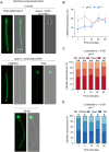
Assessment of seminal fluid extracellular vesicle (SFEV) transfer interaction with human spermatozoa. The localisation of biotinylated SFEV proteins was used as a surrogate to assess the specificity, efficacy, kinetics, and distribution of SFEV interactions with human spermatozoa. For this purpose, isolated SFEVs (i.e. EVs present in the pooled fractions 9 and 10 – see Fig. 1) were labelled with a membrane-impermeable biotin reagent, prior to quenching of the biotinylation reaction and co-incubation with spermatozoa for 60 min (with regular sampling at intervals of 1, 5, 15, 30, and 60 min). Spermatozoa were subsequently affinity-labelled with AlexaFluor 488 conjugated streptavidin. (A) Representative images indicating the localisation patterns obtained following direct biotinylation of spermatozoa (i.e. no SFEVs), sperm incubation with unlabelled SFEVs (i.e. no biotin), and sperm incubation with biotinylated SFEVs (scale bar = 10 µm). (B) The percentage of spermatozoa displaying the uptake of biotinylated proteins and the domains into which these proteins were distributed at (C) pH 5 and (D) pH 7 were determined by examining the fluorescence staining of a minimum of 100 cells per sample. All experiments were repeated using n = 5 independent biological replicates, with representative images being shown and graphical data in panels presented as (B) mean ± s.e.m., and (C and D) stacked bar chart showing mean of each group. Data were assessed by 2-way ANOVA with Tukey’s multiple comparisons test (φ, P < 0.05 comparing tail vs head labelling, ϕ, P < 0.05 comparing tail vs whole cell labelling). Differences in the overall effects of Localisation or Time are presented as text depicting the P value in each panel where a significant difference (P < 0.05) was detected.
Figure 3
Figure 3
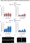
Assessment of the uptake and redistribution of biotinylated seminal fluid extracellular vesicle proteins following co-culture of human spermatozoa. Distinct from the previous experiment, proteins encapsulated within human seminal fluid extracellular vesicles (SFEVs) were labelled with a membrane-permeable derivative of biotin prior to co-incubation with spermatozoa over a time course of 3 h (with regular sampling at 1, 5, 15, 30, 60, and 180 min). Following incubation, spermatozoa were affinity-labelled with AlexaFluor 488 conjugated streptavidin to detect the uptake and fate of SFEV protein cargo. (A) Representative images illustrating the localisation patterns obtained following direct biotinylation of spermatozoa (i.e. no SFEVs), sperm incubation with unlabelled SFEVs (i.e. no biotin), and sperm incubation with SFEVs pre-labelled with membrane-permeable biotin reagent (scale bar = 10 µm). (B) The percentage of spermatozoa displaying the uptake of biotinylated proteins and the domains into which these proteins were distributed at (C) pH 5 and (D) pH 7 was determined by examining the fluorescence staining of a minimum of 100 cells per sample. All experiments were repeated using n = 5 independent biological replicates, with representative images being shown and graphical data in panels presented as (B) mean ±  s.e.m., and (C and D) stacked bar chart showing mean of each group. Data were assessed by 2-way ANOVA with Tukey’s multiple comparisons test (φ, P < 0.05 comparing sperm tail vs head labelling, δ, P < 0.05 comparing whole sperm vs head labelling). Differences in the overall effects of Localisation or Time are presented as text depicting the P value in each panel where a significant difference (P < 0.05) was detected.
Figure 4
Figure 4
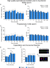
Seminal fluid extracellular vesicles (SFEVs) have minimal influence on the function of spermatozoa. Human spermatozoa were fractionated over a discontinuous Percoll density gradient and high-quality sperm were resuspended in non-capacitating Biggers, Whitten, and Whittingham medium (BWW) buffered to either (A and B) pH 5 or (C and D) pH 7 and incubated for 1, 5, 15, 30, 60, 180, or 300 min. At regular intervals throughout the extended co-incubation, a portion of the sperm suspension was sampled and assessed for (A and C) total motility and (B and D) progressive motility using objective computer-assisted sperm analysis (CASA) counting a minimum of 200 cells per sample. All experiments were repeated using n = 5 independent biological replicates. Graphical data are presented as mean ±  s.e.m. Data were assessed by 2-way ANOVA with Tukey’s multiple comparisons test. Differences in the overall effects of SFEV or time are presented as text depicting the P value in each panel where a significant difference (P < 0.05) was detected. Significant changes at individual time points are depicted using symbols (#, P < 0.05 difference within treatment group compared to earlier time points).
Figure 5
Figure 5
Seminal fluid extracellular vesicles (SFEVs) have minimal influence on spermatozoa motility parameters in a neutral pH (pH 7) environment. Human spermatozoa were fractionated over a discontinuous Percoll density gradient and high-quality sperm were resuspended in non-capacitating Biggers, Whitten, and Whittingham medium (BWW) buffered to pH 7 and incubated for 1, 5, 15, 30, 60, 180, or 300 min with SFEVs as described in Fig. 4. At regular intervals throughout the extended co-incubation, a portion of the sperm suspension was sampled and assessed for motility parameters using objective computer-assisted sperm analysis (CASA) counting a minimum of 200 cells per sample. Parameters assessed included (A) Beat cross frequency (BCF, Hz), (B) Distance straight line (DSL, µm), (C) Linearity (LIN, %), (D) Straightness (STR, %), (E) Straight-line velocity (VSL, µm/second), and (F) Wobble (WOB, %). All experiments were repeated using n = 5 independent biological replicates. Graphical data are presented as mean ±  s.e.m. Data were assessed by 2-way ANOVA with Tukey’s multiple comparisons test. Differences in the overall effects of SFEV or Time are presented as text depicting the P value in each panel where a significant difference (P < 0.05) was detected. Significant changes at individual time points are depicted using symbols (*P < 0.05 vs same time point comparing sperm: SFEV and sperm alone groups).
Figure 6
Figure 6
Seminal fluid extracellular vesicles (SFEVs) do not influence spermatozoa progressive motility parameters in a neutral pH (pH 7) environment. Human spermatozoa were fractionated over a discontinuous Percoll density gradient and high-quality sperm were resuspended in non-capacitating Biggers, Whitten, and Whittingham medium (BWW) buffered to pH 7 and incubated for 1, 5, 15, 30, 60, 180, or 300 min with SFEVs as described in Fig. 4. At regular intervals throughout the extended co-incubation, a portion of the sperm suspension was sampled and assessed for progressive motility kinetic parameters using objective computer-assisted sperm analysis (CASA) counting a minimum of 200 cells per sample. Parameters assessed included (A) Beat cross frequency (BCF, Hz), (B) Distance straight line (DSL, µm), (C) Linearity (LIN, %), (D) Straightness (STR, %), (E) Straight-line velocity (VSL, µm/second), and (F) Wobble (WOB, %). All experiments were repeated using n = 5 independent biological replicates. Graphical data are presented as mean ±  s.e.m. Data were assessed by 2-way ANOVA with Tukey’s multiple comparisons test. Differences in the overall effects of SFEVs or time are presented as text depicting the P value in each panel where a significant difference (P < 0.05) was detected.
Figure 7
Figure 7
Seminal fluid extracellular vesicles (SFEVs) do not influence capacitation or acrosome reaction rates of spermatozoa isolated from normozoospermic donors. Human spermatozoa were fractionated over a discontinuous Percoll density gradient and high-quality sperm were resuspended in non-capacitating Biggers, Whitten, and Whittingham medium (BWW) buffered to either (A and B) pH 5 or (C and D) pH 7 and incubated for 1, 60, 180, or 300 min. At regular intervals throughout the extended co-incubation, a portion of the sperm suspension was sampled and capacitation status and the ability to complete an acrosome reaction were assessed using immunofluorescence-based assays. (A and C) Sperm were immunostained with anti-phosphotyrosine antibodies to determine capacitation status, or (B and D) incubated with the calcium ionophore A23187 to assess their capacity to undergo an ionophore-induced acrosome reaction; with representative images depicted from positive and negative controls for (E) capacitation (pentoxifylline-driven) and (F) acrosome reaction (A23187-driven following capacitation). Graphical data are presented as mean ±  s.e.m. Data were assessed by 2-way ANOVA with Tukey’s multiple comparisons test. Differences in the overall effects of SFEVs or time are presented as text depicting the P value in each panel where a significant difference (P < 0.05) was detected.
Figure 8
Figure 8
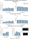
Seminal fluid extracellular vesicles (SFEVs) have minimal influence on the functional parameters of high-quality sperm collected prior to liquefaction. Human spermatozoa collected prior to liquefaction were fractionated over a discontinuous Percoll density gradient and high-quality sperm were resuspended in non-capacitating Biggers, Whitten, and Whittingham medium (BWW) buffered to pH 7 and incubated for a period of 300 min. At regular intervals throughout the extended co-incubation, a portion of the sperm suspension was sampled and assessed for motility parameters (A–D), capacitation status (E and G), and ability to complete an acrosome reaction (F and H). Assessment of sperm parameters utilised objective computer-assisted sperm analysis (CASA), counting a minimum of 200 cells per sample. Data presented are (A) Total sperm motility (%), (B) Beat cross frequency (BCF, Hz) of the motile sperm population, (C) Progressive sperm motility (%), and (D) BCF (Hz) of the progressively motile sperm population. Assessment of capacitation status and the ability to complete an acrosome reaction used immunofluorescence-based assays. (E and G) Sperm were immunostained with anti-phosphotyrosine antibodies to determine capacitation status, or (F and H) incubated with the calcium ionophore A23187 to assess their capacity to undergo an ionophore-induced acrosome reaction; with representative images depicted from positive and negative controls for (G) capacitation (pentoxifylline-driven) and (H) acrosome reaction (A23187-driven following capacitation). Graphical data are presented as mean ±  s.e.m. Data were assessed by 2-way ANOVA with Tukey’s multiple comparisons test. Differences in the overall effects of SFEV or Time are presented as text depicting the P value in each panel where a significant difference (P < 0.05) was detected. Significant changes at individual time points are depicted using symbols (#, P < 0.05 difference within treatment group compared to the 1-mintime point).
Figure 9
Figure 9
Seminal fluid extracellular vesicles (SFEVs) have minimal influence on the functional parameters of low-quality sperm collected prior to liquefaction. Human spermatozoa collected prior to liquefaction were fractionated over a discontinuous Percoll density gradient and low-quality sperm were resuspended in non-capacitating Biggers, Whitten, and Whittingham medium (BWW) buffered to pH 7 and incubated for a period of 300 min. At regular intervals throughout the extended co-incubation, a portion of the sperm suspension was sampled and assessed for motility parameters (A–D), capacitation status (E and G), and ability to complete an acrosome reaction (F and H). Assessment of sperm parameters utilised objective computer-assisted sperm analysis (CASA), counting a minimum of 200 cells per sample. Data presented are (A) Total sperm motility (%), (B) Beat cross frequency (BCF, Hz) of the motile sperm population, (C) Progressive sperm motility (%), and (D) BCF (Hz) of the progressively motile sperm population. Assessment of capacitation status and the ability to complete an acrosome reaction used immunofluorescence-based assays. (E and G) Sperm were immunostained with anti-phosphotyrosine antibodies to determine capacitation status, or (F and H) incubated with the calcium ionophore A23187 to assess their capacity to undergo an ionophore-induced acrosome reaction; with representative images depicted from positive and negative controls for (G) capacitation (pentoxifylline-driven) and (H) acrosome reaction (A23187-driven following capacitation). Graphical data are presented as mean ±  s.e.m. Data were assessed by 2-way ANOVA with Tukey’s multiple comparisons test. Differences in the overall effects of SFEV or Time are presented as text depicting the P value in each panel where a significant difference (P < 0.05) was detected.

References

    1. Aalberts M Van Dissel-Emiliani FMF Van Adrichem NPH Van Wijnen M Wauben MHM Stout TAE & Stoorvogel W. 2012Identification of distinct populations of prostasomes that differentially express prostate stem cell antigen, annexin A1, and GLIPR2 in humans. Biology of Reproduction 8682. (10.1095/biolreprod.111.095760) - DOI - PubMed
    1. Aalberts M Sostaric E Wubbolts R Wauben MWM Nolte-'T Hoen ENM Gadella BM Stout TAE & Stoorvogel W. 2013Spermatozoa recruit prostasomes in response to capacitation induction. Biochimica et Biophysica Acta 18342326–2335. (10.1016/j.bbapap.2012.08.008) - DOI - PubMed
    1. Aalberts M Stout TAE & Stoorvogel W. 2014Prostasomes: extracellular vesicles from the prostate. Reproduction 147R1–R14. (10.1530/REP-13-0358) - DOI - PubMed
    1. Aitken RJ Harkiss D & Buckingham D. 1993Relationship between iron-catalysed lipid peroxidation potential and human sperm function. Journal of Reproduction and Fertility 98257–265. (10.1530/jrf.0.0980257) - DOI - PubMed
    1. Alghamdi AS Foster DN & Troedsson MH. 2004Equine seminal plasma reduces sperm binding to polymorphonuclear neutrophils (PMNs) and improves the fertility of fresh semen inseminated into inflamed uteri. Reproduction 127593–600. (10.1530/rep.1.00096) - DOI - PubMed

LinkOut - more resources